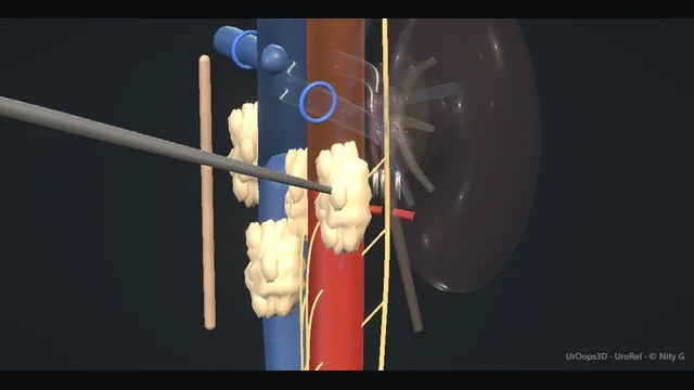
A midline exposure of the retroperitoneum with the dissection done directly on the walls of the aorta and IVC, where the short posterior lumbar veins are the named pitfall.
The sympathetic chains run longitudinal and lateral through the same field: preserving them preserves ejaculation, and clearing the residual mass clears the disease, and the operation is the discipline of doing both in one plane.
Advanced tierFRCS oncology, testicular cancerInteractive 3D
Midline retroperitoneum, the corridor between the aorta and the cava. Schematic, not to scale; relationships only.
The retroperitoneum is a corridor between two vertical vessels and a vertebral column behind them.
The aorta sits left of the midline, the cava right, and the lumbar veins bridge from the cava's posterior wall to the vertebrae at each level, tethering both the vessel and the dissection.
The sympathetic chains run parallel to the vessels, lateral and slightly posterior, their ganglia at segmental intervals, and the postganglionic fibres cross the nodal packet anteromedially toward the hypogastric plexus.
The residual mass sits where the nodes sit: paraaortic, interaortocaval and precaval, wrapped onto the vessel walls.
The ureters mark the lateral limits, the renal veins the cranial limit, and the bifurcation the caudal one.
Every step of the operation is a relationship to a vessel wall.
›Aorta and IVC - the twin columns the dissection rolls on; everything else in the field is managed relative to their walls.
›Lumbar veins - short posterior tributaries of the cava at each vertebral level; the named pitfall of the operation.
›Sympathetic chains - run longitudinal and lateral with segmental ganglia; carry the ejaculatory reflex and fertility with them.
›Residual mass - post-chemotherapy tissue, often fused to the vessel wall, containing teratoma, necrosis or viable cancer until proven otherwise.
›Ureters and gonadal vein - cross the template margins laterally; identified early and kept out of the dissection.
Midline exposureA midline laparotomy gives the working corridor, with self-retaining retraction and the small bowel packed high and wrapped out of the field. The posterior peritoneum is incised lateral to the duodenum and a Kocher-style manoeuvre with right colonic mobilisation lifts the duodenum and colon off the great vessels. The exposure is complete only when the aorta and cava stand bare from the renal veins above to the bifurcation below, with the mass visible along its whole length before any dissection starts. An exposure that stops short of the renal veins tempts the dissection to approach the mass from the side, which is where the dangerous bleeding lives. where it bites: an exposure that stops short of the renal veins tempts the dissection into the mass from the side rather than over the vessels.
Template boundariesThe template is written down before a vessel is touched, from the markers and the imaging, and its boundaries are named to the team: renal veins above, ureters laterally, bifurcation and iliac origins below. Full bilateral clearance is the default for post-chemotherapy residual masses and for bulky or bilateral disease. Modified unilateral templates, which spare the contralateral field to protect the sympathetic chains, are reserved for selected primary cases in centres that do this work regularly. The point of a fixed template is that it cannot be negotiated downward in theatre: a clearance drawn around what looks involved on the scan misses exactly the microscopic disease the template exists to clear. Before a packet is lifted, the sympathetic chains are identified running longitudinal and lateral to the great vessels, their ganglia noted, and the postganglionic fibres picked up as fine threads crossing the packet anteromedially, so that what follows rolls the nodes out from beneath them. where it bites: a template drawn around what is visible on the scan misses the microscopic disease that templates exist to clear.
Dissection on the aortaThe packet is opened on the anterior wall of the aorta and split laterally, so the nodal tissue can be rolled off the vessel in sheets under gentle traction. Each lumbar artery met on the posterior wall is clipped or ligated close to its origin before division, because an artery pulled on rather than controlled avulses at the aorta and retracts toward its foramen. The inferior mesenteric artery is preserved and confirmed pulsatile at the end of the case unless the template or the mass demands its sacrifice. Throughout, the dissection stays on the adventitia of the vessel and never in it, and anything crossing the field is identified before it is divided. where it bites: the lumbar arteries tether the aorta: an artery pulled instead of controlled avulses at its origin.
Dissection on the IVCThe cava is rolled with the same split-and-roll discipline, the packet stripped from its anterior and lateral walls with traction and sharp division of attachments. The difference is on the back wall: each lumbar vein is short, enters almost at right angles, and must be isolated and controlled with a clip or tie before any traction is put on the packet above it. The veins are divided between controlled ties, never avulsed by pulling the packet off them. This is the step where the named pitfall of the operation lives: a traction tear at the posterior cava bleeds from exactly the position where fingers and instruments reach worst. where it bites: the lumbar veins are short, posterior, and enter the cava at its back wall: traction tears a hole exactly where fingers and staples reach worst.
Sympathetic chain preservationThe chains and fibres identified at the start, before any packet was lifted, are kept undisturbed while the nodal tissue is rolled out from beneath them, freed with fine instruments where they cross the packet. Preservation is attempted field by field wherever the disease allows, and the side and status of each chain is recorded for the note. The failure mode is a chain sacrificed incidentally inside a fibrotic post-chemotherapy packet, which takes ejaculation with it, and against that the only real mitigation, sperm banking before chemotherapy, belongs months earlier on the pathway and cannot be retrofitted. where it bites: the chain sacrificed with a bulky post-chemotherapy mass takes ejaculation with it; sperm banking before chemotherapy is the mitigation that cannot be retrofitted.
Residual mass out intactThe residual mass and every nodal packet are removed as intact specimens, taken off the vessel walls in the subadventitial plane where post-chemotherapy fibrosis has fused them down. At the end of the clearance the field is checked in order: haemostasis on the vessel walls, lymphostasis with clips or energy on the divided channels at the suprarenal apex and over the vertebrae, ureters confirmed intact and peristalsing, and the bowel returned without torsion. Counts, drains and the integrity of any repaired wall are confirmed before closure. Teratoma left on a vessel wall grows slowly, transforms sometimes, and makes any second operation far harder than the first, which is why the clearance is complete or is consciously incomplete and documented as such. where it bites: residual teratoma left on a vessel wall grows slowly, transforms sometimes, and makes the second operation far harder than the first.
The spatial relationships the animation is built to make automatic. Before you read each one, ask yourself: at this point in the operation, what is about to go wrong, and what is my first move? The save for each sits in the Complications tab.
RiskLumbar veins at the cava's back wall - short, posterior, and avulsed by traction rather than division; the classic great-vessel injury of RPLND.
RiskSympathetic chains - the fertility of a young man runs through two golden threads in the same field as the cancer.
RiskMass adherent to the vessel wall - post-chemotherapy fibrosis blurs the plane; the dissection is subadventitial when it has to be, and never through the wall.
Trigger each of these live in the Animation tab, then read the save: the first move, the escalation trigger, and the line that goes in the op note.
Great vessel injury (lumbar vein tear)
a lumbar vein avulsed from the posterior cava during rolling; haemorrhage is brisk and the hole is in the least reachable part of the vessel.
The save
Immediate proximal and distal compression of the cava with swabs and the field kept filled: do not suck the vessel empty and do not attempt blind clips.first move
Tell the anaesthetist, call for vascular help, blood and autotransfusion capability, and convert or extend the incision as needed for control.
Control the vessel properly: vascular clamp or sponge-stick compression above and below the injury, then a direct sutured repair of the cava wall.
Ligate the avulsed lumbar vein stump; a lumbar vein can be sacrificed, the cava wall cannot.
Uncontrolled haemorrhage, haemodynamic collapse or a retrohepatic extension → senior vascular assistance and damage control packing before any definitive repair.escalation trigger
Op note: vessel injured, level, repair method, estimated loss and products given.document
Chylous ascites
lymphatic channels divided at the paraaortic apex and over the vertebrae leak chyle; ascites accumulates over the first weeks with distension, weight loss and low albumin.
The save
Prevention at surgery: clips or energy on the lymphatics at the suprarenal apex and over the vertebrae before division, and a low-pressure check at the end.first move
Recognise it early after surgery: increasing abdominal girth, full drains with milky fluid, falling albumin; send the fluid, which settles the diagnosis.
First-line treatment is drainage plus a fat-restricted or medium-chain triglyceride diet and nutritional support, continued patiently.
Image the drainage path and exclude obstruction; maintain drainage rather than repeated paracentesis alone.
Persistent high-output leak beyond the expected course, worsening nutrition or sepsis → radiological lymphatic intervention or re-operation for ligation.escalation trigger
Note: lymphostasis achieved at surgery, drain outputs, diet and the response to it.document
Ejaculatory loss
one or both sympathetic chains sacrificed or stripped with the packet; ejaculation is lost, and fertility with it unless sperm was banked before chemotherapy.
The save
Prevention long before theatre: sperm banking discussed at diagnosis, before chemotherapy, in every young man with testicular cancer.first move
At surgery, identify the chains early and plan the dissection to keep them: pick up the postganglionic fibres as they cross the packet.
When the mass engulfs a chain, oncological clearance comes first: the operation is for cancer, and the decision is made deliberately, not by accident.
Counsel the patient honestly afterwards about dry ejaculation and its distinction from erectile function and orgasm, both of which are usually preserved.
Desired fertility with no banked sperm → semen analysis and andrology referral for assisted reproduction or sperm retrieval.escalation trigger
Note: chains preserved or sacrificed, side, counselling given and fertility status before treatment.document
When the case turns: the problem, then the next move.
ActionThe mass is fused to the aortic wall - dissect in the subadventitial plane deliberately; a controlled, repairable adventitial tear beats a trail through tumour.
ActionA lumbar vein avulses as the cava is rolled - compression first, control above and below, then a direct sutured repair; never a blind clip into a pool.
ActionThe ureter disappears into the packet - go lateral and find it on psoas away from the disease, then follow it back; it is sturdier at a distance than in the fibrosis.
ActionMilky fluid wells up from the apex of the field - stop, suck dry, clip or energy the leak point until dry, and re-check at the end of the case with pressure down.
The examiner stems this scene prepares you for. Try to answer out loud first, then reveal, and learn the discriminator, the detail that turns a pass into a strong pass.
QWhich template would you use for which disease state?reveal answer
The template follows the disease and the treatment already given.
For primary RPLND in selected clinical stage I disease with risk factors, and for residual masses after chemotherapy, the modern default in most UK practice is chemotherapy for metastatic disease and surgery for the residue, so the operation I most often describe is the post-chemotherapy bilateral template: both paraaortic gutters, the interaortocaval and precaval zones, from the renal veins above to the bifurcation and iliac origins below, laterally to the ureters, with the contralateral boundary extended when the disease was right-sided, because right-sided tumours involve the interaortocaval and left para-aortic nodes more often than left-sided tumours cross to the right.
Modified unilateral templates, sparing the contralateral field to protect ejaculation, are legitimate in experienced centres for low-volume primary disease with negative markers.
For bulky residual masses, re-do surgery and post-chemotherapy fibrosis, the clearance is full bilateral and complete, because the cost of leaving disease behind exceeds the cost of the extra dissection.
The inferior mesenteric artery is taken if the template demands it, and the kidneys and ureters are boundaries, not targets.
The principle I would state is that the template is written before the incision, on imaging and markers, and not negotiated downward during the operation because the field looks clean.
Strong pass
Naming the boundaries in order, renal veins, ureters, bifurcation, and the left-to-right relapse logic that pushes left-sided templates across the midline.
QWhy does residual teratoma have to come out?reveal answer
Because chemotherapy does not touch it.
Teratoma is chemoresistant by nature: it is mature or immature differentiated tissue, not dividing rapidly enough for cytotoxic agents to matter, so after a cycle of chemotherapy for mixed germ cell tumour the teratoma component remains exactly as it started while the sensitive elements melt away.
The consequence is the clinical problem.
First, it grows: what was a small residual mass enlarges as the teratomatous element expands, and growing retroperitoneal tissue causes pain, obstruction of the ureter or bowel, and vascular compression.
Second, it transforms: malignant transformation into somatic cancer, sarcoma most often, is a recognised fate of untreated residual teratoma, and that disease behaves like the transformed histology, not like testicular cancer, with far worse survival.
Third, it hides: viable cancer can sit beside teratoma in the same residual mass, and imaging cannot tell necrosis, fibrosis, teratoma and viable tumour apart, which is precisely why residual masses of meaningful size are resected and examined rather than observed.
The operation is therefore not a biopsy of a lump; it is the definitive local therapy for chemotherapy-resistant disease, timed after markers have normalised.
That is the argument for complete excision with clear intent, in a centre that does them regularly.
Strong pass
Naming malignant transformation and the imaging impossibility of distinguishing necrosis from teratoma or viable tumour as the two drivers for resection.
QIs nerve-sparing RPLND feasible after chemotherapy?reveal answer
Feasible, but harder, and it depends entirely on the relationship between the mass and the chains.
In primary RPLND with low-volume disease the chains are usually identifiable, and deliberate nerve-sparing preserves ejaculation in the great majority, because the postganglionic fibres cross the nodal tissue in a plane that can be developed.
After chemotherapy the field is fibrotic: the mass and the desmoplastic reaction stick the chains, the fibres and the vessel walls together, and the dissection that spares every golden thread risks leaving disease on it.
So the decision is made at operation, field by field: where the chain can be freed without compromising clearance of the mass, it is freed, with sharp dissection directly on the vessel and the nerve;
where the mass engulfs the chain, the chain goes, because the operation is for cancer and a relapse in the retroperitoneum after failed nerve-sparing is a poor trade for dry ejaculation.
Patient factors matter as much: fertility intent, sperm banked or not, and contralateral function.
What is not acceptable is losing a chain by accident through not looking for it; the identification should be deliberate at the start, so that sacrifice, when it happens, is a decision.
And the mitigation belongs to the oncology pathway, not the operation: sperm banking before chemotherapy for every young man.
Strong pass
Say both
Framing nerve-sparing as a field-by-field intra-operative decision made after deliberate identification
With cancer clearance as the fixed constraint and sperm banking as the pathway mitigation
Examiner note
Framing nerve-sparing as a field-by-field intra-operative decision made after deliberate identification, with cancer clearance as the fixed constraint and sperm banking as the pathway mitigation.
QYou are rolling the cava and a lumbar vein avulses. Talk me through the next two minutes.reveal answer
The first move is tamponade, not sight: a swab and finger pressure over the bleeding point and the retroperitoneum kept filled, because the injury is on the posterior wall and suction simply empties the field while the patient bleeds.
I tell the anaesthetist immediately, in words, that this is a caval injury, call for help including vascular surgery, and ask for blood, products and autotransfusion capability.
With pressure holding, I improve access: extend the incision or convert, pack the small bowel fully out of the way, and get suction into the field.
Then control: sponge-stick or vascular clamp compression above and below the injury, or a side-biting clamp on the cava, exposing just enough of the posterior wall to repair.
The repair is a direct sutured closure of the cava, and the avulsed lumbar vein stump is ligated or oversewn; a lumbar vein can always be sacrificed, the cava wall cannot.
If the tear runs retrohepatic or control fails, the operation becomes damage control: packing, resuscitation, and a definitive plan with vascular help, either then or at a planned second look.
Throughout, someone records loss and products.
The op note records the vessel, the level, the repair and the physiology.
What never happens in that two minutes is a blind clip into a pool of blood.
Strong pass
The sequence
Examiner note
The sequence: tamponade with the field kept full, announce and mobilise resources, proximal and distal control, then a direct sutured repair, with ligation of the vein and sacrifice of nothing on the cava wall.
Say it aloud, then reveal. Reading is not remembering.
01The relationshipSay the critical spatial relationship out loud, then reveal.reveal
Memory frame
The retroperitoneum is a corridor between two vertical vessels and a vertebral column behind them.
Why?
The aorta sits left of the midline, the cava right, and the lumbar veins bridge from the cava's posterior wall to the vertebrae at each level, tethering both the vessel and the dissection. The sympathetic chains run parallel to the vessels, lateral and slightly posterior, their ganglia at segmental intervals, and the postganglionic fibres cross the nodal packet anteromedially toward the hypogastric plexus. The residual mass sits where the nodes sit: paraaortic, interaortocaval and precaval, wrapped onto the vessel walls. The ureters mark the lateral limits, the renal veins the cranial limit, and the bifurcation the caudal one. Every step of the operation is a relationship to a vessel wall.
02The first moveGreat vessel injury (lumbar vein tear). Your first move?reveal
First move
Immediate proximal and distal compression of the cava with swabs and the field kept filled: do not suck the vessel empty and do not attempt blind clips.
03The escalation triggerGreat vessel injury (lumbar vein tear). What makes you call for help?reveal
Escalate when
Uncontrolled haemorrhage, haemodynamic collapse or a retrohepatic extension → senior vascular assistance and damage control packing before any definitive repair.
04The discriminatorWhat turns a pass into a strong pass?reveal
Strong pass
Naming the boundaries in order, renal veins, ureters, bifurcation, and the left-to-right relapse logic that pushes left-sided templates across the midline.
05The pitfallWhere does this operation bite first?reveal
Pitfall
Lumbar veins at the cava's back wall
Why?
short, posterior, and avulsed by traction rather than division; the classic great-vessel injury of RPLND.
Revision practice with self-marked answers, not an assessment of clinical competence. Kept in
this browser only; it does not sync between devices.