UrOops3D, danger-zone atlas

Schematic model.

Operative animation, Scrotal trauma exploration

Scrotal and testicular trauma: exploration, debridement and tunical repair

Exploration of a blunt scrotal injury with a ruptured testis. Ultrasound shows a heterogeneous testis that has lost its outline.

Through a midline raphe incision the haematocele is evacuated, the testis delivered, the extruded non-viable tubules trimmed and the tunica albuginea closed. The lesson is time: a rupture explored early is usually saved, and one left for days is often lost.

Core tier FRCS emergency urology, genital trauma and testicular salvage Interactive 3D

Operative visualisation

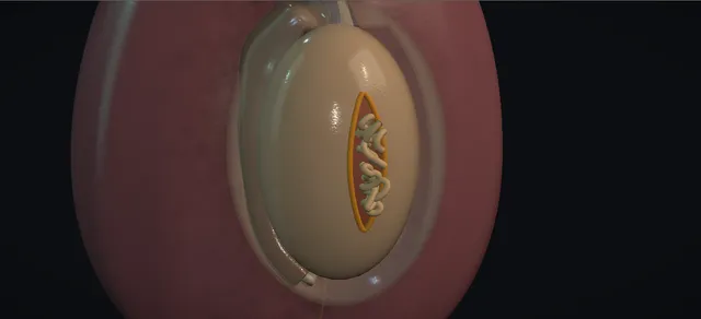
Schematic scene preview: Scrotal trauma: exploration
Preparing the 3D anatomy…

UrOops3D · UroRef · © Nity G

How to use this module. Play through the eight steps, or use Back and Next. Watch the scan, the haematocele cleared, the tear with its extruded tubules, the trim and the running closure. The triggers show a haematoma, an abscess, atrophy after delay, a testis that cannot be saved and a tunica vaginalis flap.

Click me next Section 2 of 8 Anatomy The structures this operation works between, and the one it must not cross.

The structures in the scene

Scrotal trauma exploration: how the structures relateThe ruptured testis, and what lies round it. Tear in the tunica albuginea: stand out through it; trimmed flush, no deeper Extruded seminiferous tubules. Tear in the tunica albuginea: bleeds into it; clot evacuated, flap if needed Tunica vaginalis and its haematocele. Tear in the tunica albuginea: enters from above and behind; never clamped blind Testicular vessels and spermatic cord (danger). Tear in the tunica albuginea: posterolateral; checked before the testis goes back Vas deferens and epididymis (danger). Tear in the tunica albuginea: the same blow can injure it; checked first Urethra and perineal tissues (danger). Tear in the tunica albuginea: kept if it bleeds; closed in layers over it Bruised scrotal skin margins.Tear in the tunicaalbugineaExtrudedseminiferous tubulesTunica vaginalis andits haematocele!Testicular vesselsand spermatic cord!Vas deferens andepididymis!Urethra and perinealtissuesBruised scrotal skinmarginsstand out throughit; trimmed flush,no deeperbleeds into it;clot evacuated,flap if neededenters from aboveand behind; neverclamped blindposterolateral;checked before thetestis goes backthe same blow caninjure it; checkedfirstkept if it bleeds;closed in layersover itTargetDangerLandmarkContext
The ruptured testis, and what lies round it. Schematic, not to scale; relationships only.
  • Seen from in front of a supine patient, each testis hangs in its own hemiscrotum, wrapped in the tunica vaginalis, with the epididymis on its posterolateral border and the cord entering from above and behind.
  • In this injury the right tunica vaginalis is distended with blood round the testis, and the tear in the tunica albuginea faces forward.
  • A midline raphe incision reaches the right testis and lets the left be seen through the same wound.
  • ›Tunica albuginea - the tough white capsule of the testis; its tear is the rupture, and its closure is the repair.
  • ›Seminiferous tubules - the soft parenchyma inside the tunica; they extrude through a tear and the extruded part is non-viable.
  • ›Tunica vaginalis - the serous sac round the testis; blood within it is the haematocele, and its parietal layer can be raised as a flap.
  • ›Spermatic cord - the testicular artery, the pampiniform veins and the vas, entering the testis from above and behind.
  • ›Epididymis - posterolateral on the testis, head above and tail below; it can tear with the testis.
  • ›Scrotal skin and dartos - thin, mobile and vascular; bruised after trauma, and closed in layers.
  • ›Median raphe - the midline ridge of the scrotum; an incision there reaches both hemiscrota.
  • ›Urethra and perineum - below and behind the scrotum; checked in every scrotal injury.
Click me next Section 3 of 8 Steps The operation in order, with the landmark that guards each move.

Operative steps

  1. Assessment and ultrasound A blunt blow: the right hemiscrotum is tense, swollen and bruised. The mechanism is recorded, other injuries are sought, and the meatus, perineum and voiding are checked for a urethral injury. Ultrasound shows a heterogeneous testis that has lost its outline: rupture, so he goes to theatre.
    where it bites: blood at the meatus, a perineal bruise or retention means a urethral injury as well; it changes the catheter plan.
  2. Midline raphe incision Under general anaesthesia, supine, prepared and draped with a time-out. A midline raphe incision is made through skin and dartos, and diathermy controls the edge vessels. One midline wound reaches both hemiscrota, so the other testis can be seen through it. The tunica vaginalis beneath is tense and dark with blood.
    where it bites: the dartos edges bleed; every point controlled now is a haematoma not seen tomorrow.
  3. Open the tunica vaginalis, evacuate the haematocele The tunica vaginalis is opened down the front of the testis. The haematocele is evacuated: clot is sucked and lifted out and the cavity is washed until the testis can be seen. Nothing is clamped blind in the clot.
    where it bites: the cord enters from above and behind; blind clamping in clot can take the testicular vessels.
  4. Deliver and inspect The testis is delivered through the wound on its cord and inspected all round. The rupture is a tear in the tunica albuginea with seminiferous tubules extruded through it. Its length, the colour of the parenchyma and any bleeding from it are noted.
    where it bites: the cord and the vas come out with the testis; traction on them is kept gentle.
  5. Debride non-viable tubules The extruded, non-viable tubules are trimmed with fine scissors, flush with the edge of the tear. Viable parenchyma inside the testis is kept, however untidy it looks. Bleeding points on the cut surface are controlled with gentle bipolar diathermy.
    where it bites: debridement is of extruded tissue only; overenthusiastic excision costs testicular volume.
  6. Close the tunica albuginea The tunica albuginea is closed with a fine absorbable running suture, from one end of the tear to the other, so that the testis is round and firm again. When the defect is too wide to close without tension, a flap of tunica vaginalis is sewn over it instead.
    where it bites: a closure under tension crushes the parenchyma beneath it; a flap is better than a tight closure.
  7. Check cord and epididymis, return Before the testis goes back the epididymis is checked along its length and the cord is checked for haematoma and injury to the vessels and the vas. The testis is returned to the scrotum the right way round. The tunica vaginalis is closed loosely and the other testis is inspected through the same wound.
    where it bites: an epididymal tear or a cord haematoma is easy to miss with attention on the testis.
  8. Layered closure and follow-up The dartos and then the skin are closed with absorbable sutures, and a dressing and a scrotal support are applied. No drain is used after a dry field. Follow-up includes examination for size and consistency, and a discussion of fertility and hormone function.
    where it bites: the bruised skin edges must be viable; non-viable skin is trimmed back before closure.
Click me next Section 4 of 8 Danger zones Where this operation bites, and why it bites there.
Critical relationship

Danger zones

The spatial relationships the animation is built to make automatic. Before you read each one, ask yourself: at this point in the operation, what is about to go wrong, and what is my first move? The save for each sits in the Complications tab.

  • RiskTesticular vessels and spermatic cord - above and behind the testis; clot is evacuated and never clamped blind, and traction on the delivered cord is gentle.
  • RiskVas deferens and epididymis - posterior and posterolateral; checked along their length before the testis goes back.
  • RiskUrethra and perineal tissues - a scrotal blow can injure the urethra; the meatus, perineum and voiding are checked before theatre.
  • RiskViable scrotal skin margins - bruised skin is kept if it bleeds; only non-viable skin is trimmed before closure.
  • RiskTime to exploration - a rupture or a large haematocele explored early is usually saved; delay costs testes.
Practise it Take me into the scene Opens the animation at a danger moment and asks you to predict the mechanism and your first move before it reveals.
Click me next Section 5 of 8 Complications The save for each one: the first move, the escalation trigger, the op-note line.

Complications and immediate saves

Trigger each of these live in the Animation tab, then read the save: the first move, the escalation trigger, and the line that goes in the op note.

Postoperative haematoma

  • the next day the right hemiscrotum is tense and swollen again: a bleeding point in the dartos or on the testis has bled into the scrotum.
The save
  1. Examine him: a soft, stable swelling is managed with a support, ice and analgesia, and reviewed. first move
  2. Check the blood count and any anticoagulant or antiplatelet he takes, with advice from whoever prescribed it.
  3. A tense, expanding or very painful haematoma is a senior review now; it may need re-exploration, evacuation and haemostasis. escalation trigger
  4. Record the size and progress of the swelling, the plan, and what he has been told. document

Delayed infection or abscess

  • days later the hemiscrotum is red, hot and fluctuant, with fever: an infected haematoma or an abscess.
The save
  1. Examine, take blood cultures and start antibiotics by the local protocol. first move
  2. Ultrasound to confirm a collection and to look at the testis.
  3. A collection needs drainage and washout in theatre, by or with a senior; a non-viable testis found then may need orchidectomy. escalation trigger
  4. Record the findings, the cultures sent and the plan, and tell him what the testis looked like. document

Testicular atrophy after late or no exploration

  • a rupture managed without surgery or explored late leaves a small, soft testis months on; the delayed orchidectomies in the ultrasound series were in men who presented late.
The save
  1. Prevention is the save: rupture or an inconclusive scan means exploration, without waiting. first move
  2. For the atrophic testis, examine both testes and check hormone function if there is doubt about the other.
  3. A painful atrophic testis, or a suspicious one on ultrasound, goes to a senior for a decision about orchidectomy. escalation trigger
  4. Record the timing of the injury, of presentation and of any surgery, and what he has been told. document

Testis cannot be saved

  • the testis is shattered and dusky, too little tunica is left to close it, and it is not reconstructable.
The save
  1. Confirm that the testis cannot be reconstructed, if necessary with a senior in theatre. first move
  2. Orchidectomy: the cord is clamped, transfixed and divided, and the field made dry.
  3. If he is unstable or reconstruction cannot be achieved, orchidectomy is the right operation, not a failure. escalation trigger
  4. Record the findings that made it unsalvageable, and plan to discuss a testicular prosthesis later and the other testis now. document
Click me next Section 6 of 8 Troubleshooting When the case turns: the problem, then the next move.

Intra-operative troubleshooting

When the case turns: the problem, then the next move.

  • ActionHaematocele without rupture - the EAU guideline manages a haematocele smaller than three times the other testis conservatively, and explores a larger one even without contusion or rupture, because non-operative care often fails.
  • ActionInconclusive ultrasound - an equivocal scan is not reassurance: the guideline recommends exploration when imaging cannot exclude rupture.
  • ActionDefect too wide to close - a flap of the patient's own tunica vaginalis is raised and sewn over the defect, keeping the viable testis.
  • ActionPenetrating injury - the same steps, compressed: explore, debride non-viable tissue, close the tunica or use a flap, and check both testes, as both are injured in about 30 per cent. A divided vas is tied and repaired later.
  • ActionLarge loss of scrotal skin - the scrotum is elastic and most defects close primarily after washout; extensive loss needs staged reconstruction, with the testes protected meanwhile.
  • ActionTestis dislocated out of the scrotum - a dislocated testis is replaced by hand and fixed later; if it cannot be replaced, it is explored and fixed at once.
Click me next Section 7 of 8 FRCS viva Say it out loud, then mark yourself against the model answer.

FRCS viva questions

The examiner stems this scene prepares you for. Try to answer out loud first, then reveal, and learn the discriminator, the detail that turns a pass into a strong pass.

QA young man was kicked in the scrotum. How does ultrasound help, and how accurate is it?reveal answer
  • I would examine him first, but the hemiscrotum is usually too swollen and tender to feel the testis, so ultrasound is the next step, and the EAU guideline recommends it for the diagnosis.
  • The sign I look for is a heterogeneous echo pattern of the parenchyma with loss of the testicular contour.
  • A visible break in the tunica albuginea is specific but often not seen: in the series of Guichard and colleagues the breach was seen in only 8 of 16 ruptures.
  • Buckley and McAninch reported a sensitivity of 100 per cent and a specificity of 93.5 per cent for that single sign in equivocal cases.
  • Guichard also found 100 per cent sensitivity but a specificity of only 65 per cent.
  • So it is a good test for ruling rupture out, less good at ruling it in, and I would explore any testis where the scan is inconclusive.
Strong pass

Naming the sign, heterogeneous parenchyma with loss of contour, and knowing that a visible tunical breach is often absent.

QWhy explore early? What are the salvage figures?reveal answer
  • Because delay costs testes.
  • The EAU guideline states that early surgery preserves the testis in more than 90 per cent of cases, whereas delayed surgery, beyond three days, ends in orchidectomy in 45 to 55 per cent.
  • In the series of Buckley and McAninch, 83 per cent of ruptured testes were salvaged and 92 per cent of all explored testes, and 4 of the 5 orchidectomies were in men who presented more than 48 hours after injury.
  • Early repair also avoids the late problems of a missed rupture, which are chronic pain, atrophy and delayed orchidectomy.
  • So my indications are a rupture on ultrasound, an inconclusive scan, a large haematocele and any penetrating injury, and I take him to theatre on the day.
Strong pass

Quoting the guideline figures for early and delayed surgery and the three-day threshold.

QThe scan shows a haematocele but an intact testis. What do you do?reveal answer
  • It depends on its size.
  • The EAU guideline recommends conservative management of a haematocele smaller than three times the size of the other testis, with rest, a support, analgesia and review.
  • A larger haematocele should be explored even when the testis is not ruptured, because non-operative management often fails and leads to more orchidectomies, and at the least the clot is evacuated from the tunica vaginalis.
  • If I manage him conservatively, I tell him to come back with increasing pain or swelling and I repeat the examination, and the scan if needed, because an enlarging haematocele or a scan that becomes equivocal means exploration.
Strong pass

Knowing the three-times rule and that a large haematocele is explored even without rupture.

QTalk me through the exploration of a ruptured testis.reveal answer
  • I check the urethra and perineum first.
  • Under general anaesthesia I make a midline raphe incision, so that I can see both testes, and open the tunica vaginalis over the injured one.
  • I evacuate the clot and wash the cavity, then deliver the testis and inspect it all round.
  • I trim the extruded, non-viable seminiferous tubules flush with the edge of the tear and keep the viable parenchyma, control bleeding with fine bipolar, and close the tunica albuginea with a fine absorbable running suture.
  • If the defect is too large to close without tension I raise a flap of tunica vaginalis and sew it over the defect.
  • I check the epididymis and the cord, return the testis, inspect the other side, and close the tunica vaginalis, dartos and skin in layers.
  • If the testis is shattered and cannot be reconstructed, or he is unstable, I remove it.
Strong pass

Say both

  • Debriding only the extruded tubules
  • Having the tunica vaginalis flap and orchidectomy as the planned alternatives
Examiner note
Debriding only the extruded tubules, and having the tunica vaginalis flap and orchidectomy as the planned alternatives.
QThe testis could not be saved. What do you tell him afterwards?reveal answer
  • I would tell him honestly what was found and why the testis had to be removed.
  • On fertility, I would explain that one healthy testis usually provides enough sperm and testosterone, but that I cannot promise his fertility is unchanged, and I would offer semen analysis if he is concerned or planning a family.
  • On hormones, I would arrange a testosterone level if he develops symptoms, and I would make sure the remaining testis has been examined.
  • I would offer a testicular prosthesis, usually at a later operation once the wound has healed and any infection has settled, with its own risks of infection, extrusion and an imperfect match in size and position.
  • I would acknowledge the psychological impact of losing a testis after an injury and offer support, and I would document the conversation.
Strong pass

Covering fertility, hormones, the other testis and a delayed prosthesis, and recognising the psychological impact.

Click me next Section 8 of 8 Evidence The guidance each of these answers is built on.

Evidence and guidance

Operative Dictations in Urologic Surgery
Armenakas NA, Fracchia JA, Golan R. Testis Biopsy. Chapter 45. Wiley, 2019
The book has no chapter on scrotal or testicular trauma, so the diagnosis, the indications, the repair and every figure on this page come from the EAU guideline and the two papers below. From chapter 45, printed pages 163 to 164, the page takes only the handling of the delivered testis and the closing sequence: the tunica vaginalis, the dartos and the skin closed in turn with absorbable sutures, meticulous bipolar haemostasis, and gauze with a support. General practice, not from the book: the midline raphe incision, the check of cord, epididymis and the other testis, no drain, and every first, escalate and document step.
Open guideline
EAU Guidelines
European Association of Urology. Guidelines on Urological Trauma, 2026 edition. Section 4.5, genital trauma. Read 19 October 2026
Testicular rupture is found in about 50% of direct blunt scrotal trauma; penetrating injuries involve both testes in 30%, against about 1% for blunt. Ultrasound is recommended for the diagnosis (strong); a heterogeneous echo pattern with loss of contour is highly sensitive and specific for rupture. Explore every rupture and every inconclusive scan (strong). Repair: clot and haematoma evacuated, necrotic tubules excised, tunica albuginea closed with absorbable sutures. Haematocele smaller than three times the contralateral testis: conservative; larger: explore even without rupture. Early surgery preserves the testis in more than 90%; delayed surgery, beyond three days, ends in orchidectomy in 45 to 55%. Penetrating injury: explore and debride; a tunica vaginalis flap when the albuginea is extensively destroyed; orchidectomy if unstable or not reconstructable; staged repair of a divided vas; most skin defects close primarily.
Open guideline
The Journal of Urology
Buckley JC, McAninch JW. Use of ultrasonography for the diagnosis of testicular injuries in blunt scrotal trauma. J Urol 2006;175:175-178
Abstract read 19 October 2026. 65 men with blunt scrotal trauma; 44 explored, 30 with rupture of the tunica albuginea. With the single sign of heterogeneous parenchyma and loss of contour, ultrasound had a sensitivity of 100% and a specificity of 93.5% in equivocal cases. 83% of ruptured testes and 92% overall were salvaged; 4 of the 5 orchidectomies followed presentation more than 48 hours after injury.
Open guideline
Urology
Guichard G, El Ammari J, Del Coro C, et al. Accuracy of ultrasonography in diagnosis of testicular rupture after blunt scrotal trauma. Urology 2008;71:52-56
Abstract read 19 October 2026. 33 men explored after blunt scrotal trauma, 16 with rupture. Every rupture was suspected on loss of contour and heterogeneous parenchyma; the tunica breach itself was seen in only 8 of 16. Sensitivity 100% and specificity 65% for rupture; haematocele sensitivity 87% and specificity 89%; epididymal injuries were poorly shown.
Open guideline

Curriculum. Supports learning relevant to the ISCP Urology Curriculum syllabus topics Genitourinary trauma and Andrology. Not a workplace-based assessment and not evidence of competence.

Two minutes

Close the lap: recall it before you leave.

Say it aloud, then reveal. Reading is not remembering.

  1. 01 The relationship Say the critical spatial relationship out loud, then reveal. reveal
    Memory frame

    Seen from in front of a supine patient, each testis hangs in its own hemiscrotum, wrapped in the tunica vaginalis, with the epididymis on its posterolateral border and the cord entering from above and behind.

    Why?
    In this injury the right tunica vaginalis is distended with blood round the testis, and the tear in the tunica albuginea faces forward. A midline raphe incision reaches the right testis and lets the left be seen through the same wound.
  2. 02 The first move Postoperative haematoma. Your first move? reveal
    First move

    Examine him: a soft, stable swelling is managed with a support, ice and analgesia, and reviewed.

  3. 03 The escalation trigger Postoperative haematoma. What makes you call for help? reveal
    Escalate when

    A tense, expanding or very painful haematoma is a senior review now; it may need re-exploration, evacuation and haemostasis.

  4. 04 The discriminator What turns a pass into a strong pass? reveal
    Strong pass

    Naming the sign, heterogeneous parenchyma with loss of contour, and knowing that a visible tunical breach is often absent.

  5. 05 The pitfall Where does this operation bite first? reveal
    Pitfall

    Testicular vessels and spermatic cord

    Why?
    above and behind the testis; clot is evacuated and never clamped blind, and traction on the delivered cord is gentle.

Revision practice with self-marked answers, not an assessment of clinical competence. Kept in this browser only; it does not sync between devices.