UrOops3D, danger-zone atlas

Schematic model.

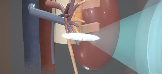
Operative animation, Shock wave lithotripsy

Extracorporeal shock wave lithotripsy

A stone in the kidney broken without an incision by shock waves made outside the body and focused on it.

The patient lies on the lithotripsy table with the treatment head coupled to the flank through gel, the stone is placed in the focal point by fluoroscopy, and the energy is stepped up from a low setting while periodic images keep the stone on target. The fragments must then pass.

The lessons are what lies on the path of the waves, the kidney that takes every shock, and the ureter the fragments travel down.

Core tier FRCS stones and endourology Interactive 3D

Operative visualisation

Schematic scene preview: Shock wave lithotripsy
Preparing the 3D anatomy…

UrOops3D · UroRef · © Nity G

How to use this module. Play through the eight steps, or use Back and Next. Watch the head couple to the flank, the stone brought into the crosshairs, the energy stepped up and the waves converge until the stone breaks. The triggers show a perirenal haematoma, steinstrasse and an obstructed infected kidney.

Click me next Section 2 of 8 Anatomy The structures this operation works between, and the one it must not cross.

The structures in the scene

Shock wave lithotripsy: how the structures relateThe stone at the focal point, and what lies on the shock path and around it. Stone in the lower-pole calyx: the focal point: the waves converge on it, coupled through gel Treatment head and water cushion. Stone in the lower-pole calyx: the focal point: every shock passes through it; a haematoma when injured Renal parenchyma and capsule (danger). Stone in the lower-pole calyx: the focal point: above the shock path; the rib shadow marks the limit Twelfth rib and pleura (danger). Stone in the lower-pole calyx: the focal point: beside and behind the kidney; kept off the path Descending colon (danger). Stone in the lower-pole calyx: the focal point: the route of the fragments; a column can obstruct it Renal pelvis and ureter (danger). Stone in the lower-pole calyx: the focal point: the skin-to-stone distance; a long one predicts failure Flank skin and body wall.Stone in the lower-polecalyx: the focal pointTreatment head andwater cushion!Renal parenchyma andcapsule!Twelfth rib andpleura!Descending colon!Renal pelvis andureterFlank skin and bodywallthe waves convergeon it, coupledthrough gelevery shock passesthrough it; ahaematoma when…above the shockpath; the ribshadow marks the…beside and behindthe kidney; keptoff the paththe route of thefragments; a columncan obstruct itthe skin-to-stonedistance; a longone predicts…TargetDangerLandmarkContext
The stone at the focal point, and what lies on the shock path and around it. Schematic, not to scale; relationships only.
  • Supine, with the treatment head beneath the left flank.
  • The shock path runs forward and inward through the posterolateral body wall, below the twelfth rib and the pleura, through the lower pole to the stone in its calyx.
  • The colon lies lateral to the kidney and behind it, off the path; the ureter runs down medially.
  • ›Stone - radiopaque, in the lower-pole calyx; seen on the plain film and the fluoroscopy screen.
  • ›Lower-pole calyx - where the stone lies and where its fragments are left; they must drain upward to the pelvis to leave.
  • ›Renal parenchyma - between the skin and the stone on the shock path; every shock passes through it.
  • ›Renal capsule and perirenal fat - where a haematoma collects, under the capsule or around the kidney.
  • ›Renal pelvis and ureter - the route of the fragments; dilated above a column of them.
  • ›Twelfth rib and pleura - above the shock path; the rib shadow on the screen marks the limit.
  • ›Posterolateral flank skin - where the water cushion is coupled through gel.
  • ›Descending colon - beside and behind the kidney; kept off the shock path.
  • ›Treatment head and water cushion - beneath the table under the flank; the waves converge from it on the focal point.
  • ›Focal zone - the region where the waves converge, placed on the stone by fluoroscopy.
Click me next Section 3 of 8 Steps The operation in order, with the landmark that guards each move.

Operative steps

  1. Confirm the stone and the patient The stone is confirmed on imaging before the time-out: here radiopaque, in the left lower-pole calyx. The patient is checked against the contraindications: pregnancy, uncorrected bleeding risk or anticoagulation, untreated urinary infection, an arterial aneurysm near the stone, and obstruction below it.
    where it bites: pregnancy, bleeding risk and untreated infection are decided here, before the patient is on the table.
  2. On the table The patient lies supine on the lithotripsy table, secured with a safety strap, the pressure points padded and pneumatic compression devices on the legs. The time-out confirms the patient, the procedure and the exact stone, with the images in the room. Analgesia, sedation or an anaesthetic follows.
    where it bites: a patient who moves or breathes deeply takes the stone out of the focal point; the analgesia is part of the targeting.
  3. Couple the head to the flank The treatment head is positioned below the patient on the side of the stone and raised until its water cushion presses on the flank. Gel couples the cushion to the skin, and the air pockets in it are swept out before the first shock.
    where it bites: an air pocket in the coupling gel deflects the shock wave away from the stone and onto the tissues around it.
  4. Localise the stone Limited fluoroscopy confirms the stone within the crosshairs of the imaging system, and the patient is moved until it sits there: the focal point is now on the stone. The path from the skin to the stone runs below the rib and the pleura, clear of bowel, and its length is the skin-to-stone distance.
    where it bites: the lung above the rib, the bowel beside the kidney and the solid organs must lie off the shock path.
  5. Ramp the energy Treatment is begun at a low energy and a slow rate. The power is then stepped up progressively to the maximum setting chosen. Each wave travels through the cushion, the skin and the kidney and converges on the focal zone at the stone.
    where it bites: every shock passes through the renal parenchyma; the slow start and the slow rate are how it is spared.
  6. Fragmentation The stone is imaged frequently in collimated mode to keep it on target as the patient breathes, and the heart rhythm is watched. It breaks into fragments over the course of the treatment, which runs to the number of shocks the manufacturer allows for the site of the stone.
    where it bites: a stone that drifts off the focal point is replaced there by kidney; targeting is checked throughout.
  7. The end check The treatment ends at the allowed number of shocks. The treatment head is lowered and the flank and abdomen are examined for a visible haematoma. The final image shows the fragments lying in the lower pole.
    where it bites: pain out of keeping with the treatment, or a falling blood pressure, is a perirenal haematoma until imaging shows otherwise.
  8. Fragments and follow-up The patient goes home with analgesia and is told to return with a fever or pain that does not settle. Imaging at follow-up decides the next step: clear, fragments still passing, a further session, or a different treatment for a stone that did not break.
    where it bites: fragments in the ureter can obstruct it, and an obstructed infected kidney is an emergency.
Click me next Section 4 of 8 Danger zones Where this operation bites, and why it bites there.
Critical relationship

Danger zones

The spatial relationships the animation is built to make automatic. Before you read each one, ask yourself: at this point in the operation, what is about to go wrong, and what is my first move? The save for each sits in the Complications tab.

  • RiskRenal parenchyma and perirenal tissues - every shock passes through them; the ramp and the slow rate limit the injury, and a haematoma is the result when they do not.
  • RiskObstructed ureter distal to fragments - a column of fragments can block the ureter; a stone with an obstruction below it is not treated this way.
  • RiskAdjacent bowel, lung and solid organs - kept off the shock path: the lung and pleura above the rib, the colon beside the kidney, the spleen and liver above.
  • RiskPregnancy, bleeding risk and untreated urinary infection - contraindications, settled before the patient is on the table.
  • RiskSkin-to-stone distance - the length of the path through the body; a long one is one reason the stone does not break.
Practise it Take me into the scene Opens the animation at a danger moment and asks you to predict the mechanism and your first move before it reveals.
Click me next Section 5 of 8 Complications The save for each one: the first move, the escalation trigger, the op-note line.

Complications and immediate saves

Trigger each of these live in the Animation tab, then read the save: the first move, the escalation trigger, and the line that goes in the op note.

Perirenal haematoma

  • the shocks injure the parenchyma and blood collects under the capsule or around the kidney; anticoagulant or antiplatelet treatment raises the risk.
The save
  1. Pain out of keeping, a falling pressure or haemoglobin: stop treating and assess the patient, then image the kidney. first move
  2. Resuscitate, group and save or crossmatch, and correct any coagulopathy; stop antithrombotic treatment with the prescribing team.
  3. Bed rest and observation for the stable patient, with serial haemoglobin.
  4. Bleeding that continues, or instability despite resuscitation: interventional radiology for angiography and embolisation. escalation trigger
  5. Record the number of shocks, the energy, the antithrombotic history and the imaging finding. document

Steinstrasse

  • a column of fragments packed in the ureter behind a lead fragment, with colic and a dilated system above it.
The save
  1. Check for fever and infection first: a column with a fever is an obstructed infected kidney, not a waiting game. first move
  2. With no infection, analgesia and expulsive therapy, and imaging to follow the column.
  3. A column that does not move, or a large lead fragment: ureteroscopy, or a further session aimed at the lead fragment.
  4. Fever, a single kidney, deteriorating function or pain that cannot be controlled: drain the kidney now. escalation trigger
  5. Record the level and length of the column, the kidney function and the plan. document

Sepsis from an obstructed infected system

  • fragments obstruct the ureter above infected urine; fever, rigors and a falling pressure, and antibiotics alone do not reach the obstructed kidney.
The save
  1. Sepsis care at once: cultures, antibiotics, fluids and lactate, and the call to drain made at the same time. first move
  2. Urgent decompression: a retrograde ureteric stent or a percutaneous nephrostomy, whichever can be done soonest and most safely. escalation trigger
  3. Urine from the drained kidney sent for culture, and the antibiotics changed to what grows.
  4. The fragments are treated only when the sepsis has resolved.
  5. Record the time of the decision and of the drainage, the culture and the antibiotic. document
Click me next Section 6 of 8 Troubleshooting When the case turns: the problem, then the next move.

Intra-operative troubleshooting

When the case turns: the problem, then the next move.

  • ActionThe stone will not hold in the crosshairs - better analgesia and a calmer breath; check the coupling again, and image more often while the energy is held.
  • ActionLittle change in the stone after many shocks - a dense stone may not break: stop at the allowed number rather than adding more, and plan ureteroscopy or percutaneous surgery.
  • ActionThe stone is faint or not seen on fluoroscopy - ultrasound targeting where the lithotripter offers it; a stone that cannot be seen cannot be treated this way.
  • ActionA patient on anticoagulants or antiplatelets - stop or bridge the treatment with the prescribing team before the session; this is a high-bleeding-risk procedure.
  • ActionA stone in the upper pole behind the rib - change the angle of the head or the position of the patient; a stone that stays under the rib shadow suits another route.
  • ActionA pacemaker or an implanted defibrillator - treat with the cardiology team; a defibrillator is reprogrammed for the session.
Click me next Section 7 of 8 FRCS viva Say it out loud, then mark yourself against the model answer.

FRCS viva questions

The examiner stems this scene prepares you for. Try to answer out loud first, then reveal, and learn the discriminator, the detail that turns a pass into a strong pass.

QA patient has a single stone in the left lower-pole calyx. Who is suitable for shock wave lithotripsy, and who is not?reveal answer
  • I would want a stone I can see on fluoroscopy or ultrasound, a system that will drain the fragments, and sterile urine.
  • The EAU urolithiasis guideline lists pregnancy, bleeding disorders, uncontrolled urinary infection, severe skeletal deformity or obesity that prevent targeting, an arterial aneurysm near the stone, and anatomical obstruction distal to the stone as contraindications.
  • A bleeding disorder is corrected 24 hours before and for 48 hours after.
  • Anticoagulation is not an absolute bar: the guideline counts this as a high-bleeding-risk procedure, with a hazard ratio for perinephric haematoma of up to 4.2 on antithrombotic treatment, so I would stop or bridge it with the prescribing team.
  • For the lower pole I would also tell the patient that endoscopic treatment is often the better choice.
Strong pass

Say both

  • Naming obstruction below the stone and the aneurysm
  • Treating anticoagulation as a risk to manage rather than an absolute bar
Examiner note
Naming obstruction below the stone and the aneurysm, and treating anticoagulation as a risk to manage rather than an absolute bar.
QWhat predicts that shock wave lithotripsy will fail?reveal answer
  • I would think of the stone, the patient and the kidney.
  • For the stone, the EAU guideline says that stones above 1,000 Hounsfield units and of high homogeneity on non-contrast CT are less likely to break, and that brushite, calcium oxalate monohydrate and cystine stones are hard.
  • For the patient, a long skin-to-stone distance and a high body mass index reduce success.
  • Perks and colleagues, in 111 patients, found that stone attenuation and skin-to-stone distance each predicted the result independently of stone size and body mass index: 91% success with both favourable and 41% with both unfavourable.
  • For the kidney, the guideline lists a steep infundibulopelvic angle, a long calyx and a narrow infundibulum as reasons lower-pole fragments stay behind.
Strong pass

Separating fragmentation, which density and distance decide, from clearance, which lower-pole anatomy decides.

QHow would you choose between shock wave lithotripsy, flexible ureteroscopy and PCNL for a renal stone?reveal answer
  • I would follow the EAU guideline.
  • It recommends PCNL first-line for stones larger than 2 cm, and for the lower pole it recommends PCNL or retrograde intrarenal surgery even for stones larger than 1 cm, because shock wave lithotripsy is limited there.
  • The randomised evidence is the Lower Pole I trial of Albala and colleagues: of 128 patients with lower-pole stones, 95% were stone-free at three months after percutaneous removal against 37% after lithotripsy, with morbidity of 22% and 11%, which did not differ significantly.
  • The guideline adds that a randomised trial found flexible ureteroscopy more effective than shock waves for small lower-pole stones, while lithotripsy was more cost-effective.
  • So I would offer it for a small stone with favourable anatomy and density, with the patient told that a second treatment is more likely.
Strong pass

Say both

  • Quoting the guideline recommendation for the lower pole and the Lower Pole I stone-free rates
  • Weighing efficacy against invasiveness
Examiner note
Quoting the guideline recommendation for the lower pole and the Lower Pole I stone-free rates, and weighing efficacy against invasiveness.
QHow do you get the most from a session of lithotripsy?reveal answer
  • I would start with the coupling: the EAU guideline notes that air pockets in the gel deflect 99% of the shock waves, so I would sweep them out.
  • I would begin at a low energy and step it up, because ramping protects the kidney; the guideline cites a trial with stone-free rates of 96% with ramping against 72% without.
  • I would keep the rate slow, since it states the optimal frequency as 1.0 to 1.5 Hz and tissue damage rises with frequency.
  • I would image the stone frequently, give enough analgesia to stop the patient moving, and give antibiotics only for infected stones or bacteriuria, not for sterile urine.
Strong pass

Knowing that technique, coupling, ramp and rate, changes the stone-free rate, not only the stone.

QTwo days after lithotripsy the patient returns with a fever, loin pain and a column of fragments in the ureter. What do you do?reveal answer
  • This is an obstructed infected kidney until proven otherwise, and I would treat it as an emergency.
  • I would start sepsis care at once, take cultures and give antibiotics, and arrange urgent decompression at the same time, by a ureteric stent or a nephrostomy;
  • the EAU guideline makes urgent decompression and delayed stone treatment strong recommendations.
  • For steinstrasse with infection or fever it prefers a nephrostomy.
  • I would send urine from the drained kidney for culture and treat the fragments only when the sepsis has resolved.
  • Steinstrasse follows about 4% of treatments in the guideline, and sepsis 0.15%.
Strong pass

Say both

  • Draining the kidney the night it is found rather than treating the fragments
  • Knowing the guideline prefers a nephrostomy here
Examiner note
Draining the kidney the night it is found rather than treating the fragments, and knowing the guideline prefers a nephrostomy here.
Click me next Section 8 of 8 Evidence The guidance each of these answers is built on.

Evidence and guidance

Operative Dictations in Urologic Surgery
Armenakas NA, Fracchia JA, Golan R. Extracorporeal shock wave lithotripsy. Chapter 126. Wiley, 2019
The page follows the indication, essential steps and template dictation of the chapter: the stone confirmed first, supine or prone, strapped and padded, the head below the patient, the stone in the crosshairs, a low setting and slow rate increased, periodic imaging, the head lowered and the flank examined. Its variations: shock number by site and manufacturer, and sonographic targeting. Its complications: haematuria, parenchymal injury or haematoma, pain, ureteral obstruction and steinstrasse, failure to fragment. Its shock counts are kept off the page. The contraindications, the ramp and rate, the coupling, the failure predictors and the comparison with other treatments are general practice taken from the EAU guideline and the papers below.
Open guideline
EAU Guidelines on Urolithiasis, 2026
Sections 3.4.5 (shock wave lithotripsy), 3.4.8 (contraindications, antithrombotic treatment, stone composition) and Table 3.9 (complications), read 18 October 2026
Contraindications: pregnancy, bleeding disorders (corrected 24 hours before and 48 hours after), uncontrolled urinary infection, skeletal deformity or obesity preventing targeting, arterial aneurysm near the stone, obstruction distal to the stone; hazard ratio for perinephric haematoma up to 4.2 on antithrombotics. Optimal frequency 1.0 to 1.5 Hz; ramping, stone-free rates 96% against 72% in one trial; air pockets deflect 99% of shock waves. Steinstrasse 4%, sepsis 0.15%, symptomatic haematoma 0.21%.
Open guideline
EAU Guidelines on Urolithiasis, 2026
Sections 3.4.2 (sepsis in the obstructed kidney), 3.4.10 (renal stones) and 3.4.12 (steinstrasse), read 18 October 2026
Strong: urgently decompress the collecting system in sepsis with obstructing stones, by percutaneous drainage or ureteral stenting, and delay definitive treatment until sepsis resolves. Strong: PCNL first-line for stones larger than 2 cm; PCNL or retrograde intrarenal surgery for the lower pole even for stones larger than 1 cm. Stones above 1,000 HU less likely to break. Weak: steinstrasse with infection or fever preferably by nephrostomy.
Open guideline
Urology
Perks AE, Schuler TD, Lee J, et al. Stone attenuation and skin-to-stone distance on computed tomography predicts for stone fragmentation by shock wave lithotripsy. Urology 2008;72:765-769
Abstract read 18 October 2026. Retrospective review of 111 patients with a solitary renal stone. Attenuation, skin-to-stone distance and composition predicted the outcome independently of size, location and body mass index; success 91% with both attenuation and distance favourable and 41% with both unfavourable.
Open guideline
Journal of Urology
Albala DM, Assimos DG, Clayman RV, et al. Lower pole I: a prospective randomized trial of extracorporeal shock wave lithotripsy and percutaneous nephrostolithotomy for lower pole nephrolithiasis. J Urol 2001;166:2072-2080
Abstract read 18 October 2026. Randomised multicentre trial, 128 patients with lower-pole stones. Stone-free at three months 95% after percutaneous removal against 37% after lithotripsy; lithotripsy results fell with stone burden; morbidity 22% and 11%, not significantly different.
Open guideline

Curriculum. Supports learning relevant to the ISCP Urology Curriculum core module Stones and endourology. Not a workplace-based assessment and not evidence of competence.

Two minutes

Close the lap: recall it before you leave.

Say it aloud, then reveal. Reading is not remembering.

  1. 01 The relationship Say the critical spatial relationship out loud, then reveal. reveal
    Memory frame

    Supine, with the treatment head beneath the left flank.

    Why?
    The shock path runs forward and inward through the posterolateral body wall, below the twelfth rib and the pleura, through the lower pole to the stone in its calyx. The colon lies lateral to the kidney and behind it, off the path; the ureter runs down medially.
  2. 02 The first move Perirenal haematoma. Your first move? reveal
    First move

    Pain out of keeping, a falling pressure or haemoglobin: stop treating and assess the patient, then image the kidney.

  3. 03 The escalation trigger Perirenal haematoma. What makes you call for help? reveal
    Escalate when

    Bleeding that continues, or instability despite resuscitation: interventional radiology for angiography and embolisation.

  4. 04 The discriminator What turns a pass into a strong pass? reveal
    Strong pass

    Say both

    • Naming obstruction below the stone and the aneurysm
    • Treating anticoagulation as a risk to manage rather than an absolute bar
    Examiner note
    Naming obstruction below the stone and the aneurysm, and treating anticoagulation as a risk to manage rather than an absolute bar.
  5. 05 The pitfall Where does this operation bite first? reveal
    Pitfall

    Renal parenchyma and perirenal tissues

    Why?
    every shock passes through them; the ramp and the slow rate limit the injury, and a haematoma is the result when they do not.

Revision practice with self-marked answers, not an assessment of clinical competence. Kept in this browser only; it does not sync between devices.