
Shock-wave lithotripsy
Non-incisional fragmentation of selected renal or ureteric calculi using externally generated shock waves focused on the stone.
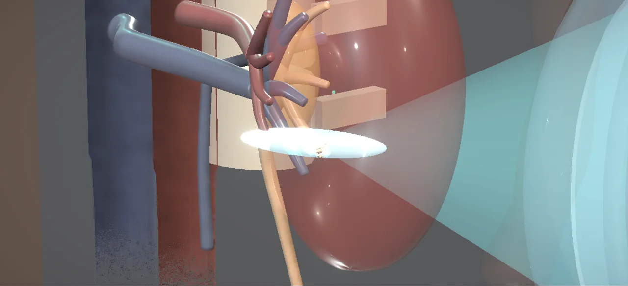 Interactive 3D Interactive shock wave lithotripsy Open the 3D animation
Teaching catalogue only. This coverage record is not a patient pathway or operative instruction.
Typical place in practice
Appropriately selected radiologically targetable stones where spontaneous passage is unlikely or active treatment is preferred.
Alternatives and neighbouring procedures
Observation, ureteroscopy, percutaneous nephrolithotomy or selected surgical stone removal according to stone and patient factors.
Same procedure family, explicit technique branches
These are variations of this canonical operation. Complication modules below identify whether they apply to all branches or only to named approaches.
Fluoroscopy-targeted SWL
Radiopaque stones are tracked with fluoroscopy while shock delivery is coupled to the flank.
Ultrasound-targeted SWL
Ultrasound localises and follows a visible stone without ionising radiation.
Stented SWL pathway
A ureteric stent is present for a selected drainage indication; it does not replace careful stone selection.
Repeat-session SWL
Fragmentation is reassessed and treatment repeated when clinically appropriate.
High-level sequence
Coverage outline only; not a step-by-step technical guide.
- Confirm stone, side, visibility and suitability for shock-wave treatment
- Position and couple the patient to the treatment head
- Localise the stone and protect non-target structures
- Deliver monitored, focused shocks while maintaining targeting
- Plan imaging and management of fragments or treatment failure
Every failure mode is its own module.
Timing and variation coverage are recorded for each module.
Steinstrasse and ureteric obstruction
A column of fragments may obstruct the ureter and cause pain, impaired renal drainage or infection; a stent does not eliminate the risk.
Applies to: Fluoroscopy-targeted SWL · Ultrasound-targeted SWL · Stented SWL pathway · Repeat-session SWLRenal or perirenal haematoma
Shock waves can injure renal tissue and vessels, producing haematuria or clinically important concealed bleeding.
Applies to: Fluoroscopy-targeted SWL · Ultrasound-targeted SWL · Stented SWL pathway · Repeat-session SWLUrinary infection or sepsis
Fragmentation may release bacteria or create obstruction in an infected system.
Applies to: Fluoroscopy-targeted SWL · Ultrasound-targeted SWL · Stented SWL pathway · Repeat-session SWLResidual fragments or treatment failure
Unfavourable composition, size, location, anatomy or targeting can leave clinically relevant stone and require another treatment.
Applies to: Fluoroscopy-targeted SWL · Ultrasound-targeted SWL · Stented SWL pathway · Repeat-session SWLPain, bruising and transient haematuria
Shock entry and fragment passage may cause flank bruising, urinary bleeding and renal colic.
Applies to: Fluoroscopy-targeted SWL · Ultrasound-targeted SWL · Stented SWL pathway · Repeat-session SWLAdjacent-organ or rhythm-related event
Poor focusing can expose neighbouring tissues, while shock delivery may interact with cardiac rhythm in susceptible circumstances.
Applies to: Fluoroscopy-targeted SWL · Ultrasound-targeted SWL · Stented SWL pathway · Repeat-session SWLStent symptoms or encrustation
An indwelling stent adds urinary symptoms, infection, migration and delayed-removal risks independent of the shock-wave session.
Applies to: Stented SWL pathwaySources for this module
SWL, ureteroscopy, RIRS, PCNL, bladder stones and decompression options.
Accessed 2026-08-03 · opens source European Association of Urology EAU Guidelines on Urological InfectionsInfection prevention, recognition and procedure-associated infection context.
Accessed 2026-08-03 · opens source British Association of Urological Surgeons Patient information leaflets by procedureUK patient-facing procedure, alternatives, recovery and complication framing.
Accessed 2026-08-03 · opens source