
Semirigid ureteroscopy and ureteric stone treatment
Retrograde direct-vision access to the ureter with a semirigid ureteroscope for stone fragmentation or extraction.
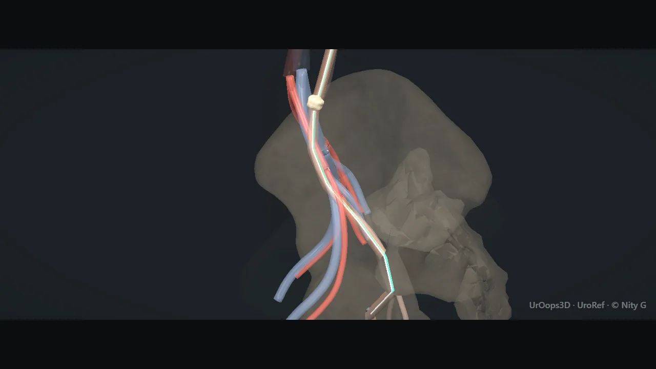 Interactive 3D Interactive operative model Open the 3D animation
Teaching catalogue only. This coverage record is not a patient pathway or operative instruction.
Typical place in practice
Active treatment of accessible ureteric calculi and selected diagnostic ureteric assessment.
Alternatives and neighbouring procedures
Observation, shock-wave lithotripsy, flexible ureterorenoscopy, antegrade access or selected surgical removal.
Same procedure family, explicit technique branches
These are variations of this canonical operation. Complication modules below identify whether they apply to all branches or only to named approaches.
Semirigid URS with basket extraction
A suitably sized mobile stone or fragment is retrieved under direct vision.
Semirigid URS with laser lithotripsy
The ureteric stone is fragmented or dusted before controlled extraction or passage.
Emergency drainage-first URS
The priority is safe drainage; definitive stone treatment is performed only when the clinical context permits.
Staged semirigid URS
Access or stone clearance is deliberately completed over more than one procedure.
High-level sequence
Coverage outline only; not a step-by-step technical guide.
- Confirm side, stone level, infection status and drainage strategy
- Gain bladder and ureteric access while preserving a safe route
- Advance under vision without force
- Fragment or retrieve stone with controlled energy and traction
- Inspect for injury and decide on drainage and follow-up imaging
Operative notes
Concise operative notes covering the key steps, landmarks and watch points, in the order you encounter them.
Ureteroscopy for ureteric stones
What it does: up the ureter with a thin rigid scope over two wires. Dust the stone.
- Cystoscopy first. See the trigone and both orifices.
- 2 wires. The first is the safety wire: up the centre, to the kidney, under screening. It stays until the end.
- Cystoscope out. Semirigid scope in with the second (working) wire through its channel. Wire 2 up the ureter through the scope. It only guides the scope.
- Hold both wires gently taut. Railroad the scope over wire 2 to the stone. Advance only under vision. See the lumen always. A semirigid scope barely bends: the ureter straightens over it. The iliac crossing is where it catches.
- Orifice too tight? Rotate the scope 180 degrees.
- Sheaths rarely help in the ureter.
- Finger on the water valve. Minimum flow. Hard flow flushes the stone back to the kidney.
- Going over the wire shields the stone.
- Stone seen: water off, pull the working wire. Keep the safety wire. Laser or pneumatic in.
- Laser tip well clear of the scope lens. Fire against the lens = cracked lens.
- Laser: dust it, do not chunk it. Paint the stone. Never leave large pieces in the ureter: they scatter and they do not pass. Dust clouds: wash out, refire.
- Let dust and fine fragments clear by gravity. A piece too big to clear: basket it, test the pull first. Resists? Release, laser it smaller. Never drag. Scope and basket come out together, fragment at the tip, wall inspected all the way down. One fragment out is not stone-free: the dust stays to pass.
- Stent after if: hole in the ureter, torn mucosa, leftover fragments, dilated stricture, 1 kidney, both sides done, or case over ~45 min.
- Simple low stone? No routine stent. (CROES data.)
- 2026 update: No change. This is still current practice.
- Watch: Scope sizes: tip 6 to 8 Fr, shaft 7.5 to 10 Fr, channel 3.6 Fr (or 2.4 and 3.4 Fr), wires 0.035 or 0.038 inch, fibre 200 to 365 micron. Owner-confirmed typical sizes.
- Wire will not pass a stone: Catheter up to just below the stone: its support often lets the sensor wire through. No? A little contrast: is the stone blocking? None passes? Terumo hydrophilic wire. Terumo up: follow with the catheter, exchange to a sensor wire. Stenting over a Terumo is hard. Still stuck: call the consultant. Semirigid scope fragments the stone so the wire passes.
- JJ over the wire: Four thin black dashes, then the thick band. Band going into the orifice: scope back to the bladder neck. Push till the golden pusher tip shows. Stop. Wire out completely. Curl in the kidney on the screen. In the bladder the stent's own end drops and curls on the base: one open coil in line with the shaft, not a separate loop out of the orifice. Lateral orifice: right side arrow to 3 o'clock, wire angles to 9; left side arrow to 9, wire to 3.
- Catheter will not go up the orifice: Guidewire through the ureteric catheter, soft tip just beyond the end. Lead the tip into the orifice. The catheter follows over it. Contrast on the screen only: it is not seen through the scope.
From the UroRef Operative Quick Guide. Open the note page →
Every failure mode is its own module.
Timing and variation coverage are recorded for each module.
Ureteric perforation or extravasation
A wire, instrument, basket or laser can breach the ureter, especially beside an impacted stone.
Applies to: Semirigid URS with basket extraction · Semirigid URS with laser lithotripsy · Emergency drainage-first URS · Staged semirigid URSUreteric avulsion
Forceful scope passage or traction on an entrapped stone or basket can cause catastrophic ureteric separation.
Applies to: Semirigid URS with basket extraction · Semirigid URS with laser lithotripsy · Staged semirigid URSUrinary infection or sepsis
Instrumentation and intrarenal pressure can disseminate infection; treatment in an inadequately drained infected system is particularly hazardous.
Applies to: Semirigid URS with basket extraction · Semirigid URS with laser lithotripsy · Emergency drainage-first URS · Staged semirigid URSStone retropulsion or residual fragments
A stone may migrate beyond semirigid reach or fragments may remain and require surveillance or another procedure.
Applies to: Semirigid URS with basket extraction · Semirigid URS with laser lithotripsy · Staged semirigid URSBasket entrapment or device failure
A basket can become trapped around a large stone or fail, creating traction risk and a need for an alternative retrieval plan.
Applies to: Semirigid URS with basket extractionPost-procedure pain, haematuria or obstruction
Oedema, fragments, clot or stent symptoms can produce colic, bleeding and impaired drainage.
Applies to: Semirigid URS with basket extraction · Semirigid URS with laser lithotripsy · Emergency drainage-first URS · Staged semirigid URSUreteric stricture
Impaction, ischaemia, perforation and thermal or mechanical injury may heal with delayed narrowing.
Applies to: Semirigid URS with basket extraction · Semirigid URS with laser lithotripsy · Emergency drainage-first URS · Staged semirigid URSSources for this module
SWL, ureteroscopy, RIRS, PCNL, bladder stones and decompression options.
Accessed 2026-08-03 · opens source European Association of Urology EAU Guidelines on Urological InfectionsInfection prevention, recognition and procedure-associated infection context.
Accessed 2026-08-03 · opens source British Association of Urological Surgeons Patient information leaflets by procedureUK patient-facing procedure, alternatives, recovery and complication framing.
Accessed 2026-08-03 · opens source