
Percutaneous nephrolithotomy
Percutaneous renal access, tract dilation and nephroscopic fragmentation or extraction of renal stone burden.
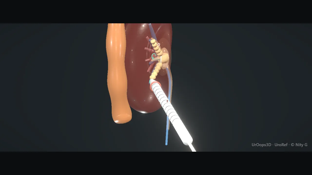 Interactive 3D Interactive operative model Open the 3D animation 3D Interactive ECIRS dual-access model
Teaching catalogue only. This coverage record is not a patient pathway or operative instruction.
Typical place in practice
Large, complex or otherwise unsuitable renal stones where percutaneous clearance offers an appropriate balance of effectiveness and risk.
Alternatives and neighbouring procedures
Flexible ureterorenoscopy, shock-wave lithotripsy, staged combined treatment, observation or selected open/laparoscopic/robotic stone surgery.
Same procedure family, explicit technique branches
These are variations of this canonical operation. Complication modules below identify whether they apply to all branches or only to named approaches.
Standard-tract PCNL
A conventional tract accommodates rigid nephroscopy and larger extraction instruments.
Miniaturised PCNL
A smaller tract and instruments may reduce tract burden while changing irrigation, fragment-extraction and operative characteristics.
Supine PCNL
Positioning supports simultaneous retrograde access and alters access geometry and anaesthetic considerations.
Prone PCNL
Posterior calyceal access is planned with the patient prone and pressure areas protected.
ECIRS
Endoscopic combined intrarenal surgery coordinates nephroscopic and flexible retrograde views for complex stone burden.
Tubeless PCNL
A nephrostomy is omitted only when haemostasis, drainage and collecting-system integrity meet the planned criteria.
High-level sequence
Coverage outline only; not a step-by-step technical guide.
- Review three-dimensional stone, organ and vascular anatomy and select position
- Gain calyceal access along a safe papillary path
- Dilate the tract while maintaining wire security
- Fragment and extract stone with pressure and energy control
- Inspect for residual stone or injury and select an exit drainage strategy
Operative notes
Concise operative notes covering the key steps, landmarks and watch points, in the order you encounter them.
PCNL
What it does: stone out through a back track, 1 cm.
- Culture urine. Treat infection 24-48 h before.
- Lithotomy. Cystoscope (17-22 Fr or flexible). 0.035 wire up. Ureteric catheter over it: 5-6 Fr open-ended, or 7 Fr occlusion balloon inflated at the PUJ. Tape it to a 14-16 Fr Foley.
- Contrast 30-50% (stone stays visible) +/- air +/- methylene blue. Air floats to the POSTERIOR calyces when prone.
- Prone. Pad all points. Drape with a pouch.
- Plan the track: straight line into the stone calyx. Below the 12th rib if you can. Pleura sits above. Do not hug under the rib. Colon lateral.
- Vessels: go posterolateral, end-on to a posterior calyx, through the papilla. Never medial. Never into the pelvis direct.
- 18G needle, 15-20 cm (or 21/22G Chiba first). Bull's-eye: hub and tip one dot. Triangulate: 0 then 20-30 degrees for depth. US option: 3.5-5 MHz, Doppler. Feel 2 gives: fascia, capsule.
- Stylet out. Urine / contrast / blue at the hub = in. Nothing dilated before this.
- Hydrophilic angled wire in. Kumpe or Cobra steers it to the pelvis and down the ureter. Occlusion balloon up at the PUJ: past the stone into a distant calyx instead. Exchange: super-stiff WORKING wire.
- 8/10 coaxial: SAFETY wire in alongside. 10 outer off. Clip the safety wire to the drape. Both wires down the ureter. Occlusion balloon up: both coiled in the kidney (pelvis or a distant calyx), never down the ureter.
- 1-1.5 cm cut (No. 11) along the wire. Open the fascia.
- 8 Fr guide. Amplatz 10 -> 30 Fr in 2s, twist-push along the wire, each to the calyx on screening, same depth mark. Alternatives: Alken metal (scarred), balloon 30 Fr (watch the waist go), one-shot 24-30 Fr, mini 16.5-22 Fr.
- Sheath (30 Fr) over the last dilator into the calyx. Dilator and guide out. Sheath + 2 wires.
- Rigid nephroscope 24-26 Fr, saline, low pressure: outflow round the scope. In line with the tract. Never lever.
- Litho: ultrasonic (soft, sucks), ballistic (hard, pushes), laser 365-550 micron. Out of line with the tract: flexible nephroscope 15-16 Fr, never lever the rigid one. Scope out: small fragments wash out. Tri-radiate grasper or basket, one piece at a time. Too big for the sheath? It jams at the mouth: release, break it again.
- Clear? Rigid then flexible, every calyx. Screen in 2 planes. One fragment out is not stone-free.
- Drain (main path): Malecot 16-22 Fr over the safety wire, flared in the pelvis. Sheath out over the tube; the tract closes round it. Occlusion balloon down, wires out. Nephrostogram through the tube: down the ureter, no leak. Stitch. Bag. Other exits: pigtail 8-10, Council-tip, re-entry Malecot, or tubeless (antegrade JJ 6-7 Fr / ureteric catheter overnight).
- Bleeding: venous = sheath back in, pause, clamp tube or Kaye balloon. Arterial (pulsatile) = stop, big tube, angioembolisation.
- 2026 update: Mini tracts (mini, ultra-mini, micro) bleed less. Same stone-free rate. Now mainstream.
- 2026 update: Supine PCNL and ECIRS (supine + scope from below at the same time) match prone results. Better ergonomics.
- Watch: Sizes and settings in this note are the owner's typical values.
From the UroRef Operative Quick Guide. Open the note page →
Every failure mode is its own module.
Timing and variation coverage are recorded for each module.
Renal haemorrhage or vascular lesion
Access, tract dilation or nephroscopic work can injure renal vessels, causing immediate bleeding or a delayed pseudoaneurysm or arteriovenous fistula.
Applies to: Standard-tract PCNL · Miniaturised PCNL · Supine PCNL · Prone PCNL · ECIRS · Tubeless PCNLPleural injury, hydrothorax or pneumothorax
An upper-pole or supracostal tract can cross pleura or diaphragm and produce fluid or air in the chest.
Applies to: Standard-tract PCNL · Miniaturised PCNL · Supine PCNL · Prone PCNL · ECIRS · Tubeless PCNLColon or solid-organ injury
Atypical anatomy or an unsafe tract can injure bowel, spleen, liver or another adjacent structure.
Applies to: Standard-tract PCNL · Miniaturised PCNL · Supine PCNL · Prone PCNL · ECIRS · Tubeless PCNLUrinary infection or sepsis
Infected stones, bacterial urine and elevated intrarenal pressure can provoke systemic infection.
Applies to: Standard-tract PCNL · Miniaturised PCNL · Supine PCNL · Prone PCNL · ECIRS · Tubeless PCNLCollecting-system perforation or urinary leak
Wire, dilator or nephroscope injury can create extravasation; inadequate drainage may lead to a persistent collection.
Applies to: Standard-tract PCNL · Miniaturised PCNL · Supine PCNL · Prone PCNL · ECIRS · Tubeless PCNLResidual stone or need for staged treatment
Complex calyceal distribution, bleeding or safety limits may prevent complete clearance in one session.
Applies to: Standard-tract PCNL · Miniaturised PCNL · Supine PCNL · Prone PCNL · ECIRS · Tubeless PCNLTract or access loss
Wire displacement or sheath movement can eliminate safe access and create parenchymal or extrarenal passage.
Applies to: Standard-tract PCNL · Miniaturised PCNL · Supine PCNL · Prone PCNL · ECIRS · Tubeless PCNLDrainage failure after tubeless exit
Omitted external drainage may expose unrecognised bleeding, oedema, obstruction or collecting-system injury.
Applies to: Tubeless PCNLSources for this module
SWL, ureteroscopy, RIRS, PCNL, bladder stones and decompression options.
Accessed 2026-08-03 · opens source European Association of Urology EAU Guidelines on Urological InfectionsInfection prevention, recognition and procedure-associated infection context.
Accessed 2026-08-03 · opens source British Association of Urological Surgeons Patient information leaflets by procedureUK patient-facing procedure, alternatives, recovery and complication framing.
Accessed 2026-08-03 · opens source