
Revision sheet
Percutaneous nephrolithotomy
11 steps, each one the authored title and caption from the interactive animation, with the frame captured from that step. Read it before the case, or print it and work through the steps in order.
Teaching only, not for diagnosis or treatment decisions. The text on this sheet is the animation's own step copy, reproduced verbatim.
UrOops3D · UroRef · © Nity G
- 01
Retrograde set-up
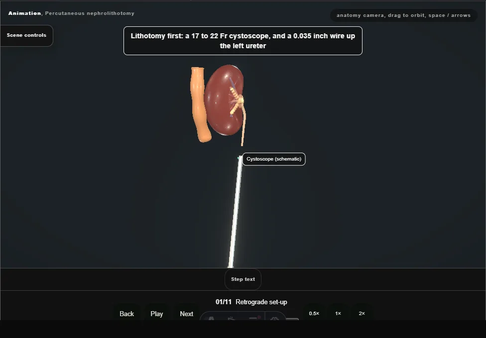
- In lithotomy a wire, then a 7 Fr occlusion balloon catheter, go up the ureter; the balloon is inflated at the PUJ, the catheter taped to a Foley, and diluted contrast fills the system.
- 02
Target calyx
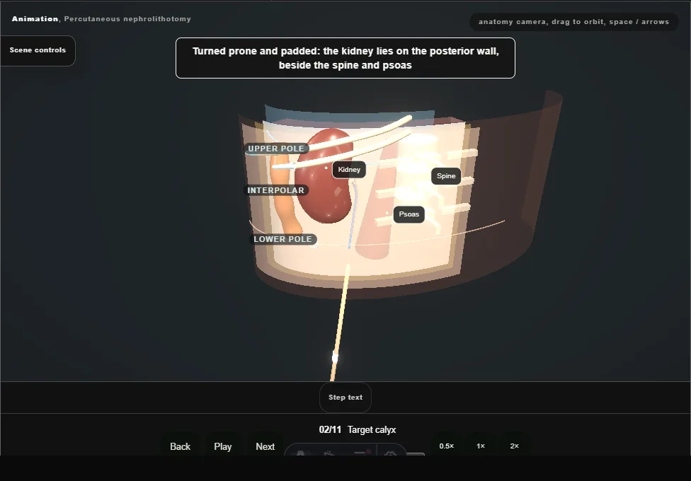
- Prone, the kidney lies on the posterior wall.
- Plan a posterior calyx that lines up with the stone bulk: its axis points back toward the skin, where an anterior calyx points away.
- 03
Trajectory
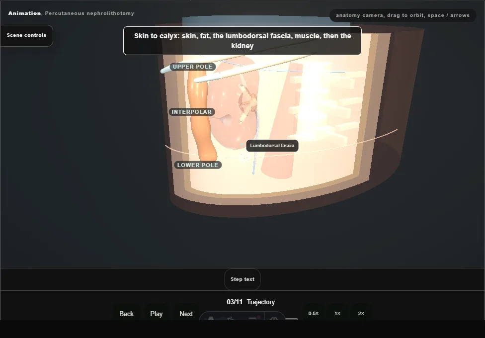
- The line from skin to calyx crosses skin, fat, the lumbodorsal fascia and muscle, and must avoid the pleura above and the colon laterally.
- 04
Calyceal puncture
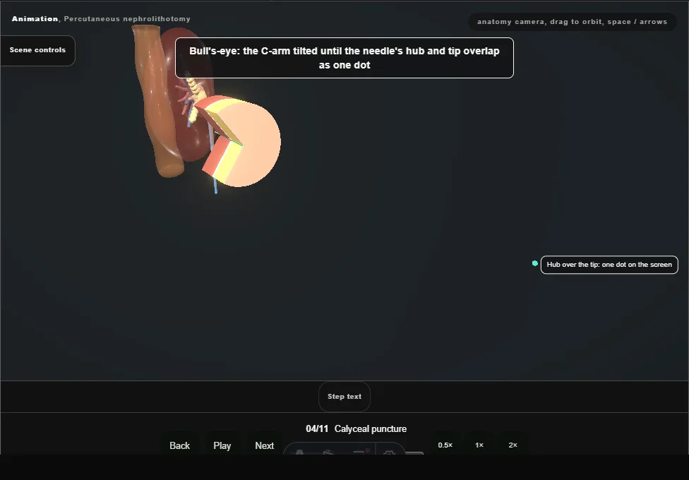
- Bull's-eye, then an oblique view for depth.
- The 18G needle gives at the lumbodorsal fascia and at the capsule, and enters the calyx through the papilla, along its axis.
- 05
Confirm access
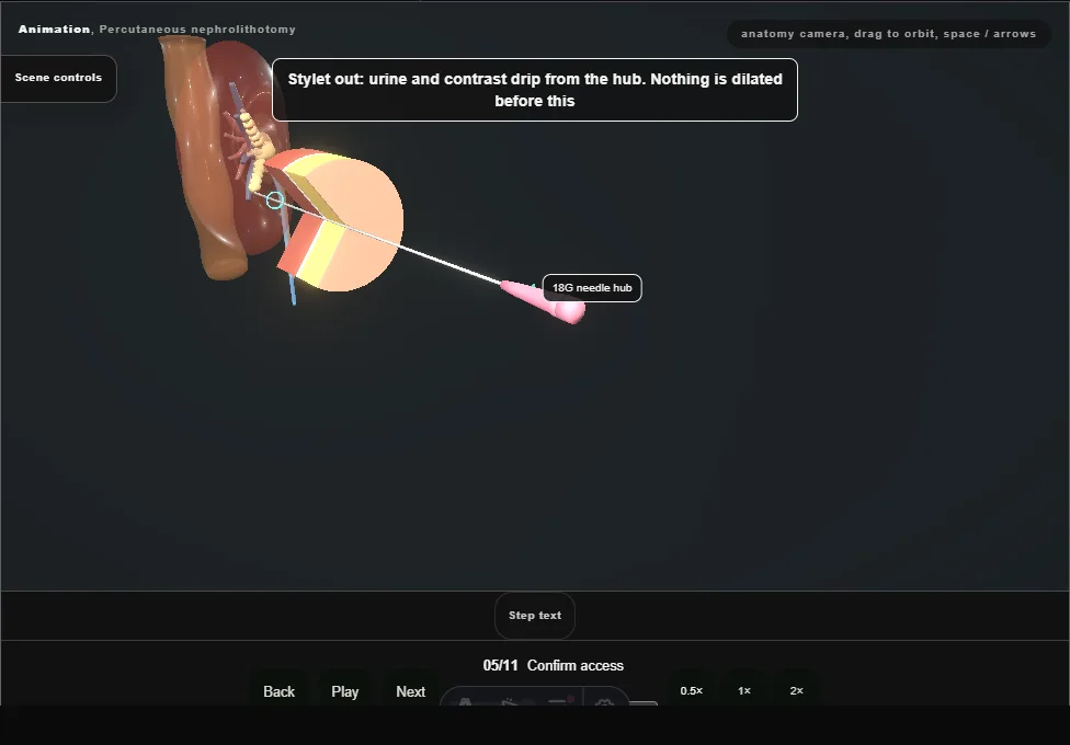
- Stylet out: urine and contrast at the needle hub confirm the tip is in the collecting system, and nothing is dilated before that.
- 06
Two wires
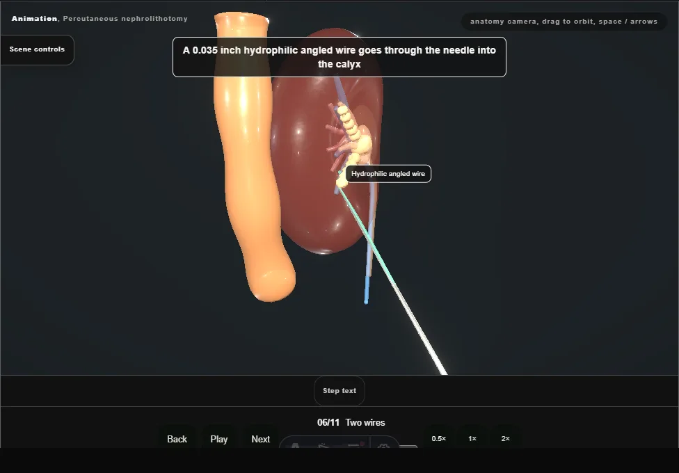
- A hydrophilic wire, steered round the stone with a Kumpe, is exchanged for a super-stiff working wire (white) coiled in the upper calyx;
- an 8/10 Fr coaxial adds the safety wire (teal), coiled in the middle calyx and clipped to the drape.
- With the balloon up at the PUJ, neither wire can go down the ureter.
- 07
Tract dilation
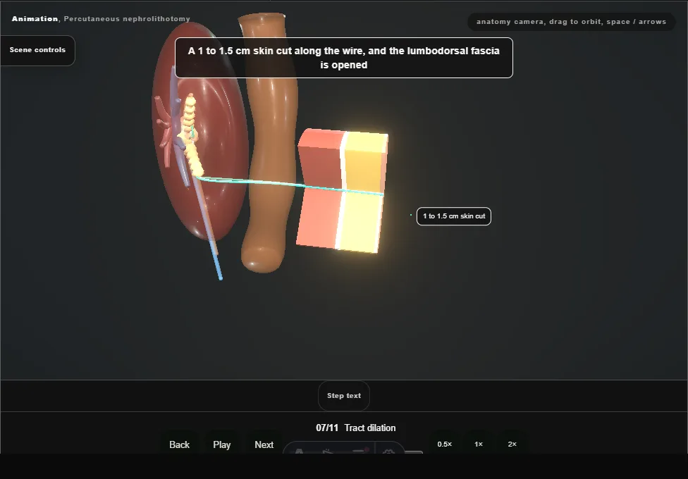
- Skin cut, fascia opened, an 8 Fr guide, then Amplatz dilators in 2 Fr steps to the calyx, each to the same depth mark and never past it.
- The chip shows the alternative: a balloon dilator over the working wire, inflated at the tract to 30 Fr in one step.
- One technique or the other, never both.
- 08
Sheath
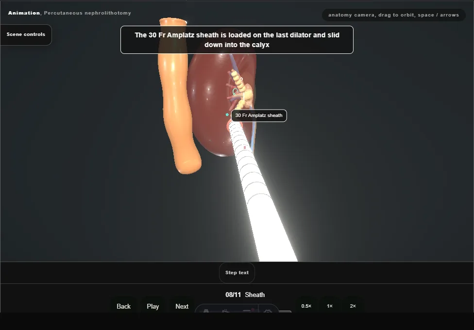
- The 30 Fr Amplatz sheath goes over the last dilator (or the inflated balloon) into the calyx; dilator and guide (or the deflated balloon) come out, and the sheath is the working channel and the tamponade.
- 09
Nephroscope
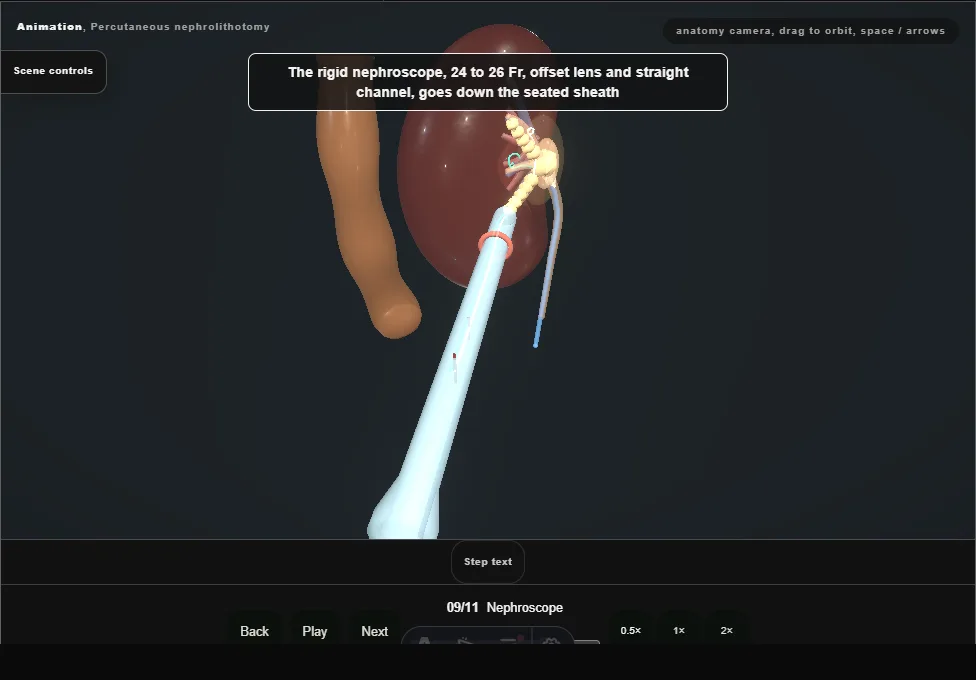
- The rigid nephroscope (24 to 26 Fr, offset lens, straight channel) goes down the sheath and stays in line with the tract, never levered.
- Saline runs in down the scope and out round it.
- A combined ultrasonic and ballistic probe takes the branch ahead; a 15 to 16 Fr flexible nephroscope and the laser break what lies out of line.
- The safety wire stays coiled as the route back in.
- 10
Stone clearance
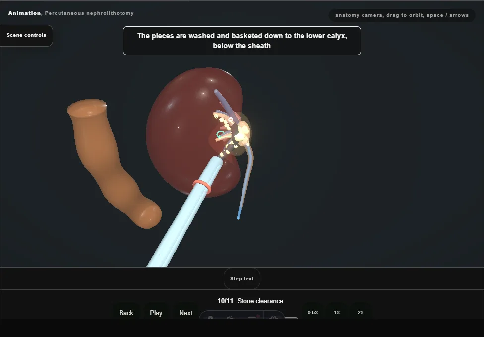
- The pieces are washed down to the calyx.
- With the scope briefly out the small ones wash out through the sheath; a tri-radiate grasper takes the larger ones one at a time.
- A piece wider than the sheath's lumen is refused at its mouth and broken again.
- Every calyx is checked, rigid, flexible and on screening: one fragment out is not stone-free.
- 11
Drainage
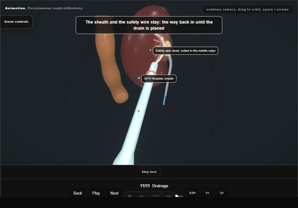
- A Malecot, 16 to 22 Fr, goes in over the safety wire and flares in the pelvis; the sheath comes out over it and the parenchyma closes round the tube.
- Balloon down, wires out, and an antegrade nephrostogram shows contrast running down the ureter with no leak.
- Pigtail, Council-tip or a tubeless JJ are the branch.
Derived from PcnlScene.tsx at build time: every step title and caption above is the authored
ProcedureDef text, reproduced verbatim, and this page shares its readable-points renderer with the animation
itself. 11 of 11 steps have a captured frame.
Text fingerprint d27b624de42fa8c3. A caption edit without a re-capture fails the production
check, so the sheet and the animation cannot drift apart silently.