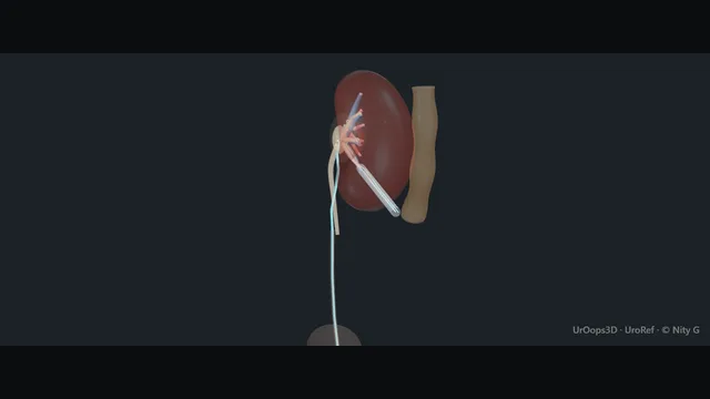
Both approaches meet the stone in one anaesthetic: a nephroscope descends from the flank into a lower-pole calyx while a flexible ureteroscope climbs from the bladder, giving two angles on one burden without turning the patient.
The supine position moves the danger: the ascending colon can lie between the abdominal wall and the kidney on the puncture corridor, so the needle advances only when ultrasound has excluded bowel from the line and fluoroscopy has confirmed the calyx.
Advanced tierFRCS endourology, stonesInteractive 3D
Supine, the flank corridor and the retrograde road meeting in the kidney. Schematic, not to scale; relationships only.
The scene is one corridor and one road meeting inside the kidney.
The corridor runs from the flank skin through the supine abdominal wall, past the ascending colon that supine gravity drops onto the line, through the avascular parenchyma between the posterior and anterior segmental arteries, and into a posterior lower-pole calyx along the infundibular axis.
The road runs from the bladder up the ureter to the same pelvis.
ECIRS works because the two instruments arrive at the stone from opposite directions: the nephroscope reaches the pelvis and lower pole in line with the tract, the flexible ureteroscope reaches the upper calyces and the angles the rigid scope cannot flex into, and the ascending colon, not the pleura, is the structure the supine position puts in the needle's way.
›Posterior lower-pole calyx - the puncture target; a tract along the infundibular axis reaches the whole collecting system and crosses parenchyma at its safest.
›Ascending colon - in supine it can lie between the anterior wall and the kidney directly on the corridor; the structure ultrasound exists to exclude.
›Posterior segmental artery - brackets the tract line with the anterior branch; the avascular plane between them is the parenchyma the needle should cross.
›Ureter - the retrograde road; marked by the catheter, then occupied by the flexible scope during the combined phase.
›Renal pelvis - the crossroads where wire, sheath, both scopes and the exit stent converge.
Supine position + retrograde catheterThe patient stays supine with the flank flush to the table edge and the hip opened, so the percutaneous corridor and the retrograde road are both live at once. The patient is secured to the table, because tilt is part of this operation and a sliding patient is a sterile-field problem. A retrograde ureteric catheter is placed first, tip in the renal pelvis: it opacifies the collecting system for the puncture, marks the ureter throughout and later carries the flexible scope. Placing it also proves the ureter is passable before any percutaneous commitment, which changes the plan early if it is not. where it bites: the position commits both approaches before draping: fluoroscopy arc clearance, table tilt and cystoscopic access are checked now, not after prep.
Combined ultrasound + fluoroscopy punctureThe needle walks from the flank to a posterior lower-pole calyx, aimed along the infundibular axis so the tract lines up with the stone bulk. Ultrasound carries the soft-tissue half of the puncture: it shows the corridor, the depth of parenchyma and, decisively, whether bowel lies in the line. Fluoroscopy with contrast running down the retrograde catheter confirms the calyx of entry and the needle tip position in two planes. Only when ultrasound has excluded bowel from the corridor and the screen has confirmed the calyx does the needle take its final increment. where it bites: in the supine position the ascending colon can lie on the corridor: bowel seen on ultrasound is excluded from the line before the needle advances, not after.
Guidewire into the pelvisThe wire is advanced through the needle under screening until it coils in the pelvis with redundant length inside the system. This is the safety wire and it stays for the entire case; every dilator, sheath and scope exchange happens over it. It is also the reason a lost tract can be regained rather than re-punctured. A wire that merely enters the infundibulum looks placed but springs out with the first dilator, so the coil in the pelvis is confirmed on the image before dilation starts. If the wire will not coil, the retrograde catheter delivers one from below to meet it rather than the needle line being forced. where it bites: a wire that merely enters the infundibulum can spring out during dilation; coil it in the pelvis and confirm on fluoroscopy before dilating.
Tract dilation + sheathDilators pass over the wire in graded steps, each to the same depth on the image, and the Amplatz sheath is railroaded and seated in the calyx under fluoroscopic control. The tract crosses parenchyma bracketed by the segmental arteries, so dilation stays on the wire and resistance is investigated rather than pushed through. The sheath tip is confirmed inside the calyx before the dilator is withdrawn, short in the parenchyma or through the far wall being the two failures to exclude. Once seated it is the working channel for the nephroscope and, when venous bleeding clouds the field, the tamponade that buys the vision back. where it bites: the tract crosses parenchyma bracketed by segmental arteries: dilation stays on the wire, resistance is investigated rather than pushed through, and bleeding means the sheath tamponades, not a deeper pass.
Dual-scope fragmentationThe rigid nephroscope works down the sheath while the flexible ureteroscope climbs from the bladder, and both views are held as one map of a single system. The nephroscope breaks the bulk in line with the tract; the retrograde scope dusts and retrieves what is dislodged into calyces the rigid scope cannot flex into. Fragments leave through the sheath with irrigation, and table tilt substitutes for the gravity drainage a prone case would have had. A fragment that flees into an upper calyx is retrieved with the flexible scope deliberately, not chased blind through a narrow infundibulum. where it bites: a fragment driven into an upper calyx the rigid scope cannot flex into is a surveillance problem; chasing it through a narrow infundibulum makes it a bleeding problem.
Exit strategyWhen the system is clear to both scopes, drainage is adequate and the field is quiet, the case ends tubeless with a JJ stent placed over the wire. The exit is decided on the field, not on preference: bleeding, suspected perforation or residual fragments each keep a nephrostomy tube in place and earn a documented second-look plan. A tubeless exit in a corridor that was never entirely trusted hides a colonic injury, so doubt about the line earns a tube and imaging instead of a clean pull-out. Whatever drains the kidney, the reason is written into the op note with the follow-up imaging attached. where it bites: a tubeless exit in a colon-injured tract hides the injury: any doubt about the corridor earns a tube and imaging, not a clean pull-out.
The spatial relationships the animation is built to make automatic. Before you read each one, ask yourself: at this point in the operation, what is about to go wrong, and what is my first move? The save for each sits in the Complications tab.
RiskAscending colon on the corridor - supine gravity drops it between wall and kidney; the commonest visceral risk of the supine puncture and the reason ultrasound is part of needle guidance, not an extra.
RiskSegmental arteries bracketing the tract - a tract off the infundibular line or dilated beyond the calyx tears a posterior branch; pulsatile bleeding that survives irrigation is arterial until proven otherwise.
RiskUpper-pole infundibulum - where lost fragments flee and where the rigid scope cannot follow; the retrograde scope is the second angle that protects it.
Trigger each of these live in the Animation tab, then read the save: the first move, the escalation trigger, and the line that goes in the op note.
Colonic injury (supine corridor)
the needle, a dilator or the sheath passes through the ascending colon lying on the supine corridor;
it announces itself with faeculent irrigation or gas along the tract, or late with peritonism after a clean-looking exit.
The save
Recognise it: faeculent debris or gas tracking along the sheath, unexplained fever or peritonism after a tubeless exit.first move
Do not simply pull the tract tube out: withdraw it into the colon and leave it as a controlled colostomy drain, or replace with a large-bore catheter in the track.
Start broad-spectrum antibiotics, keep the patient nil by mouth and resuscitate.
Image with contrast through the drain to define the injury and plan the healing check before removal.
Generalised peritonitis, uncontrolled sepsis or failure of the drain → operative repair with colorectal input.escalation trigger
Op note: corridor used, injury recognised intra-operatively or after, drainage strategy, antibiotic plan and imaging follow-up.document
Segmental arterial bleeding
dilation tears a posterior segmental branch where the tract crosses the parenchyma; venous ooze clears with irrigation, arterial bleeding is pulsatile and does not.
The save
Stop fragmenting. Seat the sheath across the bleeding segment or inflate a balloon catheter in the tract: the tamponade is the treatment.first move
Do not remove the sheath to look; hold the tamponade while the team readies blood products and imaging.
Check the whole patient: position, pressure, crossmatch; a patient drifting towards transfusion is a patient drifting towards embolisation.
Bleeding that recurs when tamponade is released → urgent angiography and selective embolisation, which preserves renal tissue better than open exploration.
Haemodynamic instability despite tamponade and transfusion → open exploration with vascular or interventional support, immediately.escalation trigger
Op note: tract site and size, tamponade method, transfusion, embolisation, and the follow-up imaging plan.document
Fragment loss into an inaccessible calyx
a fragment migrates into an upper calyx the rigid nephroscope cannot flex into; pursuit through a narrow infundibulum trades a surveillance problem for a bleeding one.
The save
Stop chasing it. Decide deliberately whether the fragment is reachable by either scope under vision.first move
Use the second angle: the retrograde flexible ureteroscope reaches the upper calyces the rigid scope cannot.
Irrigation and table tilt can float the fragment back to the pelvis and onto the sheath without any new puncture.
Dust rather than dig: a small residual under surveillance beats a torn infundibular vessel every time.
A large unreachable residual → staged second-look endoscopy rather than extending a hostile tract.escalation trigger
Op note: residual size and site, surveillance imaging plan, and the metabolic stone workup intention.document
When the case turns: the problem, then the next move.
ActionNo efflux of urine from the punctured calyx - Efflux is a comfort, not a test: an obstructed or low-pressure system may not efflux. Confirm the needle tip on two planes and aspirate; let the wire and fluoroscopy decide.
ActionWire slips down the ureter instead of coiling in the pelvis - Leave it in place as a road map, pass a second wire for safety, and use the retrograde catheter to deliver a wire up from below to meet it.
ActionSheath will not pass over the wire - Re-check the tract line on fluoroscopy, redilate one size under vision, and never force against resistance: a kinked wire or a rib in the line is the usual cause.
ActionVision lost to venous bleeding mid-fragmentation - Raise irrigation, seat the sheath to tamponade and pause; if vision does not return, leave a nephrostomy tube and plan a second look rather than fragmenting blind.
The examiner stems this scene prepares you for. Try to answer out loud first, then reveal, and learn the discriminator, the detail that turns a pass into a strong pass.
QDefend the supine position for percutaneous stone surgery. What are you trading away?reveal answer
The supine, Galdakao-modified position buys three things.
First, a single position for a combined procedure: the retrograde and percutaneous approaches run together without turning, which shortens the anaesthetic and removes the turn with its lines, airway and pressure-area risk.
Second, anaesthetic access to the airway throughout, which matters in the obese and the comorbid.
Third, surgeon ergonomics and radiation hygiene: the surgeon stands beside the patient with hands largely out of the beam, working downward rather than over a prone torso.
The trade is real.
Gravity no longer drains fragments out of the sheath, so clearance depends more on irrigation and the retrograde scope helping from below.
The sheath sits less securely and needs deliberate holding.
And the bowel behaves differently: in supine, the ascending colon can fall onto the puncture corridor between the wall and the kidney, which is why the supine puncture is a combined ultrasound and fluoroscopy puncture rather than fluoroscopy alone.
None of these trade-offs is a reason to abandon supine; they are the reason supine percutaneous surgery is deliberately guided, irrigated and staffed differently from prone.
Strong pass
Naming the colon, not the pleura, as the visceral structure the supine corridor re-orders, and gravity-dependent fragment clearance as the functional price of not turning.
QHow do you keep radiation exposure defensible in ECIRS?reveal answer
By treating screening time as a clinical variable that is planned, watched and documented rather than a background cost.
Before the case: the right imaging has already been done, so fluoroscopy confirms rather than explores;
pregnancy status is checked;
the equipment is set to pulsed acquisition at the lowest useful frame rate with tight collimation over the kidney.
During the case: the combined approach is itself protective, because ultrasound carries the soft tissue half of the puncture, deciding bowel and depth before any screening, while fluoroscopy only confirms the calyx and the wire.
Last-image hold is used instead of re-screening; the image intensifier is kept close to the patient and the beam angled once rather than repeatedly; hands stay out of the field with forceps technique for wire handling.
The patient is a child or pregnant colleague scenario only rarely, but the same discipline applies with more force.
After the case: screening time is read off the machine and recorded in the notes, because a dose that is not documented cannot be defended or learned from.
The principle is that ECIRS uses two imaging modalities precisely so that neither one has to carry the whole puncture alone.
Strong pass
Say all three
Saying that ultrasound is part of radiation protection
Not an alternative to fluoroscopy
That screening time is documented as a clinical variable
Examiner note
Saying that ultrasound is part of radiation protection, not an alternative to fluoroscopy, and that screening time is documented as a clinical variable.
QWhen does ECIRS beat a staged percutaneous and retrograde approach?reveal answer
ECIRS earns its complexity when one anaesthetic can genuinely finish the stone disease and staging cannot.
The clearest case is a combined burden: a renal stone with a ureteric stone or a stone that has migrated, where the retrograde road is needed anyway and the percutaneous tract finishes the bulk.
The second case is stone anatomy that defeats a single approach: a sharp lower-pole infundibular angle or a narrow infundibulum, where the flexible ureteroscope alone works slowly and loses fragments, and percutaneous alone over-serves a stone that also dusts well from below.
The third is the patient for whom two anaesthetics are the real risk: the comorbid, the anticoagulated needing a single interruption, the patient who will simply not come back for a second stage.
Against that, staging still wins when the urine is infected and the system obstructed: drain, treat, then treat the stone in a clean field.
Staging also wins for the full staghorn needing multiple tracts, where one long combined session serves the surgeon rather than the patient.
So the decision is made on burden distribution, collecting-system anatomy and patient fitness, in that order, before the patient is ever positioned.
Strong pass
Say both
Framing the infected obstructed system as the absolute brake on single-stage surgery
Rather than presenting ECIRS as the default for every stone
Examiner note
Framing the infected obstructed system as the absolute brake on single-stage surgery, rather than presenting ECIRS as the default for every stone.
QA patient develops peritonism on the ward the night after a tubeless supine ECIRS. What has happened and what do you do?reveal answer
A colonic injury on the supine puncture corridor has declared itself late, which is exactly how it declares itself after a tubeless exit: no drain, no faeculent return in theatre, then pain, fever and peritonism as the undrained tract communicates with the colon.
The response is sequenced and does not wait for certainty.
Resuscitation and examination first, with the senior surgeon and colorectal team informed at once, not after imaging.
Broad-spectrum antibiotics on culture logic, nil by mouth, fluids and bloods including clotting and gas, because this patient is heading towards a decision, not an observation.
Imaging next: a CT with contrast, and contrast through any tube left in situ, to define the injury, the collection and the tract anatomy.
Management follows the image and the abdomen.
A contained injury with a drainable collection is managed with a drain in or alongside the colon, antibiotics and staged imaging of healing before anything is removed.
Generalised peritonitis, uncontrolled sepsis or deterioration on resuscitation is an operative problem with colorectal support.
The late lesson is the preventive one: the supine corridor is planned with ultrasound precisely so that this night does not happen, and any corridor that worried the operating surgeon at the time earns a tube and a low threshold for imaging before discharge.
Strong pass
Say both
Naming the tubeless exit as the reason the injury stayed hidden
Getting colorectal input early rather than after imaging has finished
Examiner note
Naming the tubeless exit as the reason the injury stayed hidden, and getting colorectal input early rather than after imaging has finished.
Say it aloud, then reveal. Reading is not remembering.
01The relationshipSay the critical spatial relationship out loud, then reveal.reveal
Memory frame
The scene is one corridor and one road meeting inside the kidney.
Why?
The corridor runs from the flank skin through the supine abdominal wall, past the ascending colon that supine gravity drops onto the line, through the avascular parenchyma between the posterior and anterior segmental arteries, and into a posterior lower-pole calyx along the infundibular axis. The road runs from the bladder up the ureter to the same pelvis. ECIRS works because the two instruments arrive at the stone from opposite directions: the nephroscope reaches the pelvis and lower pole in line with the tract, the flexible ureteroscope reaches the upper calyces and the angles the rigid scope cannot flex into, and the ascending colon, not the pleura, is the structure the supine position puts in the needle's way.
02The first moveColonic injury (supine corridor). Your first move?reveal
First move
Recognise it: faeculent debris or gas tracking along the sheath, unexplained fever or peritonism after a tubeless exit.
03The escalation triggerColonic injury (supine corridor). What makes you call for help?reveal
Escalate when
Generalised peritonitis, uncontrolled sepsis or failure of the drain → operative repair with colorectal input.
04The discriminatorWhat turns a pass into a strong pass?reveal
Strong pass
Naming the colon, not the pleura, as the visceral structure the supine corridor re-orders, and gravity-dependent fragment clearance as the functional price of not turning.
05The pitfallWhere does this operation bite first?reveal
Pitfall
Ascending colon on the corridor
Why?
supine gravity drops it between wall and kidney; the commonest visceral risk of the supine puncture and the reason ultrasound is part of needle guidance, not an extra.
Revision practice with self-marked answers, not an assessment of clinical competence. Kept in
this browser only; it does not sync between devices.