
Aquablation of the prostate
Image-planned robotic waterjet resection of obstructing prostatic tissue, followed by a procedure-specific haemostasis and drainage phase.
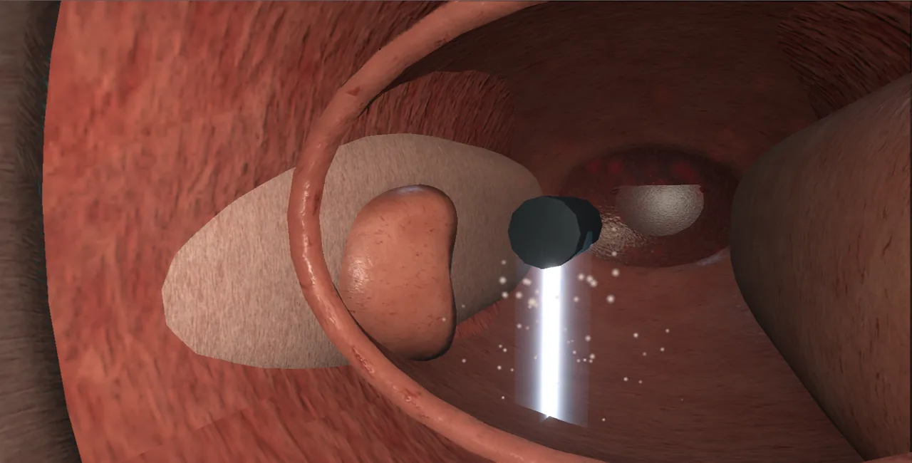 Interactive 3D Interactive planned waterjet resection Open the 3D animation
Teaching catalogue only. This coverage record is not a patient pathway or operative instruction.
Typical place in practice
Selected benign prostatic obstruction where prostate anatomy and patient priorities fit an image-guided waterjet approach.
Alternatives and neighbouring procedures
TURP, enucleation, vaporisation, simple prostatectomy, medication or other minimally invasive outlet procedures.
Same procedure family, explicit technique branches
These are variations of this canonical operation. Complication modules below identify whether they apply to all branches or only to named approaches.
Standard Aquablation
A treatment contour is planned on real-time imaging and executed by a high-velocity saline jet.
Aquablation with focal cautery haemostasis
Selected bleeding points are treated endoscopically after waterjet resection.
Aquablation with traction-based haemostasis
Catheter-based pressure contributes to postoperative haemostasis within a defined pathway.
High-level sequence
Coverage outline only; not a step-by-step technical guide.
- Confirm gland configuration and position the imaging and treatment systems
- Map bladder neck, verumontanum and intended treatment contour
- Execute waterjet passes while maintaining registration
- Inspect the resection cavity and complete the planned haemostasis strategy
- Establish drainage and monitor for bleeding, retention and systemic recovery
The operation, step by step
A step that touches one of this module's danger zones is marked and cross-linked below.
- SetupGeneral anaesthesia, lithotomy position, perineal ultrasound probe positioned and coupled, Aquablation console with handpiece and live ultrasound linked, and the table integration checked.
- AccessPass the handpiece sheath under vision and survey the bladder and outlet.
- Build the live ultrasound map and confirm the imaging and treatment views correspond before contouring, because everything depends on the imaging–treatment registration interface. danger zone: Imaging–treatment registration interface
- Identify the bladder neck and ureteric orifices on the map and down the scope. danger zone: Bladder neck and ureteric orifices
- Draw the contour from bladder neck to verumontanum, following adenoma to the capsule and stopping short of the external sphincter beyond the apical contour. danger zone: External sphincter beyond the apical contour
- Lock the map and recheck that probe, table and handpiece have not moved; re-register if anything has shifted.
- Run the waterjet passes inside the contour, watching the console and the cavity together.
- Resurvey the cavity: adenoma taken to the capsule, sphincter untouched, and nothing strayed through the prostatic capsule and venous channels. danger zone: Prostatic capsule and venous channels
- Apply the variant’s haemostasis strategy: catheter traction alone, focal cautery to named bleeders, or a traction catheter.
- Evacuate debris and confirm bladder integrity before the sheath comes out.
- ClosureNo closure: three-way catheter drainage only.
- AftercareOvernight irrigation with the catheter out within a day or two, and watch haemoglobin in the first hours because the raw-surface ooze is the main early risk.
Every failure mode is its own module.
Timing and variation coverage are recorded for each module.
Bleeding, haemoglobin drop or clot retention
Waterjet resection creates a broad raw surface, and the haemostasis strategy has variant-specific trade-offs.
Applies to: Standard Aquablation · Aquablation with focal cautery haemostasis · Aquablation with traction-based haemostasisCapsular or bladder-neck injury
Incorrect contouring, movement or loss of registration can direct the jet beyond intended adenoma.
Applies to: Standard Aquablation · Aquablation with focal cautery haemostasis · Aquablation with traction-based haemostasisThermal or ureteric-orifice injury during haemostasis
Cautery used after the waterjet phase can damage bladder neck, orifices or capsule if landmarks are unclear.
Applies to: Aquablation with focal cautery haemostasisUrinary infection
Instrumentation, tissue resection and catheterisation contribute to postoperative infection risk.
Applies to: Standard Aquablation · Aquablation with focal cautery haemostasis · Aquablation with traction-based haemostasisRetention, dysuria or persistent storage symptoms
Oedema, clot, tissue debris and baseline bladder dysfunction may impair voiding after catheter removal.
Applies to: Standard Aquablation · Aquablation with focal cautery haemostasis · Aquablation with traction-based haemostasisEjaculatory or erectile-function change
Although contouring may aim to spare ejaculation-related structures, sexual-function change remains possible.
Applies to: Standard Aquablation · Aquablation with focal cautery haemostasis · Aquablation with traction-based haemostasisUrethral stricture, bladder-neck contracture or retreatment
Instrumentation, healing or residual/progressive obstruction may lead to delayed narrowing or another outlet procedure.
Applies to: Standard Aquablation · Aquablation with focal cautery haemostasis · Aquablation with traction-based haemostasisSources for this module
Contemporary outlet procedures, selection, variants and functional trade-offs.
Accessed 2026-08-03 · opens source European Association of Urology EAU Guidelines on Urological InfectionsInfection prevention, recognition and procedure-associated infection context.
Accessed 2026-08-03 · opens source British Association of Urological Surgeons Patient information leaflets by procedureUK patient-facing procedure, alternatives, recovery and complication framing.
Accessed 2026-08-03 · opens source