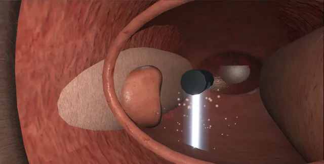
The surgeon draws the resection on the ultrasound and the robot cuts it with a jet of saline in a few minutes, the same every time, without heat.
What the robot does not do is stop the bleeding, and that is the part this page is about as much as the plan: the safe zone drawn from the veru, the neck contour drawn from the orifices, the cautery at five and seven, and the catheter on traction overnight.
Advanced tierFRCS endourology, BPH / LUTS and the alternatives to TURPInteractive 3D
Along the prostatic urethra, handpiece above and ultrasound probe in the rectum. Schematic, not to scale; relationships only.
The handpiece lies along the prostatic urethra with the veru behind its tip and the neck ahead, and the ultrasound probe lies in the rectum below the gland; the two are locked to one arm.
The plan is a contour on the sagittal image from the neck to short of the veru and a depth on each transverse slice to the capsule; the jet sweeps its arc and retracts along that contour.
Afterwards the bleeding points are at the neck at five and seven, a centimetre from the orifices.
›The probe and the handpiece on one arm - locked together; the plan is only true while neither moves.
›Verumontanum and the safe zone - drawn from the veru on the sagittal image; the jet is turned down or off beyond it.
›The surgical capsule - the depth set on each transverse slice; the jet stops at the plane the loop would.
›The bladder neck contour and the orifices - drawn to spare the trigone behind a large median lobe.
›The neck arteries at five and seven - the focal cautery, a centimetre from the orifices.
›The raw bed - bleeds for hours; the reason for the three-way catheter, the traction and the night in hospital.
›The three-way catheter - 22 French, balloon large on the neck, traction released in the morning.
Who gets the waterjetThe bigger gland, from about thirty up to 150 grams where a resection would be long and an enucleation is the usual alternative; the man who wants ejaculation preserved, which the plan can protect by sparing the tissue around the verumontanum and the ejaculatory ducts; and the surgeon who wants the resection to be reproducible and short. The trade-offs are bleeding, which is the main complication and the reason for a night in hospital with irrigation, a general or spinal anaesthetic, and the capital cost. Histology is available from the fragments, though the tissue is disrupted.
Consent and the set-upThe consent names bleeding first: transfusion in a few per cent, a return to theatre for clot or haemostasis, a catheter with irrigation overnight; then infection, retrograde ejaculation in a minority when the plan spares the veru, retention at the trial without catheter, the small risks of incontinence and stricture, and retreatment in a proportion. Lithotomy under general or spinal anaesthetic. The transrectal ultrasound probe is placed and fixed on its arm; the 24 French handpiece with its telescope is passed into the bladder under vision and locked to the same arm, so that the two cannot move against each other during the plan or the cut.
The handpieceThe handpiece sits in the prostatic urethra with the verumontanum and the sphincter behind its tip and the bladder neck ahead. The endoscopic view is checked against the ultrasound: the tip position on the screen matches what the lens sees. The probe gives a sagittal view of the whole urethra and a transverse view at any level, and the surgeon works from both. The patient is not moved, the arm is not touched, and the bladder is kept at a constant fill from now on. where it bites: the probe and the handpiece are locked to one arm; if either moves, the plan no longer describes the anatomy.
The plan on the ultrasoundOn the sagittal image the contour of tissue to remove is drawn from the bladder neck to a point short of the verumontanum, with the veru and the sphincter beyond it inside a safe zone where the jet is turned down or off. The distal limit is drawn from the veru on the image, not estimated. On transverse slices at several levels the depth is set to the surgical capsule, so that the jet stops at the same plane the loop would; laterally the contour follows the lobes. At the neck the contour is drawn to spare the trigone and the ureteric orifices, which sit behind a large median lobe. where it bites: the plan is checked against the endoscopic view before the pedal: the safe zone at the veru and the orifices out of the neck contour.
The jetWith the plan confirmed the foot goes on the pedal. A high-velocity jet of saline emerges from the handpiece at right angles and sweeps side to side through its arc while the handpiece retracts along the plan under the robot's control, cutting the adenoma without heat and sparing the capsule and the vessels of the neck to a degree, because the jet cuts soft tissue before firm. The surgeon watches the endoscopic view and the ultrasound together; the field fills with fragments and blood and the ultrasound shows the cavity forming to the contour. A hand near the pedal stops the jet at once if the field is lost or anything moves. Four to six minutes for most glands; the plan can be run twice for a very large one.
HaemostasisThe jet stops nothing, and a raw bed the size of the gland is now bleeding. The handpiece comes out and a resectoscope goes in. The cavity is inspected sector by sector with the irrigation slowed, and focal cautery is applied with the button at the bladder neck at five and seven o'clock where the arteries enter, on the points, not painted across the bed; the orifices are kept in view. The rest of the bed is left for the catheter: coagulating the whole cavity undoes the advantage of a cut made without heat. Some units use the button routinely at the neck; others rely on traction alone. Both orifices are seen again with their jets. where it bites: the neck arteries at five and seven o'clock sit a centimetre from the orifices; the button goes on the point.
The catheter and the noteA 22 French three-way catheter goes in, the balloon is filled large and pulled down onto the bladder neck for traction, and the catheter is taped to the thigh. Continuous irrigation runs overnight until the effluent is rosé and the traction is released in the morning. The haemoglobin is checked the next day. The note records the plan volume and the resection time, the cautery used and where, the orifices and the veru seen intact, the catheter, the traction and the irrigation plan, and the fragments sent for histology.
The spatial relationships the animation is built to make automatic. Before you read each one, ask yourself: at this point in the operation, what is about to go wrong, and what is my first move? The save for each sits in the Complications tab.
RiskThe plan across the veru - the jet into the sphincter zone; the safe zone is drawn from the veru on the image.
RiskThe neck contour through the trigone - the orifices in the cavity; drawn from the orifices on the transverse image.
RiskThe arm unlocked or the patient moved - the plan no longer matches the anatomy; pedal off at once.
RiskThe raw bed - the bleeding that transfuses a few per cent; traction, irrigation and the haemoglobin in the morning.
When the case turns: the problem, then the next move.
ActionThe handpiece tip on the screen does not match the endoscopic view - the arm has moved or the bladder fill has changed: re-lock, re-image, re-plan.
ActionCannot see the veru on the sagittal image - adjust the probe angle before planning; the safe zone is drawn from the veru, never estimated.
ActionThe endoscopic field is lost during the jet - pedal off; irrigate and wait; the cut resumes only with the field seen.
ActionEverything bleeds at the end - expected: the button at the neck at five and seven on the points, then the catheter on traction; do not paint the bed.
ActionThe effluent is red at two hours - the catheter is blocking or an artery is open: syringe washout, faster irrigation, haemoglobin; theatre if it will not clear.
ActionA large median lobe - the neck contour drawn from the orifices on the transverse image, and the cavity inspected at the neck before any cautery.
The examiner stems this scene prepares you for. Try to answer out loud first, then reveal, and learn the discriminator, the detail that turns a pass into a strong pass.
QWhat is Aquablation, and how does it compare with TURP and enucleation?reveal answer
Aquablation is an image-guided robotic waterjet resection of the prostate: a transrectal ultrasound probe and a transurethral handpiece are locked to one arm, the surgeon draws the resection contour on the sagittal and transverse images, and a high-velocity saline jet executes it under robot control in a few minutes, cutting without heat.
Compared with TURP it gives a similar improvement in symptoms and flow, with a shorter resection time, a lower rate of retrograde ejaculation when the plan spares the tissue around the verumontanum, and the same catheter and hospital stay, but a higher rate of bleeding, with transfusion in a few per cent, because the jet does not coagulate and haemostasis is added afterwards by focal cautery at the neck and catheter traction.
Compared with enucleation for a large gland it is quicker and needs less training, but removes less tissue and has more bleeding.
It suits glands from about thirty to 150 grams, particularly the larger gland and the man prioritising ejaculation.
Histology is available but the tissue is disrupted.
The capital and consumable cost is high.
Strong pass
Say both
Naming bleeding as the main trade-off with its cause
Ejaculation preservation as the specific advantage that depends on the plan
Examiner note
Naming bleeding as the main trade-off with its cause, and ejaculation preservation as the specific advantage that depends on the plan.
QHow do you plan the resection and what are the limits you draw?reveal answer
With the probe and the handpiece locked to one arm and the bladder at a constant fill, I would first confirm on the endoscope and the sagittal image that the handpiece tip position matches.
On the sagittal image I would draw the contour from the bladder neck to a point proximal to the verumontanum, marking the veru and placing a safe zone distal to it where the jet is reduced or off so that the sphincter and the ejaculatory ducts are protected;
this distal limit is drawn from the veru on the image, not estimated.
On transverse slices at several levels I would set the depth of the contour to the surgical capsule so that the cut stops at the plane a loop would reach, following the lateral lobes.
At the bladder neck I would draw the contour to spare the trigone and the ureteric orifices, which sit behind a large median lobe, using the transverse image at the neck.
I would then check the plan against the endoscopic view before touching the pedal, and I would not move the patient, the arm or the bladder fill afterwards.
Strong pass
Say all four
The safe zone drawn from the veru
The depth to the capsule on transverse slices
The neck contour drawn from the orifices
Each checked against the endoscope
Examiner note
The safe zone drawn from the veru, the depth to the capsule on transverse slices, and the neck contour drawn from the orifices, each checked against the endoscope.
QDescribe your approach to haemostasis after Aquablation.reveal answer
The waterjet cuts without heat, so the resection leaves a raw bed the size of the gland with no coagulation, and haemostasis is the surgeon's task afterwards.
I would remove the handpiece and pass a resectoscope, slow the irrigation and inspect the cavity sector by sector.
I would apply focal cautery with the button electrode at the bladder neck at five and seven o'clock where the prostatic arteries enter, touching the points and keeping the ureteric orifices in view, and to any other arterial jet, but I would not paint the whole bed, because that thermal injury is what the waterjet was chosen to avoid.
I would then place a 22 French three-way catheter with the balloon filled large and pulled down onto the neck for traction, taped to the thigh, with continuous irrigation overnight until the effluent is rosé, releasing the traction in the morning and checking the haemoglobin.
I would warn the ward about clot retention and the syringe washout, and I would escalate persistent bleeding to a return to theatre or embolisation.
Strong pass
Say all three
Focal cautery at the neck on the points with the orifices in view
Traction released in the morning
The reason the bed is not painted
Examiner note
Focal cautery at the neck on the points with the orifices in view, traction released in the morning, and the reason the bed is not painted.
QThe transrectal probe slips during the resection. What has happened and what do you do?reveal answer
The plan was drawn on images taken with the probe and the handpiece in a fixed relationship;
if the probe slips, the anatomy on the screen no longer describes where the jet is cutting, and the robot will continue to execute a contour that may now run through the capsule, the trigone or the sphincter zone.
I would take my foot off the pedal immediately.
I would re-lock the arm, re-image the prostate, and confirm the position of the handpiece tip against the endoscopic view.
I would then re-plan on the new images rather than resume the old plan, and before cutting again I would inspect the cavity endoscopically for any cut outside the intended contour.
If there were a capsular perforation, with fat or irrigant leaving, or a cut toward the veru, I would manage those as after any resection, with haemostasis, a catheter and senior help, and I would document what moved, what was found and the re-plan.
Prevention is checking the lock before the pedal and not moving the patient, the table or the bladder fill during the case.
Strong pass
Say both
Pedal off at once and a re-plan on new images
With an endoscopic inspection for a cut outside the contour before resuming
Examiner note
Pedal off at once and a re-plan on new images, with an endoscopic inspection for a cut outside the contour before resuming.
Say it aloud, then reveal. Reading is not remembering.
01The relationshipSay the critical spatial relationship out loud, then reveal.reveal
Memory frame
The handpiece lies along the prostatic urethra with the veru behind its tip and the neck ahead, and the ultrasound probe lies in the rectum below the gland; the two are locked to one arm.
Why?
The plan is a contour on the sagittal image from the neck to short of the veru and a depth on each transverse slice to the capsule; the jet sweeps its arc and retracts along that contour. Afterwards the bleeding points are at the neck at five and seven, a centimetre from the orifices.
02The first moveBleeding needing transfusion. Your first move?reveal
First move
Check the catheter is draining and the balloon is large and on traction; irrigation fast.
03The escalation triggerBleeding needing transfusion. What makes you call for help?reveal
Escalate when
Bleeding that will not stop in theatre: interventional radiology for embolisation of the prostatic arteries.
04The discriminatorWhat turns a pass into a strong pass?reveal
Strong pass
Say both
Naming bleeding as the main trade-off with its cause
Ejaculation preservation as the specific advantage that depends on the plan
Examiner note
Naming bleeding as the main trade-off with its cause, and ejaculation preservation as the specific advantage that depends on the plan.
05The pitfallWhere does this operation bite first?reveal
Pitfall
The plan across the veru
Why?
the jet into the sphincter zone; the safe zone is drawn from the veru on the image.
Revision practice with self-marked answers, not an assessment of clinical competence. Kept in
this browser only; it does not sync between devices.