
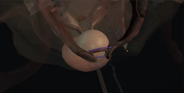
UrOops3D, danger-zone atlas
Operative animation, Simple prostatectomy

Simple prostatectomy for the large benign gland

The benign prostate over about eighty millilitres, or the gland with a bladder stone or diverticulum that needs the bladder opened as well: the adenoma is shelled out of its capsule with a finger, through the capsule from the front or through the bladder from above, leaving the peripheral zone and the sphincter behind.

This page is the choice against enucleation with the laser, the exposure of the dorsal vein, the capsulotomy, the plane, the apex, the bleeding at five and seven, and the catheter that follows.

Advanced tier FRCS core urology, BPH / LUTS and bladder outlet obstruction Interactive 3D

Operative visualisation

Schematic scene preview: Simple prostatectomy
Preparing the 3D anatomy…

UrOops3D · UroRef · © Nity G

Click me next Section 2 of 8 Anatomy The structures this operation works between, and the one it must not cross.

The structures in the scene

Simple prostatectomy: how the structures relateOpen retropubic from the patient's left, bladder retracted upward. Surgical capsule and the adenoma plane: transverse capsulotomy across the front Space of Retzius and anterior capsule. Surgical capsule and the adenoma plane: sutured above and below before the incision Dorsal venous complex and capsular vessels (danger). Surgical capsule and the adenoma plane: adenoma divided or pinched, never pulled Apex and external sphincter (danger). Surgical capsule and the adenoma plane: orifices seen before each stitch at five and seven Bladder neck, trigone and orifices (danger). Surgical capsule and the adenoma plane: a finger leaving the plane behind enters it Rectum (danger).Surgical capsule andthe adenoma planeSpace of Retzius andanterior capsule!Dorsal venouscomplex and capsularvessels!Apex and externalsphincter!Bladder neck,trigone and orifices!Rectumtransversecapsulotomy acrossthe frontsutured above andbelow before theincisionadenoma divided orpinched, neverpulledorifices seenbefore each stitchat five and sevena finger leavingthe plane behindenters itTargetDangerLandmark
Open retropubic from the patient's left, bladder retracted upward. Schematic, not to scale; relationships only.
  • Standing on the patient's left with the bladder retracted upward, the anterior capsule of the prostate is seen below the bladder neck at the bottom of the space of Retzius, with the symphysis in front of it and the dorsal vein running on its surface.
  • The capsulotomy is a transverse line across the front of the gland, and through it the finger enters the plane and sweeps around the adenoma with the rectum behind and the neurovascular bundles at the lateral limits.
  • The fossa that is left opens upward into the bladder neck, where the orifices sit on the trigone just behind, and downward to the apex where the urethra and sphincter continue.
  • ›Space of Retzius and the anterior capsule - opened with a finger off the symphysis; the capsule shown below the bladder neck with the dorsal vein branches on it.
  • ›The dorsal venous complex and capsular vessels - on and lateral to the capsule; sutured above and below the capsulotomy before it is made.
  • ›The surgical capsule - the compressed peripheral zone; the plane runs between it and the adenoma and opens to the finger.
  • ›The apex and the external sphincter - the adenoma attached by the urethral mucosa at the apex; divided under vision or pinched, never pulled.
  • ›Bladder neck, trigone and the orifices - the arteries enter at five and seven; the orifices a centimetre or two behind the neck are seen before each suture.
  • ›The rectum - behind the capsule and Denonvilliers' fascia; entered by a finger that turns outward through the posterior capsule.
  • ›The three-way catheter - 22 or 24 French, balloon in the bladder drawn onto the neck; irrigation until clear, out at five to ten days.
Click me next Section 3 of 8 Steps The operation in order, with the landmark that guards each move.

Operative steps

  1. Who: the gland too big for the scope The man with bladder outlet obstruction and a prostate over about eighty millilitres on ultrasound or MRI, where a resection would be long, bloody and incomplete. In a unit with holmium or bipolar enucleation that is the first offer, size-independent and transfusion-sparing; the open or robotic operation is chosen where enucleation is not available, where a large bladder stone or a diverticulum is dealt with at the same time, where hip disease prevents lithotomy, or where the surgeon does one operation well. Cancer is excluded as far as it can be by PSA and MRI, because the peripheral zone stays. The consent names transfusion in about one in ten, retrograde ejaculation in most, incontinence in a few per cent, bladder neck contracture and stricture, and a stay of days.
  2. Open retropubic, transvesical or robotic Three routes to the same plane. The retropubic Millin operation opens the anterior capsule of the prostate below the bladder neck and leaves the bladder closed: the best view of the apex and the fossa, the route for the large lateral lobes. The transvesical Freyer operation opens the bladder and comes at the adenoma through the bladder neck: the route when there is a stone or diverticulum, a large intravesical median lobe, or a bladder that has to be seen. The robotic operation reproduces either through ports with the advantage of sutured haemostasis and a smaller wound and the disadvantage of no finger in the plane. General anaesthesia, a lower midline or Pfannenstiel incision, extraperitoneal, the bladder emptied by a catheter that stays in the field.
  3. Exposure of the prostate and the dorsal venous complex The rectus muscles are separated and the space of Retzius opened with a finger sweeping the bladder off the back of the symphysis. The fat over the anterior prostate is swept aside to show the capsule and the superficial branch of the dorsal vein running on it toward the bladder. The endopelvic fascia is not opened as it is for a radical operation; the capsule is exposed only across its anterior surface below the bladder neck. Haemostatic sutures of 0 or 2-0 Vicryl are placed transversely through the capsule and the veins on it, one row above the planned capsulotomy just below the bladder neck and one below it, taking the capsular vessels before they are cut. The bladder neck is felt as the ridge where the catheter balloon sits.
    where it bites: the dorsal vein and its branches lie on the capsule exactly where the capsulotomy is made; they are sutured before the knife touches them, not after.
  4. The capsulotomy In the Millin operation a transverse incision is made through the anterior capsule about a centimetre and a half below the bladder neck, between the two rows of haemostatic sutures, and deepened until the adenoma bulges through; the incision is carried to the lateral margins of the gland and no further, because the neurovascular bundles and the capsular vessels lie beyond. In the Freyer operation the bladder is opened between stay sutures in the anterior midline and the incision carried down to, but not through, the bladder neck; a self-retaining retractor exposes the trigone and the orifices are seen, a centimetre or two behind the neck, and their distance noted before the mucosa is incised around the bladder neck to start the plane.
  5. The enucleation plane The plane is between the adenoma and the surgical capsule, which is the compressed peripheral zone, and it opens easily to the finger once it is found because the two have grown apart. The index finger enters through the capsulotomy, or through the bladder neck, and sweeps laterally and posteriorly around each lateral lobe with the pulp against the adenoma, not the capsule, working from the bladder neck toward the apex. A large gland is enucleated one lobe at a time; the median lobe follows the lateral lobes and is delivered through the bladder neck. Bleeding at this stage is venous and diffuse and is ignored until the adenoma is out, because pressure from the gland is the best haemostasis while the plane is being developed.
    where it bites: a finger that turns outward into the capsule tears the capsular vessels and can enter the rectum posteriorly; the pulp stays on the adenoma.
  6. The apex and the sphincter The adenoma remains attached at the apex by the urethral mucosa, and this is the moment that decides continence. The finger is not pulled hard, because the mucosa and the sphincter distal to it tear away with the apex and the tear extends into the membranous urethra. In the Millin operation the apical attachment is seen and the mucosa divided with scissors under vision, flush with the adenoma, so that the distal urethra stays attached to the capsule and the sphincter; in the Freyer operation the apex is pinched between finger and thumb, or the fingertip pushes the adenoma proximally to fracture the mucosa at the apex while the other hand supports the perineum from outside. The adenoma is delivered and weighed and sent for histology.
    where it bites: the external sphincter lies immediately distal to the apical mucosa; traction avulses it and the incontinence is permanent.
  7. Haemostasis at five and seven, and trigonisation With the fossa empty it is packed with a warm swab for a few minutes and the pack removed slowly. The main prostatic arteries enter the fossa at the bladder neck posterolaterally, at five and seven o'clock, and a figure-of-eight 2-0 Vicryl is placed at each, through neck and capsule, with the orifices seen so that neither is caught. Diffuse ooze is controlled by these sutures, the catheter balloon and time. The posterior bladder neck is then trigonised: the mucosa of the trigone is advanced into the fossa and sutured to the posterior capsule with a running 2-0 Vicryl, covering the raw floor, and a wedge of the posterior neck is taken if it stands as a high shelf, so that it does not contract to a ring.
    where it bites: a figure-of-eight at seven o'clock placed deep and medial can pick up an orifice; the jet is seen after the sutures are tied.
  8. Capsule closure, catheter, drain A 22 or 24 French three-way catheter is passed into the bladder, the balloon inflated with 30 to 40 millilitres and drawn gently onto the bladder neck; if it is used for traction it is released within hours, because a balloon pulled into the fossa bleeds and damages the neck. The capsulotomy is closed with a running 2-0 Vicryl in one or two layers, watertight, from each lateral corner to the middle; the bladder in the Freyer operation is closed in two layers and some add a suprapubic catheter. The bladder is irrigated until clear. A tube drain is left in the space of Retzius. The rectus sheath is closed with looped 1 PDS and the skin with staples or a subcuticular suture.
  9. After Continuous irrigation until the urine is pale, usually by the first morning, the haemoglobin checked, and the catheter never allowed to block: a blocked catheter over a fresh fossa is clot retention and a return to theatre. Thromboprophylaxis by the unit policy, early mobilisation, the drain out when dry, and home at three to five days. The catheter stays five to seven days after a Millin and seven to ten after a Freyer, with a cystogram only if the closure was doubtful. He is told that the urine may bleed again at ten to fourteen days when the slough separates, that ejaculation will be retrograde, and that urgency for some weeks is expected while stress leakage that persists is not. The histology is read for incidental cancer.
Click me next Section 4 of 8 Danger zones Where this operation bites, and why it bites there.
Critical relationship

Danger zones

The spatial relationships the animation is built to make automatic. Before you read each one, ask yourself: at this point in the operation, what is about to go wrong, and what is my first move? The save for each sits in the Complications tab.

  • RiskThe dorsal venous complex and the capsular vessels - on the anterior capsule and at its lateral margins; the reason for the transfusion rate and for the sutures placed before the capsulotomy.
  • RiskThe ureteric orifices and trigone - behind the bladder neck, close in a large median lobe; caught by a deep suture at five or seven or by the trigonisation stitch.
  • RiskThe external sphincter at the apex - avulsed by traction on the adenoma; divided under vision or pinched and the distal urethra left on the capsule.
  • RiskThe rectum behind the capsule - a finger that leaves the plane posteriorly enters it; recognised at the time and repaired in layers.
Practise it Take me into the scene Opens the animation at a danger moment and asks you to predict the mechanism and your first move before it reveals.
Click me next Section 5 of 8 Complications The save for each one: the first move, the escalation trigger, the op-note line.

Complications and immediate saves

Trigger each of these live in the Animation tab, then read the save: the first move, the escalation trigger, and the line that goes in the op note.

Major bleeding and clot retention

  • venous ooze from a large fossa in theatre, or clot retention on the ward behind a blocked catheter, with the transfusion rate that gives this operation its reputation.
The save
  1. In theatre: pack the fossa and wait, then the figure-of-eight sutures at five and seven under vision with the orifices seen; do not chase points in the fossa with the diathermy. first move
  2. Trigonise the posterior neck and close the capsule watertight; the closed capsule and the balloon tamponade what the sutures do not.
  3. On the ward: a blocked catheter is flushed and aspirated now, the irrigation restarted and the haemoglobin sent; group and save becomes cross-match.
  4. Bleeding that does not clear with irrigation and a washout, haemodynamic change, or transfusion beyond two units: return to theatre to evacuate clot and resuture the fossa, or embolisation with senior help. escalation trigger
  5. Op note: the estimated loss, the sutures placed, the balloon volume and whether traction was used, the irrigation plan and the haemoglobin threshold for review. document

Ureteric orifice or rectal injury

  • an orifice caught in a suture at the neck or the trigonisation, seen as no jet or a rising creatinine; a rectal tear from a finger through the posterior capsule.
The save
  1. Look for both jets after the neck sutures are tied; a suture that has caught an orifice is cut and replaced at once, and the orifice catheterised to confirm it. first move
  2. A rectal injury recognised at the time: repaired in two layers with absorbable suture, omentum or a flap of capsule laid between rectum and fossa, and the catheter left longer.
  3. Found later: loin pain, fever or a rising creatinine is an ultrasound and a nephrostomy; faeces or gas in the urine is a fistula and a CT.
  4. A large or contaminated rectal injury, or one found late: defunctioning colostomy with colorectal help, and a staged repair. escalation trigger
  5. Op note: both jets seen, the distance from the orifices to the neck, and any injury with its repair and the plan for imaging. document

Urine leak from the capsule or bladder

  • urine in the drain after the first day: a capsulotomy or bladder closure that is not watertight, or a catheter that blocked and pressurised the closure.
The save
  1. Check the catheter drains and flush it; a blocked catheter is the usual reason a closure leaks. first move
  2. Drain fluid creatinine to confirm urine; keep the drain on free drainage and the catheter in.
  3. A cystogram at a week; a small leak with good drainage closes with time and a longer catheter.
  4. A leak that grows, sepsis, or a collection on CT: drain it percutaneously, and return to theatre if the closure has given way. escalation trigger
  5. Op note: the closure technique and layers, the catheter size, and the cystogram date before removal. document

Incontinence and the apex

  • stress leakage after the catheter comes out: the sphincter avulsed with the apex, or the distal urethra torn by traction on the adenoma.
The save
  1. Prevent it at the apex: the mucosa divided under vision or pinched, the adenoma never pulled free. first move
  2. Distinguish urgency, which is expected for weeks, from stress leakage on coughing, which is not; a pad weight and a bladder diary.
  3. Pelvic floor physiotherapy from the day the catheter comes out; anticholinergics for urgency once infection is excluded.
  4. Stress incontinence persisting beyond six to twelve months: urodynamics, cystoscopy for the sphincter, and a sling or an artificial sphincter with a continence surgeon. escalation trigger
  5. Op note: how the apex was released and what was seen of the distal urethra; the discharge summary names the leakage and the physiotherapy plan. document

Bladder neck contracture and stricture

  • a slowing stream months later: the neck healed as a ring where the posterior shelf was not trigonised, or a urethral stricture from the large catheter.
The save
  1. Trigonise the posterior neck and take a wedge from a high shelf at the operation; use the smallest catheter that will irrigate. first move
  2. Flow rate and residual at review; a falling flow is a flexible cystoscopy, not another alpha blocker.
  3. A bladder neck contracture: bladder neck incision at five and seven; a urethral stricture: dilatation or optical urethrotomy.
  4. A contracture that recurs after two incisions, or a long stricture: reconstruction with a urethral surgeon. escalation trigger
  5. Note: the neck reconstruction done, the catheter size, and the flow and residual at each visit. document
Click me next Section 6 of 8 Troubleshooting When the case turns: the problem, then the next move.

Intra-operative troubleshooting

When the case turns: the problem, then the next move.

  • ActionThe plane will not open to the finger - the finger is in the capsule, not on the adenoma, or the capsulotomy is too high in the bladder neck; deepen the incision until white adenoma bulges and re-enter with the pulp on it.
  • ActionThe adenoma will not separate at the apex - do not pull; in the Millin route retract the capsule and divide the mucosa with scissors under vision, in the Freyer route pinch it off with the other hand supporting the perineum.
  • ActionThe fossa fills with blood as fast as it is emptied - pack and wait five minutes by the clock; then the figure-of-eights at five and seven with the orifices seen, then close the capsule and let the balloon and the closed space do the rest.
  • ActionNo jet from one orifice after the neck sutures - cut the suture on that side and replace it more laterally and shallow; catheterise the orifice with a 5 French catheter to prove it and stent if it is doubtful.
  • ActionThe catheter will not pass into the bladder after enucleation - a flap of posterior neck or a false passage; guide it with a finger in the fossa through the capsulotomy, or over a bougie, never by force.
  • ActionClot retention on the first night - flush and aspirate with a bladder syringe until the catheter runs, restart irrigation, send a haemoglobin and cross-match; a washout in theatre if it will not clear within the hour.
Click me next Section 7 of 8 FRCS viva Say it out loud, then mark yourself against the model answer.

FRCS viva questions

The examiner stems this scene prepares you for. Try to answer out loud first, then reveal, and learn the discriminator, the detail that turns a pass into a strong pass.

QWhen would you offer a simple prostatectomy rather than an endoscopic operation?reveal answer
  • I would offer it to a man with bladder outlet obstruction due to a benign prostate over about eighty millilitres, where a transurethral resection would be prolonged and incomplete with a high bleeding and TUR syndrome risk.
  • In a unit with holmium or bipolar enucleation available I would offer enucleation first, because the guidelines regard it as equivalent in outcome for the large gland with less blood loss, a shorter catheter time and a shorter stay.
  • The open or robotic operation remains the choice where enucleation is not available or the surgeon is not trained in it, where a large bladder stone or a diverticulum needs to be dealt with through the same opening, where hip disease prevents lithotomy, or where the man prefers a single definitive operation.
  • I would exclude prostate cancer as far as possible with PSA, examination and MRI where indicated, because the peripheral zone is left behind.
  • I would consent him for transfusion in about one in ten, retrograde ejaculation in most men, incontinence in a few per cent, bladder neck contracture and stricture, infection, venous thromboembolism, and a hospital stay of several days, and I would compare those figures honestly with enucleation.
  • The transfusion figure is mainstream teaching.
Strong pass

Say both

  • Enucleation named as the guideline first offer for the large gland
  • With the specific circumstances that still justify the open or robotic route
Examiner note
Enucleation named as the guideline first offer for the large gland, with the specific circumstances that still justify the open or robotic route.
QDescribe a retropubic simple prostatectomy.reveal answer
  • Under general anaesthesia through a Pfannenstiel or lower midline incision I would open the space of Retzius extraperitoneally and expose the anterior capsule of the prostate below the bladder neck, leaving the endopelvic fascia intact.
  • I would place transverse haemostatic sutures of 0 or 2-0 Vicryl through the capsule and the dorsal vein branches above and below the planned capsulotomy, then make a transverse capsulotomy with diathermy about a centimetre and a half below the bladder neck, carried laterally to the margins of the gland and deepened until the adenoma bulged.
  • I would develop the plane between adenoma and surgical capsule with the index finger, pulp on the adenoma, sweeping laterally and posteriorly from the bladder neck toward the apex, one lobe at a time.
  • At the apex I would divide the urethral mucosa with scissors under vision, flush with the adenoma, so the sphincter and distal urethra stay on the capsule, and deliver the median lobe through the neck.
  • I would pack the fossa, then place figure-of-eight sutures at five and seven o'clock at the bladder neck with both orifices seen, trigonise the posterior neck, pass a 22 to 24 French three-way catheter, close the capsule watertight with running 2-0 Vicryl, leave a retropubic drain, and irrigate until clear.
Strong pass

Say all three

  • The dorsal vein sutured before the capsulotomy
  • The apex divided under vision rather than pulled
  • The orifices seen before the five and seven o'clock sutures
Examiner note
The dorsal vein sutured before the capsulotomy, the apex divided under vision rather than pulled, and the orifices seen before the five and seven o'clock sutures.
QWhy does this operation bleed, and how do you control it?reveal answer
  • It bleeds from three sources, and each has its own control.
  • First the dorsal venous complex and the capsular vessels on the anterior surface and lateral margins of the gland, which are divided by the capsulotomy: they are controlled by haemostatic sutures placed through the capsule above and below the incision before it is made, and by keeping the capsulotomy within the lateral margins.
  • Second the enucleation surface itself, a large raw fossa oozing from the capsular sinuses: this is controlled by finishing the enucleation quickly rather than pausing to chase points, then packing the fossa and waiting, and finally by closing the capsule watertight so that the balloon and the closed space tamponade it.
  • Third the main prostatic arteries, which enter at the bladder neck posterolaterally at five and seven o'clock: figure-of-eight sutures are placed there through neck and capsule with the orifices seen, and trigonising the posterior neck covers the raw floor.
  • Postoperatively bleeding presents as clot retention behind a blocked catheter or as secondary haemorrhage at ten to fourteen days when the slough separates, and the immediate move is always to unblock the catheter and restart irrigation, with return to theatre or embolisation for bleeding that does not clear or needs continued transfusion.
Strong pass

Say both

  • The three sources named with their own timing and control
  • The catheter unblocked as the first move on the ward before any other decision
Examiner note
The three sources named with their own timing and control, and the catheter unblocked as the first move on the ward before any other decision.
QWhat are the complications of simple prostatectomy and how do they differ from TURP?reveal answer
  • Bleeding and transfusion are the main early complications and are more frequent than after TURP, about one in ten historically and less in robotic series;
  • clot retention follows a blocked catheter and secondary haemorrhage at about two weeks.
  • Urine leak from the capsulotomy or bladder closure, wound infection, urinary infection and venous thromboembolism reflect an open pelvic operation with a wound and days in bed, and do not occur in the same way after TURP.
  • Injury to a ureteric orifice at the neck sutures or trigonisation, and rectal injury through the posterior capsule, are specific to the finger in the plane.
  • Incontinence follows avulsion of the apex with the sphincter and is a few per cent, comparable with TURP if the apex is respected.
  • Retrograde ejaculation is near universal because the bladder neck is reconstructed widely, more than after TURP; erectile function is usually preserved.
  • Late, bladder neck contracture and urethral stricture occur at rates similar to TURP.
  • What this operation does not have is TUR syndrome, since there is no irrigant absorption, and its re-treatment rate for recurrent obstruction is the lowest of any outlet procedure because the whole adenoma is removed.
  • The figures are mainstream UK teaching.
Strong pass

Say both

  • Bleeding and the open-wound complications set against the absence of TUR syndrome and the lowest re-treatment rate
  • Rather than a flat list
Examiner note
Bleeding and the open-wound complications set against the absence of TUR syndrome and the lowest re-treatment rate, rather than a flat list.
Click me next Section 8 of 8 Evidence The guidance each of these answers is built on.

Evidence and guidance

Two minutes

Close the lap: recall it before you leave.

Say it aloud, then reveal. Reading is not remembering.

  1. 01 The relationship Say the critical spatial relationship out loud, then reveal. reveal
    Memory frame

    Standing on the patient's left with the bladder retracted upward, the anterior capsule of the prostate is seen below the bladder neck at the bottom of the space of Retzius, with the symphysis in front of it and the dorsal vein running on its surface.

    Why?
    The capsulotomy is a transverse line across the front of the gland, and through it the finger enters the plane and sweeps around the adenoma with the rectum behind and the neurovascular bundles at the lateral limits. The fossa that is left opens upward into the bladder neck, where the orifices sit on the trigone just behind, and downward to the apex where the urethra and sphincter continue.
  2. 02 The first move Major bleeding and clot retention. Your first move? reveal
    First move

    In theatre: pack the fossa and wait, then the figure-of-eight sutures at five and seven under vision with the orifices seen; do not chase points in the fossa with the diathermy.

  3. 03 The escalation trigger Major bleeding and clot retention. What makes you call for help? reveal
    Escalate when

    Bleeding that does not clear with irrigation and a washout, haemodynamic change, or transfusion beyond two units: return to theatre to evacuate clot and resuture the fossa, or embolisation with senior help.

  4. 04 The discriminator What turns a pass into a strong pass? reveal
    Strong pass

    Say both

    • Enucleation named as the guideline first offer for the large gland
    • With the specific circumstances that still justify the open or robotic route
    Examiner note
    Enucleation named as the guideline first offer for the large gland, with the specific circumstances that still justify the open or robotic route.
  5. 05 The pitfall Where does this operation bite first? reveal
    Pitfall

    The dorsal venous complex and the capsular vessels

    Why?
    on the anterior capsule and at its lateral margins; the reason for the transfusion rate and for the sutures placed before the capsulotomy.

Revision practice with self-marked answers, not an assessment of clinical competence. Kept in this browser only; it does not sync between devices.