
UrOops3D, danger-zone atlas
Operative animation, Transperineal prostate biopsy

Transperineal prostate biopsy under local anaesthetic

The prostate sampled through the perineal skin rather than the rectum, awake, guided by the ultrasound probe and by an MRI done first, with targeted cores into the lesion and systematic cores through every sector. The rectal route and its sepsis are gone;

what remains is the block, the geometry of the needle around the urethra, the anterior gland that is finally reachable, and the retention and the bleeding that follow a thorough biopsy. This page is the selection, the anaesthetic, the cores, and the results.

Core tier FRCS uro-oncology, prostate cancer diagnosis Interactive 3D

Operative visualisation

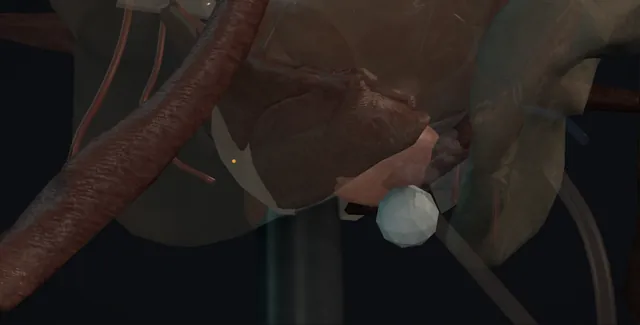
Schematic scene preview: Transperineal prostate biopsy
Preparing the 3D anatomy…

UrOops3D · UroRef · © Nity G

Click me next Section 2 of 8 Anatomy The structures this operation works between, and the one it must not cross.

The structures in the scene

Transperineal prostate biopsy: how the structures relateAlong the needle from the perineum toward the base, probe in the rectum. Perineal skin and pelvic floor: pudendal branches beside the track Perineal and periprostatic vessels (danger). Perineal skin and pelvic floor: the needle enters the gland from below Apex and external sphincter. Apex and external sphincter: immediately behind; never entered Rectal wall and Denonvilliers' fascia (danger). Apex and external sphincter: cores run along the long axis to the base Prostate on the biplanar probe. Prostate on the biplanar probe: a core through it bleeds; retention follows Urethra and transition zone (danger). Prostate on the biplanar probe: a core fired up from the base can enter it Bladder neck (danger).Perineal skinBladderPerineal skin and pelvicfloor!Perineal andperiprostaticvessels!Apex and externalsphincter!Rectal wall andDenonvilliers'fasciaProstate on the biplanarprobe!Urethra andtransition zone!Bladder neckpudendal branchesbeside the trackthe needle enters the glandfrom belowimmediately behind;never enteredcores run along the long axisto the basea core through itbleeds; retentionfollowsa core fired up from the basecan enter itTargetDangerLandmark
Along the needle from the perineum toward the base, probe in the rectum. Schematic, not to scale; relationships only.
  • From the foot of the table the perineum faces the operator with the probe in the rectum below and the gland on the screen above it, the urethra a bright midline line running from the bladder at the top of the image to the apex at the bottom.
  • The needle enters the skin on either side of the midline, passes through the pelvic floor and enters the gland from below, so that a core runs along the long axis of the gland toward the base, with the rectum behind the needle and the urethra beside it.
  • The anterior gland is reached by angling the same needle upward toward the pubis.
  • ›The prostate on the biplanar probe - sagittal and axial views; the peripheral zone posterior and lateral, the transition zone around the urethra, the anterior stroma in front.
  • ›The urethra - the echogenic midline line from bladder neck to apex; the needle is planned around it on every core.
  • ›The apex and the external sphincter - the lowest gland, sampled with the needle nearly parallel to the urethra; the sphincter just beyond it is not fired through.
  • ›The rectal wall and Denonvilliers' fascia - immediately behind the gland; the block is placed in front of it and the needle never enters it.
  • ›The neurovascular bundles - posterolateral at the apex; the site of the periprostatic block and of the pain if it is missed.
  • ›The perineal skin and pelvic floor - the puncture sites a centimetre and a half above the anus on each side of the midline; infiltrated along the track.
  • ›The bladder neck - above the base; a core fired upward from the base can enter it, so the throw is seen before the shot.
Click me next Section 3 of 8 Steps The operation in order, with the landmark that guards each move.

Operative steps

  1. Who, and the MRI first The man with a raised age-specific PSA or an abnormal prostate on examination, fit enough and willing to be treated if cancer is found, or already on active surveillance and due a repeat. Multiparametric MRI comes before any needle: a Likert or PI-RADS score of 3 or more is offered biopsy with targeted cores; a score of 1 or 2 with a low PSA density can be offered no biopsy after a discussion of what may be missed. The MRI report gives the lesion, its zone, side and size, and the prostate volume; the PSA density is calculated from it. Active surveillance, a previous negative biopsy with a rising PSA, or an equivocal MRI is a targeted and systematic biopsy with the question written on the request.
  2. Consent and preparation, no rectal route The consent names haematuria in most men for a week or two, haematospermia for weeks, perineal bruising, discomfort during the block, acute retention in a few per cent, infection in well under one per cent, a small chance of a non-diagnostic biopsy and a repeat, and that a negative biopsy does not exclude cancer. Antibiotic prophylaxis is a single dose or none according to local policy, since the transperineal route does not cross the rectal mucosa. Aspirin continues; other antiplatelets and anticoagulants follow the local bleeding policy and are stopped or bridged before the list. A urine dip that shows infection postpones the biopsy. No enema, no starvation, a chaperone and a nurse for the awake patient, who has emptied his bladder.
  3. Position, the probe and the template or freehand grid Lithotomy with the perineum at the edge of the table and the scrotum lifted out of the field with tape. A biplanar transrectal probe on a stepper or held by the assistant, with lubricant and a condom, gives the sagittal and axial views. The prostate is measured, the urethra found in the midline as the echogenic line from bladder neck to apex, and the MRI lesion located by zone and by clock position. With the freehand technique an access needle with a guide is placed through one puncture on each side of the midline, about a centimetre and a half above the anus, and angled to reach every sector; with the grid technique a brachytherapy template on the stepper gives five millimetre coordinates. The perineal skin is prepared and draped.
    where it bites: the urethra runs in the midline from bladder neck to apex; every needle path is planned around it, not through it.
  4. Local anaesthetic: skin, then the periprostatic and apical block Lidocaine 1 per cent, 10 to 20 millilitres, with the total dose calculated for the man's weight. A skin bleb at each puncture site, then the subcutaneous fat and the pelvic floor infiltrated along the intended track while watching the needle on the sagittal view. The periprostatic block is placed at the apex where the neurovascular bundles enter, between the prostate and the rectum, five to ten millilitres on each side, and seen to spread as a hypoechoic wheal that lifts the gland; some add a second deposit along the lateral border. Three to five minutes are allowed to pass. The awake man is told what he will feel: pressure, the click of the gun, an urge to pass urine when the needle is near the apex.
    where it bites: the rectal wall lies immediately behind the apex; the block needle is watched on the sagittal view so that it does not enter the rectum and carry organisms forward.
  5. Targeted cores to the lesion The MRI lesion first, while the gland is undisturbed and the ultrasound is not clouded by blood. With software fusion the MRI contour is overlaid on the ultrasound and the needle guided into it; with cognitive fusion the operator places the needle by zone, side, level and clock position from the report and from what is seen on the screen, since many lesions are visible as a hypoechoic area. The 18 gauge spring-loaded needle is advanced to the edge of the target on the sagittal view, fired, and the core inspected for length. Three to five cores are taken from each lesion, through and around it, into a pot labelled with the lesion number and side. A second lesion is sampled separately.
  6. Systematic cores, sector by sector Then the systematic cores, because a proportion of significant cancer lies outside the MRI lesion and because the systematic pattern maps the gland for surveillance and for planning. The gland is divided into sectors, base, mid and apex on each side, medial and lateral, and one or two cores are taken from each into a labelled pot, about twelve cores in a freehand biopsy and more in a template mapping biopsy with sectors five millimetres apart. The needle is angled in the sagittal plane so that each core lies along the long axis of its sector and includes the peripheral zone, and the ultrasound is watched for the needle passing the urethra or into the bladder. The count is kept aloud with the nurse.
    where it bites: a core fired toward the bladder neck from the base can pass through the bladder wall; the tip and the throw are seen on the sagittal view before every shot.
  7. The apex and the anterior gland These are the two regions the transrectal route missed and the transperineal route reaches by design. The apical cores are taken with the needle nearly parallel to the urethra, entering low and staying in the peripheral zone under the sphincter, with the block already placed there because these are the cores that hurt. The anterior gland, the anterior fibromuscular stroma and the anterior horns of the peripheral and transition zones, is sampled by angling the needle upward from the same puncture so that the core runs through the anterior third; in a large gland a longer path or a second, higher puncture is needed. The anterior lesion on MRI in the man with several previous negative biopsies is the classic finding.
    where it bites: the external sphincter sits just beyond the apex; the apical core stays within the gland and the needle is not fired through the sphincter to reach it.
  8. After: haematuria, haematospermia, retention The punctures are pressed and dressed and the man sits up when he is ready. He passes urine before he leaves, and he is told what is normal: blood in the urine for a week or two, blood in the semen for several weeks, a bruise in the perineum, and some discomfort on sitting. He is told what is not normal: fever, rigors, inability to pass urine, or heavy bleeding with clots, and who to call. Acute retention follows in a few per cent, more after a large gland or a template biopsy, and is treated with a catheter, an alpha blocker and a trial without catheter at about a week. Sepsis is rare but is treated as sepsis: cultures and intravenous antibiotics without waiting for the results.
  9. Results and the MDT The pots go to histopathology with the labelling on the form matching the labelling on the pots, the MRI lesion number, the side and the sector, so that the report can be read against the MRI. The report gives the grade group, the number of cores and the percentage involved per sector, and perineural or extraprostatic extension where seen. Every positive biopsy goes to the multidisciplinary meeting, where the grade group, the PSA, the PSA density, the MRI stage and the man's fitness and wishes decide between surveillance and treatment. A negative biopsy with a persistent MRI lesion or a rising PSA density is not discharged: it is a repeat biopsy or an MRI at a defined interval, and the plan is written to the man and his general practitioner.
Click me next Section 4 of 8 Danger zones Where this operation bites, and why it bites there.
Critical relationship

Danger zones

The spatial relationships the animation is built to make automatic. Before you read each one, ask yourself: at this point in the operation, what is about to go wrong, and what is my first move? The save for each sits in the Complications tab.

  • RiskThe urethra and periurethral transition zone - a core through the urethra bleeds into the lumen and swells the gland; retention and heavy haematuria follow.
  • RiskThe apex and the external sphincter - the cores that hurt and the region the block must cover; a needle fired beyond the apex is in the sphincter.
  • RiskThe perineal and periprostatic vessels - the pudendal branches beside the track and the venous plexus in front of the gland; a haematoma in the perineum or under the capsule.
  • RiskThe anterior gland that is undersampled - the reason for repeated negative biopsies in a man with a rising PSA; sampled deliberately, not by chance.
Practise it Take me into the scene Opens the animation at a danger moment and asks you to predict the mechanism and your first move before it reveals.
Click me next Section 5 of 8 Complications The save for each one: the first move, the escalation trigger, the op-note line.

Complications and immediate saves

Trigger each of these live in the Animation tab, then read the save: the first move, the escalation trigger, and the line that goes in the op note.

Acute urinary retention

  • the man who cannot pass urine before he leaves or that night: oedema and clot in a gland that was already obstructing, more after a large gland or a template mapping biopsy.
The save
  1. No man leaves before he has passed urine; a bladder scan if he has not, and a catheter if the volume is high and he cannot. first move
  2. A 14 or 16 French catheter passed gently, or a smaller one if the gland is large; a coude tip if it will not pass.
  3. An alpha blocker started that day and a trial without catheter at about a week, with a plan if it fails.
  4. A catheter that will not pass, or clot retention with a full bladder: a suprapubic catheter under ultrasound with senior help, or a three-way catheter and washout. escalation trigger
  5. Note: the residual, the catheter size, the alpha blocker, the trial without catheter date and the warning given about a second retention. document

Bleeding: haematuria, haematospermia, perineal haematoma

  • blood in the urine for a week or two and in the semen for weeks is expected; clots, a swelling in the perineum or a fall in haemoglobin are not.
The save
  1. Warn every man before the biopsy so that expected bleeding does not bring him back; pressure and a dressing on the punctures at the end. first move
  2. Antiplatelets and anticoagulants managed before the list by the written policy, and restarted at the agreed time after.
  3. Haematuria with clots: fluids, and if retention threatens, a three-way catheter and a washout; the perineum examined for a haematoma.
  4. A haematoma that is expanding, a falling haemoglobin or haemodynamic change: admit, cross-match, image, and senior review for embolisation or exploration. escalation trigger
  5. Note: the anticoagulant plan, the number of cores, what was said about bleeding, and the restart date. document

Infection and sepsis

  • rare by the transperineal route because the rectal mucosa is never crossed, but a man with fever and rigors after a biopsy is septic until proved otherwise.
The save
  1. Fever, rigors or feeling unwell within the first week: he is seen, not reassured over the telephone; observations, urine and blood cultures. first move
  2. Intravenous antibiotics by the local policy at once, before the cultures return; the prophylaxis given, if any, is noted.
  3. A urine dip that was positive before the biopsy should have postponed it; a catheter in situ is changed.
  4. Hypotension, a rising lactate or no response within twenty-four hours: sepsis pathway, critical care review, and imaging for an abscess of the prostate or perineum. escalation trigger
  5. Note: the prophylaxis, the cultures sent, the antibiotic started and the time, and the review plan. document

Pain, bruising and the failed block

  • the awake man who cannot tolerate the biopsy: a block that missed the apex, an anxious patient, or a large gland needing many cores.
The save
  1. Stop and top up the block at the apex under vision before continuing; the cores at the apex are the painful ones. first move
  2. Talk him through every core; entonox where available; a shorter systematic scheme in the man who is struggling rather than an incomplete one done badly.
  3. Check the total lidocaine dose against his weight before any top-up.
  4. A biopsy that cannot be completed awake: stop, record what was taken, and rebook under sedation or general anaesthetic rather than persist. escalation trigger
  5. Note: the lidocaine dose, the cores taken, the sectors not taken and the reason, and the plan for completion. document

Non-diagnostic or false-negative sampling

  • a negative biopsy in a man with a persistent lesion or a rising PSA density: the target missed, the anterior gland undersampled, or a labelling error that mislocates the cancer.
The save
  1. Read the histology against the MRI at the meeting: a negative targeted core from a high-scoring lesion is a technical question, not a reassurance. first move
  2. Check the core lengths on the report; short or fragmented cores from a sector mean that sector was not sampled.
  3. Labelling reconciled between the pots and the form at the time of the biopsy, with the nurse reading each label aloud.
  4. A persistent lesion, a rising PSA density or a suspicious digital examination with a negative biopsy: repeat targeted biopsy, with fusion and more cores, or an MRI at a defined interval. escalation trigger
  5. Note: the lesion number, the cores per target, the sectors sampled, the pot labels and the follow-up interval. document
Click me next Section 6 of 8 Troubleshooting When the case turns: the problem, then the next move.

Intra-operative troubleshooting

When the case turns: the problem, then the next move.

  • ActionThe lesion cannot be seen on ultrasound - place it cognitively by zone, side, level and clock position from the report, take five cores through that region, and say so in the note; fusion software if the unit has it.
  • ActionThe needle keeps hitting the pubic arch - the anterior gland in a large prostate; drop the entry point lower on the perineum and angle upward, or use a longer path from the contralateral puncture.
  • ActionBlood on the screen obscures the gland - the targeted cores were meant to come first; press on the perineum, wait, and change the angle so the needle track does not shadow the next sector.
  • ActionThe man cannot tolerate the apical cores - top up the periprostatic block at the apex on each side under vision, wait three minutes, and warn him before each shot.
  • ActionUrine in the needle tract or a core that is bladder mucosa - the needle passed the base into the bladder; redirect lower and shorten the throw, and expect haematuria.
  • ActionHe has not passed urine an hour after the biopsy - bladder scan; a small volume is a drink and time, a large volume with discomfort is a catheter today, not a discharge with advice.
Click me next Section 7 of 8 FRCS viva Say it out loud, then mark yourself against the model answer.

FRCS viva questions

The examiner stems this scene prepares you for. Try to answer out loud first, then reveal, and learn the discriminator, the detail that turns a pass into a strong pass.

QA 64 year old man has a PSA of 6.8. How do you decide whether he needs a biopsy, and which biopsy?reveal answer
  • I would repeat the PSA if the first was taken during an infection or soon after instrumentation, examine the prostate and estimate his fitness and his wishes, since a man who would not accept treatment does not benefit from diagnosis.
  • If the PSA is confirmed raised I would arrange multiparametric MRI before any biopsy, in line with NICE guidance, reported with a Likert or PI-RADS score and a volume so that a PSA density can be calculated.
  • A score of 3 or more is offered biopsy with targeted cores to the lesion and systematic cores of the rest of the gland.
  • A score of 1 or 2 with a PSA density below about 0.15 can be offered no biopsy after a discussion of the small proportion of significant cancer that will be missed, with PSA follow-up;
  • an abnormal examination or a high density argues for biopsy anyway.
  • The biopsy would be transperineal under local anaesthetic, because it has a lower infection rate than the transrectal route, samples the apex and the anterior gland better, and can be done awake as a day case.
  • I would consent him for haematuria, haematospermia, retention, infection and the chance of a negative biopsy that does not exclude cancer.
Strong pass

Say all three

  • MRI before biopsy with the density threshold quoted
  • The transperineal route justified by both infection
  • Sampling of the anterior gland
Examiner note
MRI before biopsy with the density threshold quoted, and the transperineal route justified by both infection and sampling of the anterior gland.
QDescribe a local anaesthetic transperineal prostate biopsy.reveal answer
  • With the man in lithotomy after a urine dip and a check of his anticoagulants, I would place a biplanar transrectal probe, measure the gland, identify the urethra in the midline and locate the MRI lesion by zone, side and level.
  • I would infiltrate lidocaine 1 per cent into the skin about a centimetre and a half above the anus on each side of the midline, along the track through the pelvic floor, and then place a periprostatic block at the apex on each side under sagittal ultrasound vision, watching the wheal spread and keeping the needle in front of the rectum.
  • After a few minutes I would place an access needle through each puncture and take the targeted cores first, three to five through and around the lesion, into a labelled pot, and then systematic cores from each sector, base, mid and apex, medial and lateral on each side, about twelve in all, angling the needle so that each core runs along its sector and includes the peripheral zone, with the apex sampled nearly parallel to the urethra and the anterior gland by angling upward.
  • I would watch the needle tip and throw on every shot, keep the count aloud, reconcile the pot labels with the form, and make sure he passes urine before he leaves.
Strong pass

Say all three

  • The block placed at the apex under vision before the apical cores
  • The targeted cores taken before the gland is clouded
  • The labels reconciled at the time
Examiner note
The block placed at the apex under vision before the apical cores, the targeted cores taken before the gland is clouded, and the labels reconciled at the time.
QWhat are the complications of transperineal biopsy, and how does it compare with the transrectal route?reveal answer
  • The common complications are bleeding and voiding: haematuria in most men for up to two weeks, haematospermia for several weeks, perineal bruising and discomfort, and acute urinary retention in a few per cent, higher after a large gland or a template mapping biopsy, treated with a catheter, an alpha blocker and a trial without catheter at about a week.
  • Infection is the complication the route was chosen to avoid: because the needle does not cross the rectal mucosa, sepsis after transperineal biopsy is well under one per cent, compared with about one to three per cent after transrectal biopsy, and antibiotic prophylaxis can be a single dose or omitted according to local policy.
  • Rarer problems are a perineal or periprostatic haematoma, a vasovagal episode during the block, local anaesthetic toxicity if the dose is not calculated, and pain from a block that missed the apex.
  • The diagnostic complication is the false negative, and here the transperineal route has the advantage of reaching the anterior gland and the apex, which the transrectal needle undersamples.
  • The trade-off is a longer procedure, more punctures and more local anaesthetic, and the need for a probe holder or template.
  • These figures are mainstream teaching.
Strong pass

Say both

  • The sepsis comparison quoted with numbers and the anterior gland named as the sampling advantage
  • Not just the infection advantage
Examiner note
The sepsis comparison quoted with numbers and the anterior gland named as the sampling advantage, not just the infection advantage.
QThe biopsy is negative but the MRI showed a PI-RADS 4 lesion. What now?reveal answer
  • I would not discharge him.
  • A negative targeted biopsy from a high-scoring lesion is a discordant result, and the first question is technical: were the targeted cores truly in the lesion, how many were taken, were they of adequate length, and were the pots labelled correctly against the MRI.
  • I would review the histology alongside the MRI at the multidisciplinary meeting with the radiologist, and reconsider the lesion itself, since some PI-RADS 4 lesions are benign mimics such as prostatitis, a benign nodule or the anterior fibromuscular stroma.
  • I would calculate the PSA density and look at the PSA trend.
  • If the density is high, the examination is suspicious or the lesion is anterior or apical where sampling is hardest, I would repeat the targeted biopsy with software fusion if available, taking more cores through the lesion and including the anterior gland deliberately.
  • If the density is low and the radiologist downgrades the lesion on review, I would offer a repeat MRI and PSA at a defined interval, usually six to twelve months, with the man told plainly that a negative biopsy does not exclude cancer.
  • Either way the plan, the interval and who is responsible for it are written to him and his general practitioner.
Strong pass

Say all three

  • Treating discordance as a technical question first
  • Naming the benign mimics
  • A written surveillance plan rather than a discharge
Examiner note
Treating discordance as a technical question first, naming the benign mimics, and a written surveillance plan rather than a discharge.
Click me next Section 8 of 8 Evidence The guidance each of these answers is built on.

Evidence and guidance

Two minutes

Close the lap: recall it before you leave.

Say it aloud, then reveal. Reading is not remembering.

  1. 01 The relationship Say the critical spatial relationship out loud, then reveal. reveal
    Memory frame

    From the foot of the table the perineum faces the operator with the probe in the rectum below and the gland on the screen above it, the urethra a bright midline line running from the bladder at the top of the image to the apex at the bottom.

    Why?
    The needle enters the skin on either side of the midline, passes through the pelvic floor and enters the gland from below, so that a core runs along the long axis of the gland toward the base, with the rectum behind the needle and the urethra beside it. The anterior gland is reached by angling the same needle upward toward the pubis.
  2. 02 The first move Acute urinary retention. Your first move? reveal
    First move

    No man leaves before he has passed urine; a bladder scan if he has not, and a catheter if the volume is high and he cannot.

  3. 03 The escalation trigger Acute urinary retention. What makes you call for help? reveal
    Escalate when

    A catheter that will not pass, or clot retention with a full bladder

    More detail
    A catheter that will not pass, or clot retention with a full bladder: a suprapubic catheter under ultrasound with senior help, or a three-way catheter and washout.
  4. 04 The discriminator What turns a pass into a strong pass? reveal
    Strong pass

    Say all three

    • MRI before biopsy with the density threshold quoted
    • The transperineal route justified by both infection
    • Sampling of the anterior gland
    Examiner note
    MRI before biopsy with the density threshold quoted, and the transperineal route justified by both infection and sampling of the anterior gland.
  5. 05 The pitfall Where does this operation bite first? reveal
    Pitfall

    The urethra and periurethral transition zone

    Why?
    a core through the urethra bleeds into the lumen and swells the gland; retention and heavy haematuria follow.

Revision practice with self-marked answers, not an assessment of clinical competence. Kept in this browser only; it does not sync between devices.