
Transperineal prostate biopsy
Ultrasound-guided sampling of the prostate through the perineal skin, using targeted, systematic or mapping strategies according to the diagnostic question.
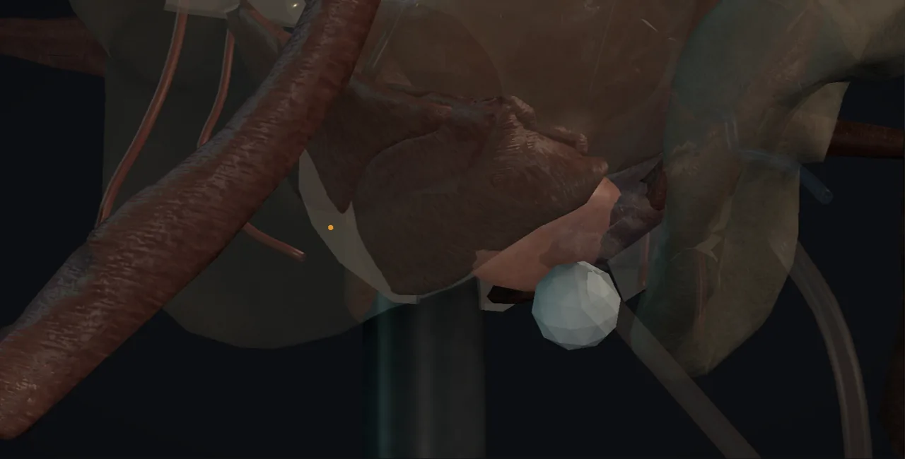 Interactive 3D Interactive transperineal biopsy, core by core Open the 3D animation
Teaching catalogue only. This coverage record is not a patient pathway or operative instruction.
Typical place in practice
Histological diagnosis, risk stratification or surveillance when prostate cancer is suspected or already under active monitoring.
Alternatives and neighbouring procedures
MRI-led surveillance or other biopsy routes may be considered in selected pathways, but tissue is required for many definitive decisions.
Same procedure family, explicit technique branches
These are variations of this canonical operation. Complication modules below identify whether they apply to all branches or only to named approaches.
Freehand transperineal biopsy
Targeted and systematic cores are obtained through one or more perineal access points without a fixed grid.
Grid-based template biopsy
A brachytherapy-style grid supports reproducible sampling across defined prostate sectors.
MRI-targeted biopsy
Suspicious MRI targets are sampled with image registration or cognitive targeting, usually alongside planned systematic cores.
Transperineal mapping biopsy
More extensive sectoral sampling maps disease distribution for selected complex decisions.
High-level sequence
Coverage outline only; not a step-by-step technical guide.
- Confirm the diagnostic question, MRI targets and sampling plan
- Position and prepare the perineum with ultrasound visualisation
- Define prostate, urethra and target sectors
- Obtain labelled targeted and systematic cores
- Assess voiding and bleeding before discharge and reconcile pathology labels
Every failure mode is its own module.
Timing and variation coverage are recorded for each module.
Acute urinary retention
Prostatic swelling, bleeding and baseline outlet obstruction can impair voiding, with risk increasing as sampling becomes more extensive.
Applies to: Freehand transperineal biopsy · Grid-based template biopsy · MRI-targeted biopsy · Transperineal mapping biopsyHaematuria, haematospermia or perineal bleeding
Needle tracks commonly produce self-limited urinary, seminal or perineal bleeding but may occasionally form a significant haematoma.
Applies to: Freehand transperineal biopsy · Grid-based template biopsy · MRI-targeted biopsy · Transperineal mapping biopsyUrinary infection or sepsis
The transperineal route reduces rectal contamination but urinary and systemic infection remain possible.
Applies to: Freehand transperineal biopsy · Grid-based template biopsy · MRI-targeted biopsy · Transperineal mapping biopsyPain, bruising or sensory disturbance
Multiple perineal punctures and positioning may cause short-term discomfort, bruising or altered sensation.
Applies to: Freehand transperineal biopsy · Grid-based template biopsy · MRI-targeted biopsy · Transperineal mapping biopsyNon-diagnostic or false-negative sampling
Targeting error, inadequate core distribution or specimen-labelling problems can miss or mislocalise clinically important disease.
Applies to: Freehand transperineal biopsy · Grid-based template biopsy · MRI-targeted biopsy · Transperineal mapping biopsySources for this module
Radical prostatectomy, lymph-node dissection and selected focal-treatment context.
Accessed 2026-08-03 · opens source European Association of Urology EAU Guidelines on Urological InfectionsInfection prevention, recognition and procedure-associated infection context.
Accessed 2026-08-03 · opens source British Association of Urological Surgeons Patient information leaflets by procedureUK patient-facing procedure, alternatives, recovery and complication framing.
Accessed 2026-08-03 · opens source