UrOops3D, danger-zone atlas

Schematic model.

Operative animation, Transplant lymphocele

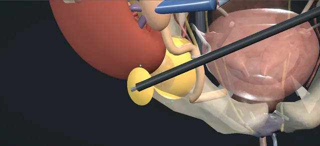
Treatment of a transplant lymphocele: drainage and the laparoscopic peritoneal window

A collection of lymph around a renal graft, from lymphatics divided over the iliac vessels or at the graft hilum, that presses on the transplant ureter, the graft or the iliac vein.

The fluid is first proved to be lymph, by a creatinine close to the serum value, and then drained under ultrasound. A lymphocele that comes back is opened into the peritoneal cavity through a wide laparoscopic window.

The lesson is the transplant ureter, which runs along the wall of the collection and is found before anything is cut.

Advanced tier FRCS transplant and specialist urology, transplant surgical complications Interactive 3D

Operative visualisation

Schematic scene preview: Transplant lymphocele
Preparing the 3D anatomy…

UrOops3D · UroRef · © Nity G

How to use this module. Play the nine steps, or use Back and Next. Watch the needle reach the collection and the drain empty it, then the ports, the ureter marked on the medial wall, the window cut and lifted out, its edges clipped. The triggers show a recurrence, a bowel injury, a venous bleed and a cut ureter.

Click me next Section 2 of 8 Anatomy The structures this operation works between, and the one it must not cross.

The structures in the scene

Transplant lymphocele: how the structures relateThe lymphocele in the right iliac fossa, and what lies against its wall. Lymphocele: a thin wall, no lining: medial to its lower pole; the needle stays clear Renal graft and its hilum (danger). Lymphocele: a thin wall, no lining: over its medial wall; found before any cut Transplant ureter (danger). Lymphocele: a thin wall, no lining: deep and medial; compressed, a deep cut tears it External iliac vein and artery (danger). Lymphocele: a thin wall, no lining: medial to it; the ureter joins its wall Bladder. Lymphocele: a thin wall, no lining: covers its dome; cut with it as one window Peritoneum over the dome. Lymphocele: a thin wall, no lining: in front; ports under vision, the tilt moves it Small bowel and caecum (danger).Lymphocele: a thinwall, no lining!Renal graft and itshilum!Transplant ureter!External iliac veinand arteryBladderPeritoneum over thedome!Small bowel andcaecummedial to its lowerpole; the needlestays clearover its medialwall; found beforeany cutdeep and medial;compressed, a deepcut tears itmedial to it; theureter joins itswallcovers its dome;cut with it as onewindowin front; portsunder vision, thetilt moves itTargetDangerLandmark
The lymphocele in the right iliac fossa, and what lies against its wall. Schematic, not to scale; relationships only.
  • The graft lies under the wall of the right iliac fossa, hilum medial, its vessels on the external iliacs.
  • The lymphocele sits medial to the lower pole, between the graft and the bladder, on the external iliac vein.
  • The transplant ureter runs over its medial wall to the bladder; peritoneum and bowel lie in front.
  • ›The renal graft - in the right iliac fossa, superficial under the wall; its lower pole lies against the collection.
  • ›The graft hilum - medial to the graft, with its vein and artery joined end to side to the external iliac vessels.
  • ›The transplant ureter - from the graft pelvis to the bladder; stretched over the medial wall of the collection and compressed by it.
  • ›The external iliac vein - deep and medial to the collection; flattened under it, which is one cause of the swollen leg.
  • ›The external iliac artery - beside the vein, carrying the graft artery; away from the window but in the field.
  • ›The lymphocele wall - a thin fibrous wall without an epithelial lining; in the window it is cut with the peritoneum over it as one layer.
  • ›The bladder - medial to the collection; the reimplanted ureter joins its anterolateral wall.
  • ›The peritoneum - over the front of the collection; the window opens the cavity into the peritoneal space.
  • ›The small bowel and the caecum - in front of the field; the tilt moves them away, and they must not seal the window.
Click me next Section 3 of 8 Steps The operation in order, with the landmark that guards each move.

Operative steps

  1. The collection Some weeks after the transplant, the creatinine rises, the leg on the side of the graft swells, or there is a fullness over the graft. A thin-walled lymphocele lies between the lower pole and the bladder, on the external iliac vein, with the transplant ureter stretched over its medial wall.
    where it bites: a collection that compresses the ureter costs the graft nephrons for as long as it is left.
  2. Ultrasound and the differential Ultrasound shows the graft pelvis and calyces dilated and an echo-free collection beside the graft, pressing on the ureter, with the vein flattened under it on the Doppler. A collection around a graft is a lymphocele, a urinoma, a haematoma or an abscess until the fluid says which.
    where it bites: a urinary leak that is drained as if it were lymph goes on leaking; the fluid is analysed before anything definitive is done.
  3. Aspiration and the fluid creatinine Under ultrasound a needle is passed onto the dome of the collection from in front and lateral, away from the ureter, the vein and the bowel. Clear straw fluid is sent for creatinine, with a serum creatinine taken at the same time, and for cells and culture. Close to the serum value means lymph; far above it, urine.
    where it bites: the needle line passes lateral to the ureter on the medial wall and clear of the graft and the iliac vessels.
  4. Percutaneous drain A drain is left in the cavity along the same track. The collection empties, the ureter falls back and the graft decompresses. Where drainage persists, sclerotherapy through the drain is an option under the unit protocol for the agent. A collection that comes back after the drain is removed goes to a peritoneal window.
    where it bites: every day the drain stays in is another day for infection to reach the graft.
  5. Ports and the bulge Under general anaesthetic with a catheter in the bladder, the camera port is placed at the umbilicus and two working ports on the left, each after the first under vision. Head down and tilted to the left, the bowel falls away and the collection is seen bulging under the peritoneum lateral to the bladder.
    where it bites: bowel can lie across the fossa or be stuck to the collection; a port placed without vision can enter it.
  6. The ureter first Before anything is cut, the transplant ureter is identified on the medial wall of the collection, with the external iliac vein deep and medial to it. The window is planned on the dome of the bulge, as wide as the dome allows, away from the ureter, the graft and the vessels.
    where it bites: the transplant ureter runs along the medial wall of the collection, stretched and thinned over it; it is the structure at risk in this operation.
  7. The window The dome is held up in a grasper and the wall is cut round with scissors, peritoneum and lymphocele wall together as one layer. The disc is lifted out in the grasper and sent for histology, and the lymph drains into the peritoneal cavity, which absorbs it.
    where it bites: a window carried round onto the medial wall goes across the ureter; a cut taken deep goes into the iliac vein.
  8. Edges secured and the check Clips are placed on the cut edge so that the window stays open and the bowel cannot seal it. The cavity is inspected: the ureter whole on the medial wall, no urine in the field, and no bleeding when the insufflation pressure is lowered.
    where it bites: only this inspection finds a ureteric or venous injury before the patient leaves theatre.
  9. After The ports come out under vision and the wounds are closed. Ultrasound before discharge and at follow-up shows no collection and the graft pelvis back to normal, and the creatinine falls. A recurrence after a wide window is uncommon and is imaged again.
    where it bites: a creatinine that does not fall needs imaging: an obstructed or injured ureter is found by ultrasound, not by waiting.
Click me next Section 4 of 8 Danger zones Where this operation bites, and why it bites there.
Critical relationship

Danger zones

The spatial relationships the animation is built to make automatic. Before you read each one, ask yourself: at this point in the operation, what is about to go wrong, and what is my first move? The save for each sits in the Complications tab.

  • RiskTransplant ureter - on the medial wall of the collection, stretched over it; identified before the window is cut and inspected at the end.
  • RiskExternal iliac artery and vein - deep and medial to the collection; a deep cut or a blind dissection on the medial wall reaches the vein.
  • RiskSuperficial transplant kidney and hilum - lateral and cranial to the collection, under the wall; the needle and the window stay clear of it.
  • RiskBowel overlying the collection - across the fossa or adherent to the bulge; ports under vision and the tilt keep it out of harm.
Practise it Take me into the scene Opens the animation at a danger moment and asks you to predict the mechanism and your first move before it reveals.
Click me next Section 5 of 8 Complications The save for each one: the first move, the escalation trigger, the op-note line.

Complications and immediate saves

Trigger each of these live in the Animation tab, then read the save: the first move, the escalation trigger, and the line that goes in the op note.

Recurrence after drainage alone

  • aspiration and drainage do not remove the wall that secretes and holds the lymph; the collection fills again after the drain comes out and the graft is obstructed once more.
The save
  1. Image it again: confirm a collection, the hydronephrosis and the creatinine trend. first move
  2. Drain it again for the graft's sake, and send the fluid once more to exclude a urinary leak.
  3. Sclerotherapy through the drain may be tried under the unit protocol for the agent.
  4. A second recurrence, or persistent drainage, is an indication for a laparoscopic window: discussed with the transplant team. escalation trigger
  5. Record the dates, the fluid results, the volumes drained and the plan. document

Bowel injury on entry

  • a trocar placed without vision enters a loop of bowel lying across the fossa or stuck to the collection.
The save
  1. Leave the trocar where it is, so that the hole can be found. first move
  2. Place another port under vision and inspect the loop on both sides of the trocar.
  3. Call a general or colorectal surgeon to theatre: the repair is decided with them, and converted to open if it cannot be closed safely. escalation trigger
  4. The window is completed or abandoned depending on the contamination; an infected field around a graft is not left undrained.
  5. Op note: the port, the loop, the size of the hole, the repair and who did it; the patient is told. document

Bleeding from the iliac vein

  • dissection on the medial wall taken too deep, or a cut through the base of the wall, tears the external iliac vein beneath it.
The save
  1. Pressure at once with a grasper and a swab through a port; tell the anaesthetist. first move
  2. Raise the insufflation pressure briefly, add suction, and see the tear before any clip or suture goes on.
  3. Call the vascular or transplant surgeon; convert to open if the bleeding is not controlled at once. escalation trigger
  4. Check the graft perfusion and the leg once it is controlled.
  5. Op note: the site, the size, the repair, the blood loss and the transfusion. document

Transplant ureter injury

  • the window is carried round onto the medial wall and cuts across the transplant ureter stretched over it; urine appears in the field.
The save
  1. Stop cutting. Recognise the injury now: urine in the field, the cut ends of the ureter on the medial wall. first move
  2. Call the transplant surgeon to theatre: the repair is planned with them. escalation trigger
  3. The ureter is repaired over a stent, by a primary repair or a reimplant into the bladder depending on its length and its blood supply.
  4. A catheter stays in; the drain fluid creatinine and the graft ultrasound are followed afterwards.
  5. Op note: where the ureter was cut, how it was repaired, the stent and who did it; the patient is told. document
Click me next Section 6 of 8 Troubleshooting When the case turns: the problem, then the next move.

Intra-operative troubleshooting

When the case turns: the problem, then the next move.

  • ActionThe fluid creatinine is far above the serum value - it is urine, not lymph: a urinary leak from the ureter or the reimplant. Drainage, a catheter, a nephrostomy and imaging of the ureter follow, not a peritoneal window.
  • ActionThe fluid is turbid, or the patient is febrile - treat it as an abscess or an infected lymphocele: culture, antibiotics and external drainage. An infected collection is not opened into the peritoneal cavity.
  • ActionThe collection cannot be found laparoscopically - intraperitoneal ultrasound shows the collection and the structures round it; filling it through a drain left in place makes it bulge.
  • ActionThe ureter cannot be seen on the medial wall - a ureteric stent or catheter placed beforehand makes it easier to find. A window is not cut where the ureter has not been identified.
  • ActionThe collection lies lateral or deep to the graft - it may not be reachable from the peritoneal cavity without risk to the graft; drainage or an open approach is the safer choice.
  • ActionA small collection found on a routine scan - most need no treatment: a lymphocele is treated when it obstructs the graft, compresses the vein, causes symptoms or becomes infected.
Click me next Section 7 of 8 FRCS viva Say it out loud, then mark yourself against the model answer.

FRCS viva questions

The examiner stems this scene prepares you for. Try to answer out loud first, then reveal, and learn the discriminator, the detail that turns a pass into a strong pass.

QSix weeks after a renal transplant, a routine ultrasound shows a collection beside the graft and a dilated graft pelvis. What is the differential, and how do you tell them apart?reveal answer
  • A collection around a graft is a lymphocele, a urinoma, a haematoma or an abscess, and the timing helps: a haematoma is usually in the first days and a urinoma in the first weeks, while a lymphocele usually appears over weeks.
  • I would look at the creatinine, the haemoglobin, the temperature and the white count, and on ultrasound at the graft pelvis, the ureter and the iliac vein.
  • Then I would aspirate it under ultrasound and send the fluid for creatinine with a serum creatinine taken at the same time, for cells and for culture.
  • A fluid creatinine close to the serum value means lymph; far above it, urine from a leak; blood means a haematoma; and pus, or organisms on culture, an abscess.
  • The guideline says a urinary leak can be suspected from the creatinine in drain fluid.
  • The distinction decides the treatment, because a urinoma needs the urinary tract drained and imaged, whereas a lymphocele needs the cavity drained or opened.
Strong pass

Say both

  • Comparing the fluid creatinine with a serum creatinine taken at the same time
  • Letting the fluid decide the treatment
Examiner note
Comparing the fluid creatinine with a serum creatinine taken at the same time, and letting the fluid decide the treatment.
QWhen does a lymphocele need treatment, and what are the options?reveal answer
  • Lymphocele is common; the EAU guideline gives an incidence of 1 to 26 per cent, associated with diabetes, mTOR inhibitors and acute rejection, and most are small and need nothing.
  • I would treat one that obstructs the graft, compresses the iliac vein, causes symptoms or becomes infected.
  • The guideline recommends percutaneous drainage as the first treatment for a large symptomatic lymphocele, and fenestration when percutaneous treatment fails, both as strong recommendations.
  • On the figures it gives, aspiration alone can recur in up to 95 per cent and carries a risk of local infection of 6 to 17 per cent, a pigtail drain succeeds in up to 50 per cent, and sclerosants reduce recurrence compared with aspiration.
  • In the systematic review of Lucewicz and colleagues, 52 retrospective series and 1,113 lymphoceles, 141 of 218 treated by aspiration alone recurred, 59 per cent, against 8 per cent after laparoscopic and 16 per cent after open surgery, with conversion from laparoscopic to open in 12 per cent.
  • There were no randomised trials.
Strong pass

Treating the collection for what it does to the graft and the vein, not for its size, and knowing that the evidence is retrospective.

QTalk me through a laparoscopic peritoneal window for a transplant lymphocele.reveal answer
  • I would confirm beforehand that the fluid is lymph, and I would consider a ureteric stent if the ureter is likely to be hard to find.
  • Under general anaesthetic with a catheter in the bladder, I place the camera port at the umbilicus and two working ports on the opposite side, each after the first under vision, and tilt the patient head down and away from the graft so that the bowel falls away.
  • The collection bulges under the peritoneum lateral to the bladder.
  • Before I cut, I identify the transplant ureter on the medial wall and the iliac vessels, and if the bulge is not clear I use intraperitoneal ultrasound or fill the cavity through a drain.
  • I hold up the dome and cut a wide window round it, peritoneum and lymphocele wall together, away from the ureter, the graft and the vessels, and send the disc for histology.
  • I secure the edges so that the window stays open; Taweemonkongsap and colleagues used clips for that.
  • I finish by inspecting the ureter, looking for urine and checking for bleeding at a lower pressure.
Strong pass

Identifying the transplant ureter before the window is cut, and making the window wide and keeping it open.

QWhy is the transplant ureter at particular risk, and what do you do if you cut it?reveal answer
  • The collection lies medial to the lower pole, and the transplant ureter runs from the graft pelvis to the bladder over its medial wall, stretched and thinned, so it can look like part of the wall.
  • Its only blood supply comes from the graft hilum, so a ureter that is injured or devascularised does not heal as a native ureter would.
  • The injury is in the series: Lima and colleagues had one ureteric injury among 25 laparoscopic fenestrations, and Taweemonkongsap and colleagues one allograft ureteric injury among nine.
  • If I saw urine in the field or the cut ends of the ureter, I would stop and call the transplant surgeon to theatre.
  • The ureter would be repaired over a stent, by primary repair or reimplantation into the bladder depending on the length and the blood supply that is left, with a catheter left in and the graft followed with ultrasound and the creatinine.
  • I would record it and tell the patient.
Strong pass

Say both

  • Knowing that the ureter lies on the wall of the collection and has a single blood supply from the graft
  • Recognising an injury in theatre
Examiner note
Knowing that the ureter lies on the wall of the collection and has a single blood supply from the graft, and recognising an injury in theatre.
Click me next Section 8 of 8 Evidence The guidance each of these answers is built on.

Evidence and guidance

Operative Dictations in Urologic Surgery
Armenakas NA, Fracchia JA, Golan R. Operative Dictations in Urologic Surgery. Wiley, 2019
The book has no chapter on the treatment of a transplant lymphocele, and nothing on this page is taken from it. The order of the steps follows the hub data for this operation (differentiate the fluid, map the graft, ureter, bladder, bowel and iliac vessels, drain by the safest route, create durable internal drainage, follow up) and the EAU guideline. The fluid analysis by a creatinine compared with a serum creatinine, the timing of the differential collections, the first moves in each complication, the troubleshooting lines and the danger line on each step are general practice. The scene is schematic: the collection is drawn as a smooth sac medial to the lower pole, the bowel is drawn only in the laparoscopic steps, the clip is the variation of Taweemonkongsap and colleagues, and no omental flap is drawn because none of the sources read describes one.
Open guideline
European Association of Urology
EAU Guidelines on Renal Transplantation, 2026 edition: section 3.1.7, recipient complications, lymphocele and urinary leakage, read 18 October 2026
Read through a fetch tool that returns a summary with quotations. Lymphocele 1 to 26 per cent, associated with diabetes, mTOR inhibitor therapy and acute rejection. Percutaneous aspiration with recurrence up to 95 per cent and local infection 6 to 17 per cent; a pigtail drain with success up to 50 per cent; sclerosants (ethanol, fibrin sealant, gentamicin, octreotide) reduce recurrence compared with aspiration; laparoscopic fenestration with the lowest recurrence (8 per cent) and complication (14 per cent) rates for a large symptomatic lymphocele. Recommendations, both strong: percutaneous drainage as the first treatment for a large symptomatic lymphocele; fenestration when percutaneous treatments fail. A urinary leak can be suspected from the creatinine in drain fluid.
Open guideline
Transplantation
Lucewicz A, Wong G, Lam VWT, Hawthorne WJ, Allen R, Craig JC, Pleass HCC. Management of primary symptomatic lymphocele after kidney transplantation: a systematic review. Transplantation 2011;92:663-673
Abstract read 18 October 2026. 52 retrospective case series, 1,113 primary lymphoceles; no randomised or prospective studies. Aspiration 218, sclerotherapy 155, drainage 219, laparoscopic 333, open 188. Aspiration alone recurred in 141 of 218 (59 per cent, 95% CI 52 to 67); laparoscopic surgery 8 per cent (6 to 12), open 16 per cent (10 to 24); conversion from laparoscopic to open 12 per cent (8 to 16). The authors find the evidence for laparoscopic surgery as first-line treatment weak.
Open guideline
International Brazilian Journal of Urology
Lima ML, Cotrim CAC, Moro JC, Miyaoka R, D'Ancona CAL. Laparoscopic treatment of lymphoceles after renal transplantation. Int Braz J Urol 2012;38:215-221
Abstract read 18 October 2026. 991 transplants, 25 symptomatic lymphoceles (2.52 per cent) treated by laparoscopic fenestration, a wide drainage into the abdominal cavity by excising the wall; indications graft dysfunction (84 per cent) and local symptoms; mean 14.2 weeks to surgery. Complications in 2 patients (8 per cent): one ureteral injury and one incisional hernia, both reoperated. One symptomatic recurrence at a mean follow-up of 36.2 months.
Open guideline
Journal of Endourology
Taweemonkongsap T, Srinualnad S, Nualyong C, Tantiwong A, Soontrapa S. Novel technique to prevent lymphocele recurrence after laparoscopic lymphocele fenestration in renal transplant patients. J Endourol 2006;20:654-658
Abstract read 18 October 2026. 9 symptomatic lymphoceles among 144 transplants (6.2 per cent), a mean 55.5 days after transplantation, treated laparoscopically; one converted to open; one allograft ureteral injury. Non-absorbable polymer ligating clips were used to keep the peritoneal window open: the first two patients, treated without clips, recurred, and none with clips recurred at a mean follow-up of 42.3 months.
Open guideline

Curriculum. Supports learning relevant to the ISCP Urology Curriculum syllabus topic Renal transplantation. Not a workplace-based assessment and not evidence of competence.

Two minutes

Close the lap: recall it before you leave.

Say it aloud, then reveal. Reading is not remembering.

  1. 01 The relationship Say the critical spatial relationship out loud, then reveal. reveal
    Memory frame

    The graft lies under the wall of the right iliac fossa, hilum medial, its vessels on the external iliacs.

    Why?
    The lymphocele sits medial to the lower pole, between the graft and the bladder, on the external iliac vein. The transplant ureter runs over its medial wall to the bladder; peritoneum and bowel lie in front.
  2. 02 The first move Recurrence after drainage alone. Your first move? reveal
    First move

    Image it again: confirm a collection, the hydronephrosis and the creatinine trend.

  3. 03 The escalation trigger Recurrence after drainage alone. What makes you call for help? reveal
    Escalate when

    A second recurrence, or persistent drainage, is an indication for a laparoscopic window: discussed with the transplant team.

  4. 04 The discriminator What turns a pass into a strong pass? reveal
    Strong pass

    Say both

    • Comparing the fluid creatinine with a serum creatinine taken at the same time
    • Letting the fluid decide the treatment
    Examiner note
    Comparing the fluid creatinine with a serum creatinine taken at the same time, and letting the fluid decide the treatment.
  5. 05 The pitfall Where does this operation bite first? reveal
    Pitfall

    Transplant ureter

    Why?
    on the medial wall of the collection, stretched over it; identified before the window is cut and inspected at the end.

Revision practice with self-marked answers, not an assessment of clinical competence. Kept in this browser only; it does not sync between devices.