UrOops3D, danger-zone atlas

Schematic model on the radical pelvis atlas.

Operative animation, Transureteroureterostomy

Transureteroureterostomy: one ureter joined to the other

The operation for a mid-ureteric injury when the bladder cannot be mobilised to reach it: the ureter is divided above the disease, taken across the midline in front of the great vessels and above the inferior mesenteric artery, and joined end to side to the other ureter.

The lesson is the route it takes, why it must lie without a kink or tension, and who should not have one, because both kidneys end up on one ureter.

Advanced tier FRCS reconstruction, ureter, ureteric injury Interactive 3D

Operative visualisation

Schematic scene preview: Transureteroureterostomy
Preparing the 3D anatomy…

UrOops3D · UroRef · © Nity G

How to use this module. Play through the eight steps, or use Back and Next. Watch the donor freed on its lateral side, the tunnel made above the artery, the ureter drawn across on its tag and the two mouths joined end to side. The triggers show a short donor, a kink at the artery, a failing join and a late stone.

Click me next Section 2 of 8 Anatomy The structures this operation works between, and the one it must not cross.

The structures in the scene

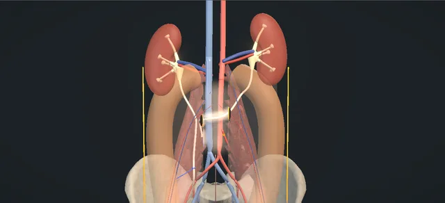
Transureteroureterostomy: how the structures relateBoth gutters open, colons lifted: the left ureter crossing to the right. Donor ureter crossing above the artery: sewn end to side, no tension Recipient ureter, anteromedial ureterotomy. Donor ureter crossing above the artery: passes through it, no kink Tunnel behind the root of the mesocolon. Donor ureter crossing above the artery: never below it Inferior mesenteric artery: cross above it (danger). Donor ureter crossing above the artery: crosses in front of both Aorta and cava under the tunnel (danger). Donor ureter crossing above the artery: left on the donor Medial periureteric tissue: the blood supply (danger). Donor ureter crossing above the artery: divided above it Scarred lower left ureter, tied and left.Donor ureter crossingabove the arteryRecipient ureter,anteromedialureterotomyTunnel behind theroot of themesocolon!Inferior mesentericartery: cross aboveit!Aorta and cava underthe tunnel!Medial periureterictissue: the bloodsupplyScarred lower leftureter, tied andleftsewn end to side,no tensionpasses through it,no kinknever below itcrosses in front ofbothleft on the donordivided above itTargetDangerLandmarkContext
Both gutters open, colons lifted: the left ureter crossing to the right. Schematic, not to scale; relationships only.
  • Seen from the front with both colons lifted medially, the two ureters lie on the psoas either side of the great vessels and cross the iliac vessels at the bifurcation.
  • The inferior mesenteric artery leaves the front of the aorta above the bifurcation and runs down and to the left.
  • The donor crosses above that origin, in front of the aorta and the cava and behind the root of the mesocolon, then comes down the medial side of the right ureter to meet its anteromedial face.
  • ›Left ureter: the donor - injured in its middle third; divided above the disease, freed cephalad on its lateral side and taken across the midline.
  • ›Right ureter: the recipient - normal, and left in its bed; opened on its anteromedial surface between two stays.
  • ›Bifurcation of the common iliac vessels - where each ureter is found and slung; the iliac vessels lie directly beneath it.
  • ›Aorta and inferior vena cava - the floor of the tunnel: the donor crosses in front of both.
  • ›Inferior mesenteric artery - leaves the front of the aorta above the bifurcation and runs down and to the left in the mesocolon; the donor crosses above its origin.
  • ›Root of the mesocolon and the posterior peritoneum - the window is made through the posterior peritoneum above the artery; the donor passes behind the root from the left retroperitoneum to the right.
  • ›Periureteric adventitia and medial tissue - carry the blood supply of the upper ureter; left on the donor, and left undisturbed on the recipient.
  • ›Gonadal vessels - cross in front of each ureter on the psoas above the brim; the donor is freed clear of them.
  • ›Colon and the white line of Toldt - both colons are mobilised along the white line and lifted medially to open the two gutters.
  • ›Both kidneys - the left now drains across the join into the right ureter; both are imaged in follow-up.
Click me next Section 3 of 8 Steps The operation in order, with the landmark that guards each move.

Operative steps

  1. Both ureters at the bifurcation An adult with a left mid-ureteric injury, a bladder that cannot be mobilised enough to reach it, and a normal right ureter and kidney. Supine, a urethral catheter, a midline incision from above the umbilicus to the pubis. The ascending and descending colon are mobilised along the white line of Toldt, the bowel is packed and a self-retaining retractor placed. Both ureters are identified and exposed where they cross the bifurcation of the common iliac vessels, and each is slung.
    where it bites: the iliac vessels lie directly under the ureter at the bifurcation and the gonadal vessels cross in front of it higher up; the right ureter is handled as little as possible from the start.
  2. Donor mobilised and divided The left ureter is dissected proximal to the diseased portion with its adventitia left on it. It is tied below, divided above the disease, and the upper end is tagged with a long fine stitch; urine is seen to run from it. Cephalad dissection then frees enough length to cross, leaving the periureteric tissue on the medial side intact so that the blood supply is not compromised. The scarred lower ureter stays where it lies.
    where it bites: above the brim the ureter takes its supply from its medial side; stripping the adventitia or the medial tissue buys length at the cost of the end that has to heal.
  3. The window above the artery The inferior mesenteric artery is identified on the front of the aorta. A window is made in the posterior peritoneum above it and in front of the great vessels, which gives a tunnel behind the root of the mesocolon from the left retroperitoneum to the right. A long curved clamp is passed through the tunnel from right to left to receive the tag.
    where it bites: the aorta and the cava are the floor of the tunnel and the inferior mesenteric artery lies just below it; a tunnel made below the artery can catch the ureter in the angle between the artery and the aorta.
  4. Donor across The clamp takes the tag and the mobilised ureter is brought gently through the window, above the inferior mesenteric artery, without kinking or angulation. It is laid beside the right ureter and must reach it with length to spare. If it does not lie there on its own, more length is freed or the plan is changed before any stitch is placed.
    where it bites: a kink at the edge of the mesocolon or at the artery obstructs the left kidney; tension at the far end pulls the join apart and takes the right ureter with it.
  5. Recipient ureterotomy The right ureter is mobilised as little as possible. Two fine stay sutures are placed in its adventitia, above and below the intended site of the join, and a vertical ureterotomy is made between them on the anteromedial surface, preserving the ureteric blood supply. The slit gapes and the lumen is seen.
    where it bites: the recipient has to stay healthy: its adventitia is kept, it is not lifted from its bed, and the ureterotomy is no longer than the mouth of the donor.
  6. Donor spatulated The donor end is spatulated until its opened mouth matches the ureterotomy. The spatulation is made on the side that will lie against the slit, so that the two meet without the ureter being twisted.
    where it bites: a mouth shorter than the slit narrows the join; a spatulation on the wrong side can twist the donor as it is laid down.
  7. End-to-side join An end-to-side, tension-free ureteroureteric anastomosis with fine absorbable sutures, interrupted or running, mucosa to mucosa: the superior and inferior apices first, then the posterior wall, then the anterior wall. (The book names a stent in the recipient ureter as a variation, taken out once the anastomosis is complete or kept; none is drawn here.)
    where it bites: every bite on the recipient narrows its lumen; the bites are small and the lumen is kept open over the stent.
  8. Close and drain The peritoneal window is loosely approximated with a running absorbable stitch on each side, so that it does not grip the ureter. A closed suction drain is placed away from the anastomosis and brought out through a separate stab incision. The field is irrigated and inspected for bleeding, the retractor removed and the abdomen closed in layers.
    where it bites: a drain lying on the join can cause a leak; a window closed tight round the ureter obstructs it.
Click me next Section 4 of 8 Danger zones Where this operation bites, and why it bites there.
Critical relationship

Danger zones

The spatial relationships the animation is built to make automatic. Before you read each one, ask yourself: at this point in the operation, what is about to go wrong, and what is my first move? The save for each sits in the Complications tab.

  • RiskThe inferior mesenteric artery - the landmark for the window: the donor crosses above it. Below it the ureter can be caught between the artery and the aorta and angulated.
  • RiskThe great vessels under the tunnel - the peritoneum is opened in front of the aorta and the cava; the clamp and the scissors work on their anterior surface.
  • RiskThe donor blood supply - the adventitia stays on and the medial periureteric tissue is left; the freed end is the part that must heal.
  • RiskThe recipient ureter - not mobilised, opened on its anteromedial face only as far as the donor mouth, and sewn with small bites, so that its lumen is not narrowed.
  • RiskTension - the donor must lie beside the recipient on its own before any stitch is placed, because both kidneys end up on one ureter.
  • RiskThe patient who should not have one - a stone former, urothelial cancer, retroperitoneal fibrosis, any disease of the recipient ureter, or a donor too short to cross without tension.
Practise it Take me into the scene Opens the animation at a danger moment and asks you to predict the mechanism and your first move before it reveals.
Click me next Section 5 of 8 Complications The save for each one: the first move, the escalation trigger, the op-note line.

Complications and immediate saves

Trigger each of these live in the Animation tab, then read the save: the first move, the escalation trigger, and the line that goes in the op note.

Donor too short

  • the donor does not reach: it bowstrings across the great vessels and its end stops short of the right ureter.
The save
  1. Stop before any stitch is placed. first move
  2. Check the route for a loop or an edge it is caught on before freeing more.
  3. Free more of the donor cephalad, on its lateral side, with the adventitia and the medial tissue left on it.
  4. Still short: abandon the crossing for another reconstruction, or drain the kidney and plan a second stage. escalation trigger
  5. Op note: the length mobilised, the route above the inferior mesenteric artery, and that the donor lay without tension before the join. document

Kink at the artery

  • the window was made below the inferior mesenteric artery: the donor is caught in the angle between the artery and the aorta and angulated.
The save
  1. Recognise it before the join: look along the whole donor, from the kidney to the recipient, for an angle. first move
  2. Take the donor back through the tunnel on its tag.
  3. Open the peritoneum above the artery and pass the donor again in a smooth curve in front of the great vessels.
  4. Found afterwards as an obstructed left kidney: nephrostomy to that kidney and a planned revision. escalation trigger
  5. Op note: the artery identified, the window above it, and the donor seen to lie without a kink or angulation. document

Leak or obstruction at the join

  • urine in the drain and a urinoma at the join, or one or both kidneys swelling: a single join drains both sides.
The save
  1. Keep the drain and the catheter, send the drain fluid for creatinine, and image both kidneys. first move
  2. A leak with both kidneys draining is given time with the drain and an empty bladder.
  3. An obstructed kidney is drained by nephrostomy, on one side or on both.
  4. Anuria, sepsis or bilateral hydronephrosis: decompress now. Anuria after this operation, once a blocked catheter is excluded, is obstruction at or below the join until proven otherwise. escalation trigger
  5. Op note: whether a stent was left in the recipient, the drain and that it was placed away from the join. document

Late stone in the shared ureter

  • a stone in the common lower ureter obstructs both kidneys at once; stones formed in a minority of the reported long-term series.
The save
  1. Treat it as two obstructed kidneys: renal function, cultures and imaging of both sides. first move
  2. A small stone may pass; in the reported series the others were treated by ureteroscopy or percutaneous surgery, or watched.
  3. Infection with obstruction, or anuria: drain both systems without delay. escalation trigger
  4. Retrograde access to the left kidney has to cross the join and is often not possible; plan an antegrade approach.
  5. Record at the time of the operation that stone disease was asked about, and why this reconstruction was chosen. document
Click me next Section 6 of 8 Troubleshooting When the case turns: the problem, then the next move.

Intra-operative troubleshooting

When the case turns: the problem, then the next move.

  • ActionThe donor will not reach without pulling - free more length cephalad on the lateral side; if it still pulls, change the plan to a Boari flap with a psoas hitch, an ileal ureter or autotransplantation, with a nephrostomy while it is arranged.
  • ActionThe inferior mesenteric artery cannot be seen - find it on the front of the aorta above the bifurcation before any peritoneum is opened, and place the window by the artery.
  • ActionThe recipient lower ureter needs tapering or reimplanting - do that first, before the ureteroureteric anastomosis is made (book variation, not drawn here).
  • ActionProtecting the join - an omental or peritoneal flap can be laid over it (book variation); the EAU principles of repair also list isolation with peritoneum or omentum.
  • ActionStent or no stent - the book places none in its dictation and lists a stent in the recipient ureter as a variation, taken out once the anastomosis is complete or kept. Burks and Santucci stent the donor through the join; Richter and colleagues, after leaks and a stenosis in unstented patients, recommend stents in both ureters.
  • ActionA low crossing - select distal transureteroureterostomies can be done through a retroperitoneal approach alone (book variation, not drawn here).
  • ActionWho should not have one - a stone former, a patient with urothelial cancer or retroperitoneal fibrosis, a diseased recipient ureter, or a donor that cannot cross without tension.
Click me next Section 7 of 8 FRCS viva Say it out loud, then mark yourself against the model answer.

FRCS viva questions

The examiner stems this scene prepares you for. Try to answer out loud first, then reveal, and learn the discriminator, the detail that turns a pass into a strong pass.

QA patient has a left mid-ureteric injury and the bladder cannot be mobilised to reach it. What are the options, and when is a transureteroureterostomy the right one?reveal answer
  • I would start from the shortest repair that bridges the gap.
  • A ureteroneocystostomy with a psoas hitch or a Boari flap is my first choice when the bladder will reach, but it needs a bladder that can be mobilised, and this one cannot.
  • An end-to-end ureteroureterostomy is for a short gap.
  • That leaves crossing to the other ureter or replacing the ureter.
  • The EAU guideline on urological trauma lists transureteroureterostomy as a valid option in extensive ureteral loss.
  • I would offer it when the donor ureter is long enough to reach the other side without tension and the recipient ureter and kidney are normal.
  • I would not offer it to a stone former, to a patient with urothelial cancer or retroperitoneal fibrosis, or when the recipient ureter is diseased, because the operation puts both kidneys on one ureter.
  • The alternatives I would discuss are an ileal ureter and renal autotransplantation, with a long-term nephrostomy for the patient who is fit for neither.
  • The book also uses the operation for bilateral injuries, one side taking a flap or a hitch and the other crossing to it, and at undiversion.
Strong pass

Saying that it places both kidneys on one ureter, and naming who is excluded

Examiner note
Saying that it places both kidneys on one ureter, and naming who is excluded: the stone former, urothelial cancer, retroperitoneal fibrosis and a diseased recipient.
QTalk me through a left-to-right transureteroureterostomy.reveal answer
  • Supine, a catheter, a midline incision.
  • I mobilise both colons along the white line of Toldt, pack the bowel and expose both ureters where they cross the bifurcation of the common iliac vessels.
  • I dissect the left ureter above the diseased part with its adventitia on, divide it, tag the upper end with a long stitch and see urine run.
  • I free it cephalad on its lateral side and leave the medial periureteric tissue alone.
  • I find the inferior mesenteric artery on the front of the aorta and open the posterior peritoneum above it, in front of the aorta and the cava, to make a tunnel.
  • I bring the ureter through on its tag without a kink or angulation and lay it beside the right ureter, where it must lie without tension.
  • I place two stays on the right ureter without mobilising it and make a vertical ureterotomy on its anteromedial surface.
  • I spatulate the donor to match and sew an end-to-side anastomosis with fine absorbable sutures, the apices first, then the posterior wall and the anterior wall.
  • A stent in the right ureter while I sew is an option, taken out at the end or kept.
  • I close the window loosely, leave a drain away from the join and close.
Strong pass

Say all three

  • Above the inferior mesenteric artery and in front of the great vessels
  • No kink and no tension
  • A recipient that is opened without being mobilised
Examiner note
Above the inferior mesenteric artery and in front of the great vessels, no kink and no tension, and a recipient that is opened without being mobilised.
QWhy does the ureter cross above the inferior mesenteric artery, and what limits how far you mobilise it?reveal answer
  • The artery leaves the front of the aorta and runs down and to the left in the mesocolon.
  • If I took the ureter across below it, the ureter would be caught in the angle between the artery and the aorta and angulated as it crossed, so I make the window above the artery and bring the ureter through in a smooth curve in front of the great vessels.
  • The limit on mobilisation is the blood supply.
  • Above the brim the ureter is supplied from its medial side, so I dissect on the lateral side, keep the adventitia on it and leave the medial tissue.
  • I take only the length I need to reach the other side without tension.
  • I mobilise the recipient as little as possible: I place stays in its adventitia and open it where it lies.
Strong pass

Say both

  • The medial periureteric tissue is left on the donor
  • The recipient is opened in its bed
Examiner note
The medial periureteric tissue is left on the donor, and the recipient is opened in its bed.
QWhat are the results and the late problems, and how do you follow the patient up?reveal answer
  • The results in selected patients are good.
  • In the single-centre series of 63 patients reported by Iwaszko and colleagues, the join was patent in 54 of the 56 who had follow-up imaging.
  • Stones developed in 8 patients, and 6 needed a later intervention for obstruction or a revision.
  • The early problems are a urine leak and obstruction at the join, which may be on one side or both, and the late ones are stricture and stones.
  • What makes them serious is that one anastomosis and one lower ureter now drain both kidneys.
  • So I follow both kidneys with renal function and imaging once the drain is out and again later.
  • If the patient becomes anuric or both kidneys swell, I treat it as obstruction at the join and drain the kidneys first.
Strong pass

One anastomosis and one lower ureter drain both kidneys: the stone risk and the follow-up of both sides follow from that.

Click me next Section 8 of 8 Evidence The guidance each of these answers is built on.

Evidence and guidance

Operative Dictations in Urologic Surgery
Armenakas NA, Fracchia JA, Golan R. Transureteroureterostomy. Chapter 52. Wiley, 2019
Indications, the five essential steps, the variations (a retroperitoneal approach for select distal cases, tapering or reimplanting the recipient first, an omental or peritoneal flap, a stent in the recipient) and the complication list. Book sizes kept off the scene: the length mobilised, the lengths of the spatulation and the ureterotomy, the catheter and the suture gauges. The dictation places no stent, and none is drawn. The chapter does not describe a tie on the lower stump, vessel loops, the instrument that leads the ureter through the window, or the gonadal vessels being swept laterally; these are drawn from general practice. It lists no contraindications; those on this page are from Burks and Santucci.
Open guideline
EAU Guidelines
Urological Trauma: ureteral trauma, proximal and mid-ureteral injury (section 4.2.4.b.2.a) and the principles of surgical repair (Table 4.2.2), read 13 October 2026
Transureteroureterostomy is a valid option in extensive ureteral loss; a stenosis rate of 4 per cent and reintervention or revision in 10 per cent are quoted, figures that match the Iwaszko series below. Principles of repair: debridement, spatulation, a watertight tension-free mucosa-to-mucosa anastomosis with absorbable sutures, an internal stent, an external drain, and isolation with peritoneum or omentum.
Open guideline
Journal of Urology
Iwaszko MR, Krambeck AE, Chow GK, Gettman MT. Transureteroureterostomy revisited: long-term surgical outcomes. J Urol 2010;183:1055-1059. doi 10.1016/j.juro.2009.11.031 (abstract read 13 October 2026)
63 patients over 22 years, both directions used, 16 with radiation beforehand. Of the 56 with follow-up imaging the join was patent in 54 and obstructed in 2. Stones developed in 8 (two percutaneous operations, one ureteroscopy, two passed, three watched). Six needed a later intervention for obstruction or revision.
Open guideline
Therapeutic Advances in Urology
Burks FN, Santucci RA. Management of iatrogenic ureteral injury. Ther Adv Urol 2014;6:115-124. doi 10.1177/1756287214526767 (PMC4003841), read 13 October 2026
Contraindications: a donor too short to cross without tension, and disease of the recipient ureter such as urothelial carcinoma, urolithiasis or retroperitoneal fibrosis. The donor is passed under the sigmoid colon through the mesentery, very little of the recipient is mobilised, and the donor is stented with the stent passing down the recipient to the bladder (their practice; the dictation in the book places no stent, and none is drawn here).
Open guideline
International Journal of Colorectal Disease
Richter S, Kollmar O, Lindemann W, Schilling MK. Transureteroureterostomy allows renal sparing radical resection of advanced malignancies with rectosigmoid invasion. Int J Colorectal Dis 2007;22:949-953. doi 10.1007/s00384-006-0235-1 (abstract read 13 October 2026)
Fifteen transureteroureterostomies after multivisceral resection. The three patients operated on without double J stents needed a percutaneous nephrostomy: two for a leak at the join and one for bilateral hydronephrosis from a stenosis. The authors recommend double J stents in both ureters at the operation.
Open guideline

Curriculum. Supports learning relevant to the ISCP Urology Curriculum syllabus topics Ureteric reconstruction and Ureteric injury. Not a workplace-based assessment and not evidence of competence.

Two minutes

Close the lap: recall it before you leave.

Say it aloud, then reveal. Reading is not remembering.

  1. 01 The relationship Say the critical spatial relationship out loud, then reveal. reveal
    Memory frame

    Seen from the front with both colons lifted medially, the two ureters lie on the psoas either side of the great vessels and cross the iliac vessels at the bifurcation.

    Why?
    The inferior mesenteric artery leaves the front of the aorta above the bifurcation and runs down and to the left. The donor crosses above that origin, in front of the aorta and the cava and behind the root of the mesocolon, then comes down the medial side of the right ureter to meet its anteromedial face.
  2. 02 The first move Donor too short. Your first move? reveal
    First move

    Stop before any stitch is placed.

  3. 03 The escalation trigger Donor too short. What makes you call for help? reveal
    Escalate when

    Still short: abandon the crossing for another reconstruction, or drain the kidney and plan a second stage.

  4. 04 The discriminator What turns a pass into a strong pass? reveal
    Strong pass

    Saying that it places both kidneys on one ureter, and naming who is excluded

    Examiner note
    Saying that it places both kidneys on one ureter, and naming who is excluded: the stone former, urothelial cancer, retroperitoneal fibrosis and a diseased recipient.
  5. 05 The pitfall Where does this operation bite first? reveal
    Pitfall

    The inferior mesenteric artery

    Why?
    the landmark for the window: the donor crosses above it. Below it the ureter can be caught between the artery and the aorta and angulated.

Revision practice with self-marked answers, not an assessment of clinical competence. Kept in this browser only; it does not sync between devices.