
UrOops3D, danger-zone atlas
Operative animation, TURP, beat by beat

Transurethral resection of the prostate: the resection through the scope

The operation for the man whose prostate has beaten the tablets: retention, a catheter he cannot lose, or symptoms that have failed alpha-blockers and 5-alpha-reductase inhibitors.

A channel is cut through the adenoma from inside the urethra with a wire loop, working from the bladder neck to the verumontanum and from the mucosa down to the surgical capsule, and no further in either direction.

You will be asked to run the irrigation, empty the Ellik, count the chips and set up the catheter; this page tells you what the surgeon is looking at while you do it.

Core tier FRCS endourology, BPH / LUTS Interactive 3D

Operative visualisation

Schematic scene preview: TURP: the resection, beat by beat
Preparing the 3D anatomy…

UrOops3D · UroRef · © Nity G

Click me next Section 2 of 8 Anatomy The structures this operation works between, and the one it must not cross.

The structures in the scene

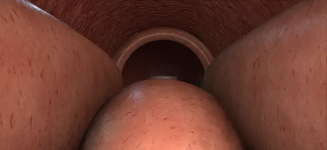
TURP, beat by beat: how the structures relateThrough the resectoscope, the tunnel from the trigone down to the veru. External urethral sphincter: no stroke starts or finishes beyond the veru Verumontanum (danger). Verumontanum: the distal limit of every cut Median lobe and two lateral lobes. Median lobe and two lateral lobes: stop at white fibres; fat is a perforation Surgical capsule, thinnest at nine and three (danger). Median lobe and two lateral lobes: cuts run from the neck toward the veru Bladder neck, arteries at five and seven. Bladder neck, arteries at five and seven: over-resection undermines the trigone Trigone and ureteric orifices (danger).Proximal: bladderDistal: penile end!Trigone and uretericorifices!Bladder neck, arteries atfive and sevenMedian lobe and twolateral lobes!Surgical capsule,thinnest at nineand threeVerumontanum!External urethralsphincterno stroke starts or finishesbeyond the veruthe distal limit of every cutstop at whitefibres; fat is aperforationcuts run from the neck towardthe veruover-resection undermines thetrigoneTargetDangerLandmark
Through the resectoscope, the tunnel from the trigone down to the veru. Schematic, not to scale; relationships only.
  • Through the scope the field is a tunnel.
  • The verumontanum is a mound at six o'clock just below the lens, the external sphincter a ring behind it, and the bladder neck a ring ahead with the trigone and both orifices beyond.
  • The median lobe fills the floor between the lens and the neck; the lateral lobes press in from either side and meet at the roof.
  • Every cut runs from the neck toward the veru and stops at the veru; every trench deepens until the white fibres of the surgical capsule appear and stops there.
  • ›Verumontanum - the mound on the floor at six o'clock where the ejaculatory ducts open; the distal limit of every cut.
  • ›External urethral sphincter - the ring two centimetres distal to the veru that opens as the scope approaches; damaged by any stroke that goes past the veru.
  • ›Surgical capsule - the compressed peripheral zone around the adenoma, seen as white criss-crossing fibres at the floor of a trench; the depth limit. Venous sinuses and fat lie beyond it.
  • ›Bladder neck - the transverse ring of fibres at the top of the median trench; over-resection at five and seven o'clock goes under the trigone and into the vessels.
  • ›Ureteric orifices - at the corners of the trigone, sometimes within a centimetre of the neck behind a large median lobe.
  • ›The lobes - a median lobe on the floor at the neck and two lateral lobes meeting at the roof; each lateral lobe is released along its roof and drops before it is taken.
  • ›The resectoscope - 26 French continuous-flow sheath, visual obturator, working element with a 30 degree telescope, wire loop and button electrode; Ellik evacuator and Toomey syringe for the chips.
Click me next Section 3 of 8 Steps The operation in order, with the landmark that guards each move.

Operative steps

  1. Before the scope: consent, position, the stack The consent conversation names the specific things: retrograde ejaculation in most men, bleeding that may need transfusion or a return to theatre, infection, a catheter for a day or two, the small risks of incontinence and stricture, the chance of needing a second procedure years later, and TUR syndrome if the resection is monopolar. An MSU is clear or treated, the anticoagulation plan is written down, and antibiotics go in at induction. The patient is in lithotomy in Lloyd-Davies supports with the buttocks just over the end of the table, the legs padded at the peroneal nerve, the diathermy plate on the thigh. On the stack: the camera and light lead, the irrigation line from a bag hung no higher than sixty centimetres above the patient, the outflow to a bucket, and the generator set for the electrode in use. A rectal examination under anaesthetic tells the surgeon the size of the gland before the scope goes in.
  2. The instrument: what a resectoscope is A 26 French continuous-flow sheath with an inner and outer channel, so irrigant runs in through one and out through the other and the bladder never fills. Into the sheath goes either the visual obturator for passing it, or the working element: a spring-loaded trigger that moves the loop in and out along a rack, carrying a 30 degree telescope and the electrode. The loop is a fine wire on two supports; when the trigger is squeezed the loop comes toward the sheath, and cutting happens on that pull, never on the push. A bipolar system cuts in normal saline, with the return electrode on the loop itself; a monopolar one needs a non-conducting irrigant, 1.5 per cent glycine, and the plate on the thigh. On the trolley beside it: the Ellik evacuator, a Toomey syringe, a 22 French three-way catheter with a 30 ml balloon, and a specimen pot for the chips.
  3. Urethroscopy and cystoscopy The sheath is passed under vision with the visual obturator, following the lumen and not the resistance: the bulbar urethra, then the external sphincter as a closed ring that opens as the scope approaches, then the verumontanum at six o'clock and the prostatic urethra with the lateral lobes kissing in the midline and the median lobe as a mound on the floor at the bladder neck. Into the bladder: the air bubble sits at twelve o'clock and gives orientation, the trigone is the raised triangle on the floor, and both ureteric orifices are found at its corners and their distance from the bladder neck noted. The walls are surveyed for stones, tumour and diverticula. Only when the surgeon has seen the sphincter, the veru, the neck and both orifices does the working element go in.
    where it bites: a ureteric orifice that sits close behind a large median lobe is the one that gets resected; find it before the first cut, not after.
  4. The landmark hold The scope rests with the lens just on the bladder side of the verumontanum and the loop parked at six o'clock. From here the surgeon can see the veru below the lens, the bladder neck as a ring ahead, and the lateral lobes either side. This is the reference position; every time orientation is lost, the scope comes back to it. The rule that runs the whole operation is that nothing distal to the veru is cut, because the external sphincter lies two centimetres beyond it, and nothing deeper than the surgical capsule is cut, because the venous sinuses and the periprostatic fat lie beyond that.
  5. The median lobe, from the neck toward the veru The first cuts go into the median lobe at six o'clock, starting at the bladder neck and working back toward the verumontanum in short strokes, each one adjacent to the last so the bed stays flat. Each stroke is the same: the loop is pushed out beyond the tissue on the bladder side, current comes on, and the trigger draws it back through the adenoma toward the sheath and the veru; the chip lifts off and floats away. The capsule does not move: each chip takes a layer of adenoma off it, and it is uncovered where the tissue has gone. The tissue changes as the depth increases: the adenoma is granular and pale, and when the loop reaches the surgical capsule the bed shows criss-crossing white fibres like the inside of a coconut. That is the floor. The lobe is levelled to it from the neck to a point just proximal to the veru, and the bladder neck fibres are seen as a transverse ring at the top of the trench.
    where it bites: a deep central trench with two shoulders is the commonest mistake; keep the bed flat so that both lateral lobes have a defect to fall into.
  6. The right lobe: release along eleven, ten, nine A lateral lobe comes out in two moves. First it is released: a trench is cut from the bladder neck to the level of the veru along the roof of the lobe at eleven o'clock, and deepened to the capsule, then the same at ten and at nine. As the trench reaches the capsule the lobe, no longer held at its roof, drops into the median defect. Second it is taken: the loop is passed behind the dropped lobe at about eight o'clock and hooked into it so that each stroke takes a full-length chip from the neck to the apex, and the lobe comes away in adjacent flat chips until the capsule shows across the whole sector. The apical part of the lobe, beside the veru, is left for the apical step.
    where it bites: the capsule is thinnest at the apex and laterally at nine and three o'clock; fat seen at the bottom of a trench means the loop has gone through it.
  7. The left lobe: the mirror at one, two, three The same two moves on the other side: release along one, two and three o'clock from the neck to the veru, let the lobe drop, hook it at four o'clock and take it in adjacent chips. Before starting the second lobe the surgeon comes back to the landmark hold and looks again at the veru and the neck, because a bleeding field and a changed shape make it easy to lose the level. One sector is finished, and its bleeding controlled, before the next is opened.
  8. The apex, from twelve o'clock, and the tissue beside the veru What remains is a shelf of tissue at twelve o'clock, where the two lobes met at the roof, and the apical tissue on either side of the verumontanum. The roof is taken with the loop turned over, its bend at twelve o'clock, and short strokes drawn back toward the veru. The apical tissue is taken the same way as every other part of the gland: the loop is put out over the tissue on the bladder side and each stroke is drawn back toward the veru, ending at it and never carried beyond it. A small apical remnant is accepted; a sphincter is not. The sphincter is checked without turning the view round: the scope is withdrawn into the bulbar urethra, still looking forward, and the sphincter is seen to close as a ring ahead of the lens with the veru beyond it.
    where it bites: the loop never goes distal to the verumontanum; the external sphincter is the next structure.
  9. Chips out: the Ellik The telescope and working element come out of the sheath and the Ellik evacuator goes on, its bulb full of irrigant with no air. Squeeze and release: the chips are drawn out of the bladder into the glass chamber and settle, and the process is repeated until the return is clear. A Toomey syringe does the same for a few remaining chips. The scope goes back in to check every chip is out and to look at the bed and the bladder for a chip lodged behind the neck. The chips are drained, weighed, and sent for histology in formalin; the house officer writes the weight on the form and the op note.
  10. Haemostasis With the resection finished the loop is exchanged for the button or roller electrode, or the loop is used on coagulation. The irrigation is slowed and the bed inspected sector by sector. Arterial bleeders spurt in a jet across the field and are coagulated at their point, not sprayed across the bed; venous sinuses ooze dark blood and bleed more when the pressure falls, and are not chased, because coagulating a sinus opens it further. The irrigant is turned off for a moment and the field watched: what fills it now is what the catheter has to control. Bleeding at the bladder neck at five and seven o'clock, where the arteries enter, is checked last.
  11. The catheter, and what to write A 22 French three-way catheter goes in over the bed with the balloon filled to 30 ml in the bladder, irrigation connected and running until the effluent is rosé rather than red. Traction, with the balloon pulled down onto the bladder neck and the catheter taped to the thigh, is used for venous ooze and released within a few hours. The op note records the resection weight, the resection time, the irrigant used and the deficit, the electrode used, whether the sphincter and orifices were seen intact, the catheter size and balloon volume, and the irrigation plan. On the ward the house officer watches the colour of the bag, the bladder for clot retention, and the sodium if the resection was monopolar.
Click me next Section 4 of 8 Danger zones Where this operation bites, and why it bites there.
Critical relationship

Danger zones

The spatial relationships the animation is built to make automatic. Before you read each one, ask yourself: at this point in the operation, what is about to go wrong, and what is my first move? The save for each sits in the Complications tab.

  • RiskDistal to the verumontanum - the sphincter. No stroke starts or finishes beyond the veru; every stroke, the apical ones included, is drawn back toward the veru and stops at it.
  • RiskThe capsule at nine and three, and at the apex - thinnest here. Fat at the floor of a trench is a perforation; venous sinuses opened here absorb irrigant.
  • RiskThe bladder neck at five and seven o'clock - the arteries enter here and the trigone lies beneath; over-resection undermines the trigone and perforates extraperitoneally.
  • RiskThe ureteric orifices - close behind a big median lobe. Found at cystoscopy before the first cut; the loop is angled away from them when the neck is defined.
Practise it Take me into the scene Opens the animation at a danger moment and asks you to predict the mechanism and your first move before it reveals.
Click me next Section 5 of 8 Complications The save for each one: the first move, the escalation trigger, the op-note line.

Complications and immediate saves

Trigger each of these live in the Animation tab, then read the save: the first move, the escalation trigger, and the line that goes in the op note.

Capsular perforation

  • fat visible, irrigant extravasates; most are small and extraperitoneal and settle with a catheter.
The save
  1. Stop resecting in that zone; keep the scope still and re-orient. first move
  2. Secure haemostasis at the breach.
  3. A small extraperitoneal perforation is the common one: complete or curtail the resection sensibly, place a catheter and observe; most settle on drainage.
  4. A large intraperitoneal perforation (abdominal distension, peritonism, big irrigant deficit) needs imaging, an abdominal or suprapubic drain, and possibly laparotomy. escalation trigger
  5. Op note: site, extra- versus intraperitoneal, irrigant deficit, catheter and drainage plan. document

TUR syndrome

  • Systemic effects of irrigant absorption, including hyponatraemia and fluid overload.
  • Assess symptoms, sodium and serum osmolality together.
The save
  1. Stop resecting and stop the irrigant inflow; secure haemostasis. first move
  2. Tell the anaesthetist immediately; assess airway, breathing and circulation, and send urgent serum sodium and osmolality.
  3. Assess symptoms and fluid balance. Do not assume all neurological symptoms are due to hypotonic hyponatraemia; absorbed glycine can complicate the picture.
  4. Severe or moderately severe symptomatic hyponatraemia needs urgent senior-led treatment in a monitored setting, using the local hypertonic saline protocol when indicated. escalation trigger
  5. Monitor sodium and urine output closely during treatment. Avoid excessive correction and seek specialist help promptly if sodium rises too quickly.
  6. Op note: resection time, irrigant used, sodium trend and treatment given. document

Bleeding and clot retention

  • wash out, coagulate at source, catheter traction and fast irrigation.
The save
  1. Wash the bladder out and evacuate clot to restore vision. first move
  2. Find the source: arterial spurters are coagulated at their point; venous ooze is controlled by the catheter, not by chasing sinuses.
  3. Coagulate the arterial bleeder under clear vision.
  4. Three-way catheter, traction on the balloon at the bladder neck, fast continuous irrigation until the effluent is rosé, not red.
  5. Bleeding that will not clear, or significant blood loss: return to theatre, transfuse, senior help. escalation trigger
  6. Op note: estimated blood loss, source controlled, catheter size and irrigation plan. document

External sphincter injury

  • a stroke carried distal to the verumontanum, or an apical remnant chased into the membranous urethra; stress incontinence that may not recover.
The save
  1. Stop. Withdraw the scope into the bulbar urethra and, still looking forward, watch the sphincter ring; if it still closes as a ring around the lumen the injury is partial. first move
  2. No further cutting or coagulation at the apex in that sector; accept the remnant.
  3. Catheter as planned, and say what happened to the patient before the trial without catheter, with pelvic floor physiotherapy from the start.
  4. Incontinence persisting beyond six to twelve months needs urodynamics and a continence surgeon: sling or artificial sphincter. escalation trigger
  5. Op note: the sector, what was seen of the ring afterwards, and the plan for review. document

Ureteric orifice or trigone injury

  • the loop taken through an orifice sitting close behind the median lobe, or the neck resected at five and seven o'clock under the trigone.
The save
  1. Recognise it: the orifice is not where it was, or efflux stops, or a trench opens toward the trigone. first move
  2. Stop resecting that sector; coagulate only what bleeds and only at the point.
  3. A resected orifice is stented for four to six weeks if it can be found; if it cannot, an ultrasound at two weeks and at three months looks for obstruction.
  4. Loin pain, fever or a rising creatinine after the operation: image the kidney and drain it by nephrostomy or stent. escalation trigger
  5. Op note: which orifice, whether cut or coagulated, stent in or not, and the imaging plan. document
Click me next Section 6 of 8 Troubleshooting When the case turns: the problem, then the next move.

Intra-operative troubleshooting

When the case turns: the problem, then the next move.

  • ActionLosing orientation - return to the landmark hold: veru below the lens, neck ahead, before resecting further.
  • ActionThe loop will not cut, or drags - check the generator and the return: plate contact for monopolar, saline running for bipolar, the loop not coated with char; a bent loop is changed, not persisted with.
  • ActionPersistent bleeding obscuring the field - evacuate chips with the Ellik, slow the irrigation, then find and coagulate the arterial source rather than the whole field.
  • ActionResection time approaching one hour (monopolar) - consider stopping, send a sodium, and stage the procedure: absorption is time-dependent.
  • ActionCapsular fibres or fat appearing - you are at or beyond the limit: stop deepening here and move on.
  • ActionThe bladder is distending - the outflow is blocked with a chip or clot; stop, empty the bladder through the sheath, clear the outflow.
Click me next Section 7 of 8 FRCS viva Say it out loud, then mark yourself against the model answer.

FRCS viva questions

The examiner stems this scene prepares you for. Try to answer out loud first, then reveal, and learn the discriminator, the detail that turns a pass into a strong pass.

QWhat are your landmarks during a TURP and why?reveal answer
  • Two landmarks set the limits of the resection and I confirm both before current flows.
  • The verumontanum is the distal limit: the external sphincter lies about two centimetres beyond it, so no stroke starts or finishes distal to the veru, and every stroke, the apical ones included, is drawn back toward the veru and stops at it.
  • The surgical capsule is the depth limit: it is the compressed peripheral zone around the adenoma and shows at the floor of a trench as pale criss-crossing fibres;
  • beyond it are the venous sinuses that absorb irrigant and the periprostatic fat that means a perforation.
  • The bladder neck and both ureteric orifices are the proximal references; I find the orifices at cystoscopy because a large median lobe can sit within a centimetre of them.
  • Whenever the field or the shape confuses me I go back to the landmark hold, with the veru below the lens and the neck ahead.
Strong pass

Two limits, and what each one protects

  • Veru The distal limit: name the structure beyond it
  • Capsule The depth limit: name the structure beyond it
  • Orifices Checked before resection starts
Examiner note
Giving the veru and the capsule as the two limits with the structure beyond each, and the orifices as a pre-resection check, rather than listing structures without saying what each limit protects.
QDescribe how you would take a lateral lobe.reveal answer
  • I take a lateral lobe in two moves.
  • First I release it: with the median lobe already levelled to the capsule from the neck to the veru, I cut a trench along the roof of the lobe at eleven o'clock from the bladder neck to the level of the veru and deepen it until the capsule shows, then repeat at ten and at nine.
  • Once the roof attachment is divided the lobe drops into the median defect.
  • Second I resect it: I pass the loop behind the dropped lobe at about eight o'clock and take it in adjacent full-length chips from the neck toward the apex, keeping the bed flat, until the capsule shows across the sector.
  • I leave the apical tissue beside the veru for the apical step, control the bleeding in that sector, return to the landmark hold, and then mirror the sequence on the left at one, two and three o'clock.
Strong pass

Say all three

  • Release then resect
  • With the roof trench deepened to the capsule so the lobe falls
  • Rather than nibbling at the lobe from the front
Examiner note
Release then resect, with the roof trench deepened to the capsule so the lobe falls, rather than nibbling at the lobe from the front.
QHow do you reduce the risk of TUR syndrome intra-operatively?reveal answer
  • TUR syndrome is dilutional hyponatraemia from absorption of hypotonic irrigant through opened venous sinuses, so my measures target absorption.
  • The single biggest is to use bipolar resection in normal saline, which abolishes the dilutional risk.
  • If I am using monopolar with glycine, I limit absorption: keep the resection time short, under about an hour, keep the irrigant bag low, around sixty centimetres, to minimise hydrostatic pressure, resect within the surgical capsule so I do not open large sinuses, and achieve good haemostasis.
  • I stop and reassess, with a sodium if needed, for a large gland or a long resection, and I keep the anaesthetist informed so absorption is recognised early.
Strong pass

Say all three

  • Leading with bipolar-in-saline as the definitive preventive
  • The monopolar package (time, bag height, stay within the capsule)
  • Not just "be quick"
Examiner note
Leading with bipolar-in-saline as the definitive preventive, then the monopolar package (time, bag height, stay within the capsule), not just "be quick".
QYou see fat during a TURP. What has happened and what do you do?reveal answer
  • Seeing periprostatic fat means I have resected through the surgical capsule, a capsular perforation.
  • I would stop resecting in that area, keep the scope still, and secure haemostasis at the breach.
  • Most are small and extraperitoneal: I would complete or curtail the resection sensibly, place a catheter, and observe, as the majority settle with drainage.
  • What I am watching for is a large intraperitoneal perforation, with abdominal distension, peritonism and a big discrepancy between irrigant in and out, which would prompt imaging, a drain, and possibly laparotomy with senior input.
  • I would document the site, the type, the fluid deficit, and the catheter plan.
Strong pass

Say both

  • Distinguishing the common small extraperitoneal perforation (catheter, observe) from the rare intraperitoneal one (imaging, drain, laparotomy)
  • Not "abandon every time"
Examiner note
Distinguishing the common small extraperitoneal perforation (catheter, observe) from the rare intraperitoneal one (imaging, drain, laparotomy), not "abandon every time".
QA patient is confused and bradycardic forty minutes into a monopolar TURP. Manage this.reveal answer
  • I would suspect irrigant absorption and TUR syndrome, stop resection and irrigant inflow, and call the anaesthetist to assess airway, breathing and circulation.
  • I would check sodium, serum osmolality and fluid balance while considering other causes of deterioration.
  • Glycine absorption can produce neurological symptoms without straightforward hypotonic hyponatraemia.
  • Severe or moderately severe symptomatic hyponatraemia requires urgent senior-led treatment in a monitored setting under the local emergency protocol.
  • Hypertonic saline is used when indicated, with repeated sodium measurements and close urine-output monitoring.
  • I would watch for overcorrection rather than dismissing it because symptoms appeared intra-operatively.
  • Once stabilised, I would secure haemostasis, catheterise and stage residual resection.
Strong pass

Distinguishing symptom severity from the sodium value alone, checking tonicity and monitoring correction even when the presentation is acute.

Click me next Section 8 of 8 Evidence The guidance each of these answers is built on.

Evidence and guidance

Two minutes

Close the lap: recall it before you leave.

Say it aloud, then reveal. Reading is not remembering.

  1. 01 The relationship Say the critical spatial relationship out loud, then reveal. reveal
    Memory frame

    Neck to veru, down to the white fibres

    • Veru A mound at six o'clock, just below the lens
    • Sphincter A ring behind the veru
    • Neck A ring ahead, trigone and orifices beyond

    Every cut stops at the veru; every trench stops at the capsule.

    Why?
    Through the scope the field is a tunnel. The verumontanum is a mound at six o'clock just below the lens, the external sphincter a ring behind it, and the bladder neck a ring ahead with the trigone and both orifices beyond. The median lobe fills the floor between the lens and the neck; the lateral lobes press in from either side and meet at the roof. Every cut runs from the neck toward the veru and stops at the veru; every trench deepens until the white fibres of the surgical capsule appear and stops there.
  2. 02 The first move Capsular perforation. Your first move? reveal
    First move

    Stop resecting in that zone

    • Keep the scope still
    • Re-orient
    More detail
    Stop resecting in that zone; keep the scope still and re-orient.
  3. 03 The escalation trigger Capsular perforation. What makes you call for help? reveal
    Escalate when

    A large intraperitoneal perforation

    • Abdominal distension
    • Peritonism
    • Big irrigant deficit

    Imaging, an abdominal or suprapubic drain, possibly laparotomy.

    More detail
    A large intraperitoneal perforation (abdominal distension, peritonism, big irrigant deficit) needs imaging, an abdominal or suprapubic drain, and possibly laparotomy.
  4. 04 The discriminator What turns a pass into a strong pass? reveal
    Strong pass

    Two limits, and what each one protects

    • Veru The distal limit: name the structure beyond it
    • Capsule The depth limit: name the structure beyond it
    • Orifices Checked before resection starts
    Examiner note
    Giving the veru and the capsule as the two limits with the structure beyond each, and the orifices as a pre-resection check, rather than listing structures without saying what each limit protects.
  5. 05 The pitfall Where does this operation bite first? reveal
    Pitfall

    No stroke starts or finishes beyond the veru

    Every stroke, the apical ones included, is drawn back toward the veru and stops at it.

    Why?
    Distal to the verumontanum: the sphincter. No stroke starts or finishes beyond the veru; every stroke, the apical ones included, is drawn back toward the veru and stops at it.

Revision practice with self-marked answers, not an assessment of clinical competence. Kept in this browser only; it does not sync between devices.