
UrOops3D, danger-zone atlas
Operative animation, Ureteric injury

Iatrogenic ureteric injury: recognition and repair

The ureter is cut, tied, clipped, crushed or burnt in somebody else's operation, and the urologist is called, now or a week later when the patient has a fever, an ileus and urine in the drain.

This page is where it happens and to whom, how it is proved at the time with dye and a retrograde, what is done with the clip, the tie and the burn, the injury found late, and the repair chosen by level, length and timing.

Advanced tier FRCS emergency urology and trauma, ureteric injury Interactive 3D

Operative visualisation

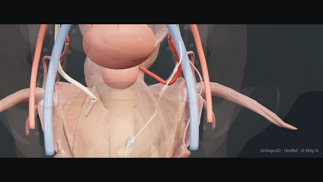
Schematic scene preview: Ureteric injury: recognition and repair
Preparing the 3D anatomy…

UrOops3D · UroRef · © Nity G

Click me next Section 2 of 8 Anatomy The structures this operation works between, and the one it must not cross.

The structures in the scene

Ureteric injury: how the structures relateFrom the surgeon's side, the ureter down the pelvic sidewall to the bladder. Ureter at the pelvic brim: crosses them; the gonadal vein tears Iliac vessels and gonadal vein (danger). Ureter at the pelvic brim: runs down the sidewall; the first clamp site Uterine artery crossing. Uterine artery crossing: stripped, the ureter necroses Adventitial blood supply (danger). Uterine artery crossing: passes beneath the artery beside the cervix Distal ureter and bladder base (danger). Distal ureter and bladder base: three quarters of injuries are here Vaginal fornix and cardinal ligament (danger). Distal ureter and bladder base: turns medially into the bladder base Bladder: psoas hitch or Boari flap. Bladder: psoas hitch or Boari flap: stent runs through the repair to the kidney Stent and drain.Pelvic brimBladder!Ureter at the pelvic brim!Iliac vessels andgonadal veinUterine artery crossing!Adventitial bloodsupply!Distal ureter and bladderbaseVaginal fornixand cardinalligamentBladder: psoas hitch orBoari flapStent and draincrosses them; thegonadal vein tearsruns down the sidewall; thefirst clamp sitestripped, theureter necrosespasses beneath the arterybeside the cervixthree quarters ofinjuries are hereturns medially into thebladder basestent runs throughthe repair to thekidneyTargetDangerLandmarkContext
From the surgeon's side, the ureter down the pelvic sidewall to the bladder. Schematic, not to scale; relationships only.
  • From the surgeon's side the ureter runs down the sidewall from the pelvic brim, crosses the iliac vessels, passes beneath the uterine artery and turns medially into the bladder base, with the gonadal vessels lateral to it above and the vaginal fornix beside it below.
  • The bladder sits in front and can be drawn up toward the psoas, and the sigmoid and rectum lie behind.
  • An injury at the brim is a ureteroureterostomy field, an injury below the vessels is a reimplant field, and the stent runs through both to the kidney.
  • ›The ureter at the pelvic brim - crossing the iliac vessels beneath the infundibulopelvic ligament and the gonadal vessels; the first place a clamp catches it.
  • ›The uterine artery crossing - water under the bridge: the ureter passes beneath the artery a centimetre or two lateral to the cervix.
  • ›The distal ureter and the bladder base - the lateral fornix, the cardinal ligament and the intramural ureter; three quarters of injuries are here.
  • ›The adventitial blood supply - longitudinal vessels in the adventitia, medial above the brim and lateral below it; stripped, the ureter necroses.
  • ›The iliac vessels and the gonadal vein - beside every mobilisation of the ureter; the gonadal vein is the vessel most often torn in a hurried dissection.
  • ›The bladder - mobile enough to reach the brim on a psoas hitch and higher as a Boari flap; the partner for every distal repair.
  • ›The stent and the drain - a 6 French stent for four to six weeks with a removal date recorded; a drain beside every anastomosis until it is dry.
Click me next Section 3 of 8 Steps The operation in order, with the landmark that guards each move.

Operative steps

  1. Where the ureter is injured, and by whom Most ureteric injuries are made by a surgeon, and most of those are gynaecological: hysterectomy in about one in a hundred to one in two hundred, more with a large uterus, endometriosis, bleeding or previous surgery, then colorectal resection, vascular surgery on the iliacs, and the urologist's own ureteroscope. The distal third takes about three quarters of them, at four points: the pelvic brim where the infundibulopelvic ligament and the gonadal vessels are clamped, the crossing beneath the uterine artery, the lateral vaginal fornix and cardinal ligament where the clamp is placed blind in bleeding, and the bladder base. External trauma is rare, a stab or a gunshot in the retroperitoneum, and is found at the laparotomy for everything else.
  2. Recognition at the time: look, dye, retrograde Only about a third are recognised at the time, and those do best. The ureter is traced from the brim to the bladder and watched for peristalsis, for a clip or a tie across it, for a dusky segment and for urine in the field. Where there is doubt, intravenous methylene blue or indocyanine green with a near-infrared camera shows a leak or a blocked ureter within minutes; rigid cystoscopy shows whether the jets are present, and a 5 French ureteric catheter to the kidney or a retrograde pyelogram proves the ureter intact or shows the level of the stop or the extravasation. A catheter that passes freely does not exclude a crush or a burn that declares itself later.
    where it bites: a clean cut is the easy injury; the crush and the diathermy burn look intact today and necrose in a week.
  3. The clip, the tie, the burn: immediate management A clip or a tie found within minutes is removed and the segment watched: pink, peristalsing and not narrowed, it is stented for four to six weeks and left; dusky, flattened or narrowed, it is excised to bleeding tissue and repaired. A crush from a clamp is treated as a devascularised segment. A diathermy burn is excised with a margin beyond the visible white because thermal spread is wider than it looks. A partial cut, less than half the circumference, is closed with interrupted 4-0 or 5-0 absorbable over a stent. A complete transection is repaired by level. Every repair is mucosa to mucosa, spatulated, tension-free, watertight, over a stent, with a drain and, where there is bowel or a vaginal cuff beside it, omentum or peritoneum between.
  4. The injury found late The unrecognised ureter presents in the first week or two: loin pain or a tender mass over an obstructed kidney, fever, an ileus that will not settle, urine from the wound, the drain or the vagina, and a creatinine that rises as the urinoma is reabsorbed across the peritoneum. Anuria after a pelvic operation is bilateral ureteric injury until proven otherwise. Drain fluid creatinine well above serum is urine. The first move is drainage of the kidney and of the collection, not a repair: an inflamed, infected pelvis is not the place to sew a ureter that will not stay sewn.
    where it bites: the missed injury is the one that costs the kidney; a rising creatinine, an ileus or a drain that will not dry up after pelvic surgery is a ureter until proven otherwise.
  5. Imaging the leak A CT urogram with a delayed phase at ten to fifteen minutes shows contrast outside the ureter, the level, the urinoma and the state of the kidney, and whether the other ureter is intact; a portal venous phase alone misses it. A retrograde pyelogram at cystoscopy defines the level and length of the gap and may allow a stent to be passed, which is treatment as well as diagnosis. Where nothing passes, a nephrostomy under ultrasound decompresses the kidney and an antegrade study from above, with a retrograde from below, measures the defect exactly, which is what the reconstruction will be planned on. A renogram is for later, to document what function the kidney has kept.
  6. Principles of repair The same six rules whatever the level: debride to bleeding tissue; mobilise the ureter with its adventitia and periureteric fat intact, because the blood supply runs longitudinally in the adventitia, from the medial side above the brim and the lateral side below it; spatulate the ends so the anastomosis is oblique and wide; join mucosa to mucosa with fine absorbable suture, interrupted or running, without tension; stent it, with a drain beside it; and isolate it from any associated bowel, vascular or vaginal repair with omentum or peritoneum. Tension is the enemy that cannot be sutured away: if the ends will not meet without pull, the plan changes to a hitch, a flap or a substitution rather than a tighter knot.
  7. Repair by level Above the iliac vessels a short defect is a ureteroureterostomy over a stent. Below them, the distal ureter is reimplanted into the bladder, directly if it reaches, with a psoas hitch for a gap of a few centimetres, and a Boari flap for ten to fifteen centimetres; the distal stump is not used because its blood supply is doubtful and reimplantation is more reliable than a low ureteroureterostomy. A long or mid-ureteric loss beyond a flap is a transureteroureterostomy, an ileal ureter or an autotransplant, and in a poorly functioning kidney, a nephrectomy. An injury at the pelviureteric junction is treated as a pyeloplasty.
    where it bites: the distal stump below a pelvic injury is not a partner for anastomosis; reimplant the ureter, hitch or flap the bladder, and leave the stump.
  8. Nephrostomy and delayed reconstruction An injury found late, a septic patient, an inflamed field or a surgeon without the experience to hand: nephrostomy and, if it passes, an antegrade or retrograde stent, drainage of the urinoma, antibiotics and time. A stented partial injury or a released tie often heals without an operation and is re-imaged at four to six weeks when the stent comes out. A defect that does not heal is reconstructed once the inflammation has settled, classically at about three months, with the defect remeasured on the antegrade and retrograde studies just before. Early reconstruction within the first week or two is reasonable if the patient is well and the tissues are clean; between then and three months the field is at its worst.
  9. Documentation and after The note records who called, what was found, the mechanism, the level and length of the injury, what was done to prove it, the repair and its suture, the stent and its planned removal date, the drain, and what the patient has been told. A stent is a device with a removal date in a register, not a memory. The catheter stays a week to ten days after a reimplant and the drain comes out when it is dry. The stent comes out at four to six weeks, and an ultrasound at three months, with a renogram if there is doubt, shows what function the kidney has kept. Duty of candour: the patient is told plainly that the ureter was injured, with the other surgeon present where possible.
Click me next Section 4 of 8 Danger zones Where this operation bites, and why it bites there.
Critical relationship

Danger zones

The spatial relationships the animation is built to make automatic. Before you read each one, ask yourself: at this point in the operation, what is about to go wrong, and what is my first move? The save for each sits in the Complications tab.

  • RiskThe segmental blood supply - stripped adventitia, diathermy on the wall and a long skeletonised segment give a repair that necroses at ten days.
  • RiskThe iliac and gonadal vessels - the ureter is mobilised off them in a scarred or inflamed pelvis; the gonadal vein tears and the external iliac vein is beneath.
  • RiskBowel and mesentery - the sigmoid and its mesentery sit on the left ureter; a repair against a bowel anastomosis without omentum between becomes a fistula.
  • RiskThe contralateral ureter - in a transureteroureterostomy, or in the anuric patient with a bilateral injury, the healthy side is the one that must not be harmed.
Practise it Take me into the scene Opens the animation at a danger moment and asks you to predict the mechanism and your first move before it reveals.
Click me next Section 5 of 8 Complications The save for each one: the first move, the escalation trigger, the op-note line.

Complications and immediate saves

Trigger each of these live in the Animation tab, then read the save: the first move, the escalation trigger, and the line that goes in the op note.

Urine leak and urinoma

  • urine in the drain, the wound or the vagina, a collection on CT, and a creatinine that rises as it is reabsorbed; a repair under tension, a devascularised end or an unrecognised second injury.
The save
  1. Check the stent and catheter are draining; a full bladder refluxes up the stent and out through the anastomosis. first move
  2. Drain fluid creatinine to confirm urine, and a CT urogram with a delayed phase to find the level and the size of the collection.
  3. A drained leak with a stent in place is given time; the collection is drained percutaneously if it is large or infected.
  4. Sepsis, a leak that grows, or no stent across the defect: nephrostomy, and a return to theatre if the anastomosis has come apart. escalation trigger
  5. Note: the site of the leak, the drains and stents in place, the antibiotic, and the date of the repeat imaging. document

Stricture and recurrent obstruction

  • hydronephrosis on the ultrasound after the stent comes out, or months later: ischaemia, thermal spread, a tensioned join or scar in an inflamed field.
The save
  1. Ultrasound at three months after every repair and a renogram if the pelvis is dilated; the kidney does not complain until it is lost. first move
  2. A renogram to confirm obstruction and the function on that side before any decision.
  3. A short stricture is dilated or incised endoscopically and stented; a long one is not.
  4. A long or recurrent stricture, or a kidney losing function: revision of the repair, a hitch or flap, or a substitution. escalation trigger
  5. Note: the length of the stricture, the split function, the treatment and the surveillance interval. document

Infected urinoma and sepsis

  • the missed injury a week on: fever, an ileus, a tender loin, a collection of infected urine in the retroperitoneum or the pelvis, and an obstructed infected kidney above it.
The save
  1. Sepsis six, cultures, intravenous antibiotics and a nephrostomy under ultrasound to decompress the kidney before anything else. first move
  2. Drain the collection percutaneously and send the fluid for creatinine and culture.
  3. No reconstruction while the patient is septic; a stent antegrade if it passes, otherwise the nephrostomy alone.
  4. Sepsis not settling with drainage of kidney and collection: critical care, and a laparotomy only for a source that cannot be drained percutaneously. escalation trigger
  5. Note: the organism, the drains and their outputs, the nephrostomy, and the plan for the delayed repair. document

Loss of renal function

  • the silent kidney: an obstruction not relieved for weeks, or a repair that strictured and was not imaged, until the renogram shows a kidney that is not worth saving.
The save
  1. Every obstructed kidney is decompressed within days, not weeks; obstruction is lost function by the week. first move
  2. A renogram after the repair to document what was kept; below about 15 to 20 per cent the kidney is a source of trouble rather than function.
  3. A kidney with poor function and a long defect: nephrectomy is the honest option rather than a complex reconstruction.
  4. Bilateral injury or a solitary kidney with a rising creatinine: nephrology, dialysis if needed, and urgent decompression of whichever side can be reached. escalation trigger
  5. Note: the split function, the decision about the kidney, and that the patient was told the reason. document

Injury to vessels, bowel or the other ureter at the repair

  • a reoperative pelvis with scar, inflammation and a urinoma: the gonadal vein torn, the external iliac vein opened, the sigmoid entered or the healthy ureter caught in a transureteroureterostomy.
The save
  1. Pressure with a swab and a call for senior or vascular help before any clamp; bleeding is controlled by pressure while the field is prepared. first move
  2. A venous tear is repaired with fine Prolene under proximal and distal control; the gonadal vein is ligated.
  3. A bowel injury is closed in two layers and kept away from the ureteric repair with omentum; a colostomy is considered with colorectal help in a contaminated field.
  4. A contralateral ureter injured or the only ureter at risk: stop, drain both kidneys, and reconstruct another day with a second surgeon. escalation trigger
  5. Op note: the structure injured, the repair, who helped, and what the patient was told. document
Click me next Section 6 of 8 Troubleshooting When the case turns: the problem, then the next move.

Intra-operative troubleshooting

When the case turns: the problem, then the next move.

  • ActionCalled to a hysterectomy: is the ureter intact? - trace it from the brim to the bladder and watch for peristalsis; give methylene blue or indocyanine green, and cystoscope for the jets; pass a ureteric catheter or do a retrograde if any doubt remains.
  • ActionA tie has been across the ureter for an hour - remove it and watch the segment for five minutes: pink and peristalsing, stent it; dusky, flattened or narrowed, excise and repair.
  • ActionThe retrograde stops at the brim and nothing passes - do not force a wire through a fresh injury; nephrostomy, antegrade study, and the defect measured from both ends before the repair is planned.
  • ActionThe ends will not meet without tension - stop pulling; mobilise the bladder for a hitch or a flap for a distal defect, and above the vessels mobilise the kidney downward or plan a substitution.
  • ActionThe patient is septic and the pelvis is a mess - nephrostomy, a drain in the collection, antibiotics, and no anastomosis today; reconstruct at three months on fresh imaging.
  • ActionAnuria after a pelvic operation - catheter first, then ultrasound of both kidneys; bilateral hydronephrosis is bilateral injury and both kidneys are decompressed tonight.
Click me next Section 7 of 8 FRCS viva Say it out loud, then mark yourself against the model answer.

FRCS viva questions

The examiner stems this scene prepares you for. Try to answer out loud first, then reveal, and learn the discriminator, the detail that turns a pass into a strong pass.

QYou are called to a hysterectomy because the gynaecologist is worried about the ureter. What do you do?reveal answer
  • I would go to theatre myself and take a history from the surgeon: what was clamped, tied or coagulated, where, and how long ago.
  • I would examine the ureter directly from the pelvic brim to the bladder, because the four points of injury are the brim beneath the infundibulopelvic ligament, the crossing under the uterine artery, the lateral fornix and cardinal ligament, and the bladder base.
  • I would look for peristalsis, a clip or tie, a dusky or flattened segment and urine in the field.
  • If there was doubt I would give intravenous methylene blue or indocyanine green with a near-infrared camera to show extravasation or a blocked ureter, and I would perform a rigid cystoscopy to see the jets and pass a 5 French ureteric catheter or a retrograde pyelogram to prove the ureter is intact or show the level of injury.
  • I would remember that a catheter that passes does not exclude a crush or a diathermy burn, which necrose later, so any segment that has been clamped or coagulated is stented or excised.
  • A recognised injury is repaired at the time, by level, over a stent, and the patient is told.
Strong pass

Say both

  • The four anatomical points of injury named with their reason
  • The crush and thermal injury treated as devascularised despite a catheter that passes
Examiner note
The four anatomical points of injury named with their reason, and the crush and thermal injury treated as devascularised despite a catheter that passes.
QDescribe the principles of ureteric repair and how the operation is chosen by level.reveal answer
  • The principles are the same at every level: debridement to bleeding tissue, mobilisation with the adventitia and periureteric fat intact because the blood supply is longitudinal and adventitial, spatulation, a mucosa-to-mucosa anastomosis in fine absorbable suture without tension, a stent, a drain, and isolation from any bowel or vascular repair with omentum or peritoneum.
  • Tension is not corrected by suture but by changing the operation.
  • Above the iliac vessels a short defect is a ureteroureterostomy.
  • Below them I would reimplant the ureter into the bladder rather than join it to the distal stump, whose blood supply is unreliable: directly if it reaches, with a psoas hitch for a gap of a few centimetres, and with a Boari flap for ten to fifteen centimetres.
  • A longer mid or upper ureteric loss is a transureteroureterostomy, an ileal ureter or an autotransplant, and nephrectomy is the honest answer for a kidney with poor function.
  • A pelviureteric junction injury is managed as a pyeloplasty.
  • I would measure the gap on the antegrade and retrograde studies in centimetres before choosing, and I would write that measurement in the note.
Strong pass

Say both

  • Tension managed by changing the operation rather than the suture
  • The distal stump abandoned in favour of reimplantation below the vessels
Examiner note
Tension managed by changing the operation rather than the suture, and the distal stump abandoned in favour of reimplantation below the vessels.
QA patient presents ten days after a colorectal resection with fever, ileus and urine in the drain. How do you manage her?reveal answer
  • This is a ureteric injury until proven otherwise, and the first priorities are sepsis and drainage, not repair.
  • I would resuscitate, culture and give intravenous antibiotics, check the creatinine and send the drain fluid for creatinine, which is diagnostic if it is well above serum.
  • I would image with a CT urogram with a delayed phase at ten to fifteen minutes, which shows the level of the leak, the urinoma, whether the kidney is obstructed and whether the other ureter is intact.
  • I would decompress the kidney with a nephrostomy under ultrasound and drain the collection percutaneously, and if an antegrade stent passes across the defect that is treatment as well as diagnosis.
  • I would not attempt a reconstruction in an infected, inflamed pelvis with a bowel anastomosis beside it.
  • When she has recovered I would define the defect with antegrade and retrograde studies and a renogram for function, and reconstruct at about three months, by level.
  • If the stented injury has healed at the four to six week check the stent comes out and she is followed with ultrasound.
  • I would tell her what has happened and record it, and inform the colorectal team.
Strong pass

Say both

  • Drain fluid creatinine and the delayed-phase CT as the diagnostic pair
  • The refusal to reconstruct in a septic field with the reason stated
Examiner note
Drain fluid creatinine and the delayed-phase CT as the diagnostic pair, and the refusal to reconstruct in a septic field with the reason stated.
QWhen should a ureteric injury be repaired immediately and when should it be delayed?reveal answer
  • An injury recognised at the time is repaired at the time, provided the patient is stable, the field is clean and a surgeon with the experience is present; that gives the best result and the fewest kidneys lost.
  • An injury found in the first week or two can still be repaired early if the patient is well, the tissues are not inflamed and the imaging is clear, and a stented partial injury or a released tie may need no operation at all.
  • Beyond that window the pelvis is oedematous, inflamed and often infected, an anastomosis there leaks and strictures, and the traditional course is drainage, a nephrostomy with or without a stent, and reconstruction at about three months when the defect has been remeasured.
  • A septic patient, an unstable patient in damage-control laparotomy, or a surgeon without the skill available are all reasons to drain now and reconstruct later;
  • a ligated ureter with a catheter passed through the tie is also drained rather than repaired.
  • In every case the stent has a removal date written in a register, the kidney is imaged at three months with a renogram if there is doubt, and the patient is told and the injury is reported through duty of candour.
Strong pass

The middle window, from about two weeks to three months, named as the worst time to operate, and the stent register and candour as part of management rather than afterthoughts.

Click me next Section 8 of 8 Evidence The guidance each of these answers is built on.

Evidence and guidance

Two minutes

Close the lap: recall it before you leave.

Say it aloud, then reveal. Reading is not remembering.

  1. 01 The relationship Say the critical spatial relationship out loud, then reveal. reveal
    Memory frame

    From the surgeon's side the ureter runs down the sidewall from the pelvic brim, crosses the iliac vessels, passes beneath the uterine artery and turns medially into the bladder base, with the gonadal vessels lateral to it above and the vaginal fornix beside it below.

    Why?
    The bladder sits in front and can be drawn up toward the psoas, and the sigmoid and rectum lie behind. An injury at the brim is a ureteroureterostomy field, an injury below the vessels is a reimplant field, and the stent runs through both to the kidney.
  2. 02 The first move Urine leak and urinoma. Your first move? reveal
    First move

    Check the stent and catheter are draining; a full bladder refluxes up the stent and out through the anastomosis.

  3. 03 The escalation trigger Urine leak and urinoma. What makes you call for help? reveal
    Escalate when

    Sepsis, a leak that grows, or no stent across the defect: nephrostomy, and a return to theatre if the anastomosis has come apart.

  4. 04 The discriminator What turns a pass into a strong pass? reveal
    Strong pass

    Say both

    • The four anatomical points of injury named with their reason
    • The crush and thermal injury treated as devascularised despite a catheter that passes
    Examiner note
    The four anatomical points of injury named with their reason, and the crush and thermal injury treated as devascularised despite a catheter that passes.
  5. 05 The pitfall Where does this operation bite first? reveal
    Pitfall

    The segmental blood supply

    Why?
    stripped adventitia, diathermy on the wall and a long skeletonised segment give a repair that necroses at ten days.

Revision practice with self-marked answers, not an assessment of clinical competence. Kept in this browser only; it does not sync between devices.