UrOops3D, danger-zone atlas

Schematic model.

Operative animation, Urethral diverticulectomy

Female urethral diverticulectomy: the sac out, three layers back

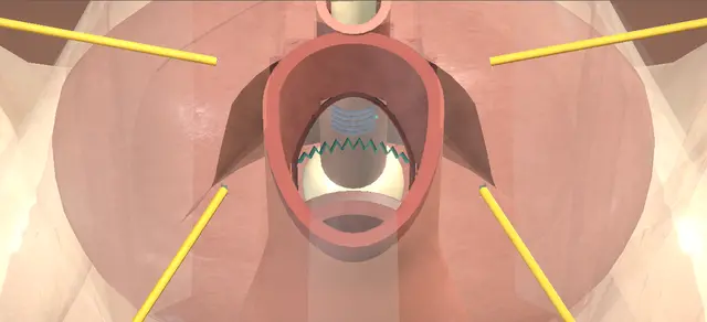
Excision of a symptomatic urethral diverticulum through the anterior vaginal wall. An inverted-U flap of vaginal wall is raised, the periurethral fascia is opened across and kept as a layer, and the sac is freed to its origin on the urethra and removed whole.

The urethra is then closed along its length, the fascia across it and the vaginal wall over both. The lesson is three layers whose suture lines do not lie on one another.

Advanced tier FRCS female and reconstructive urology, urethra Interactive 3D

Operative visualisation

Schematic scene preview: Female urethral diverticulectomy
Preparing the 3D anatomy…

UrOops3D · UroRef · © Nity G

How to use this module. Play through the nine steps, or use Back and Next. Watch the flap come off the fascia, the fascia open across, the sac come down on its neck and leave whole, and three suture lines go in at three depths. The triggers show a recurrence, a fistula and a stricture.

Click me next Section 2 of 8 Anatomy The structures this operation works between, and the one it must not cross.

The structures in the scene

Urethral diverticulectomy: how the structures relateThe sac on the ventral mid-urethra, and the layers between it and the vagina. Diverticulum: sac, neck and ostium: arises from its ventral wall; excised at its origin Urethra, with the catheter in it (danger). Diverticulum: sac, neck and ostium: lies under it; opened across, kept, closed across Periurethral fascia. Diverticulum: sac, neck and ostium: lifts it into a swelling; raised as a flap Anterior vaginal wall: inverted-U flap. Diverticulum: sac, neck and ostium: the flap apex lies a short way proximal to it Meatus. Diverticulum: sac, neck and ostium: proximal to it; checked by cystourethroscopy Bladder neck and ureteric orifices (danger). Diverticulum: sac, neck and ostium: three offset suture lines lie between them Vagina: where a fistula would open (danger).Diverticulum: sac, neckand ostiumUrethra, with thecatheter in itPeriurethral fasciaAnterior vaginalwall: inverted-UflapMeatus!Bladder neck andureteric orifices!Vagina: where afistula would openarises from itsventral wall;excised at its…lies under it;opened across,kept, closed acrosslifts it into aswelling; raised asa flapthe flap apex liesa short wayproximal to itproximal to it;checked bycystourethroscopythree offset suturelines lie betweenthemTargetDangerLandmarkContext
The sac on the ventral mid-urethra, and the layers between it and the vagina. Schematic, not to scale; relationships only.
  • From the foot of the table in lithotomy the urethra runs back from the meatus on the anterior vaginal wall, with the catheter in it.
  • The sac hangs from the ventral mid-urethra and lifts the vaginal wall into a swelling.
  • Between the sac and the vaginal wall lies the periurethral fascia.
  • The three closures stack in that order: the urethra along its length, the fascia across it, and the vaginal flap round its edge.
  • ›Urethra - short, running from the bladder neck to the meatus on the anterior vaginal wall; the catheter marks it throughout.
  • ›Diverticulum - a thin-walled sac on the ventral mid-urethra, between the urethra and the anterior vaginal wall.
  • ›Neck and ostium - the urethral origin of the sac; the dissection goes round the sac to here and the excision is made here.
  • ›Periurethral fascia - the layer between the vaginal wall and the sac; incised transversely, kept, and closed transversely.
  • ›Anterior vaginal wall - raised as an inverted-U flap with its base at the level of the proximal urethra; closed as the third layer.
  • ›Meatus - the apex of the flap lies a short distance proximal to it.
  • ›Proximal urethra and bladder - inspected at the cystourethroscopy once the repair is complete.
  • ›Ureteric orifices - efflux from both is seen before the vaginal wall is closed.
Click me next Section 3 of 8 Steps The operation in order, with the landmark that guards each move.

Operative steps

  1. Catheter, speculum, infiltration Antibiotics, then dorsal lithotomy in padded stirrups; the imaging is in the room. A urethral catheter is passed to drain the bladder. A weighted speculum is placed on the posterior vaginal wall and a self-retaining retractor is set for exposure. The anterior vaginal wall is infiltrated with local anaesthetic and adrenaline, which opens the plane for the dissection and helps haemostasis.
    where it bites: the bladder is emptied before any incision is made.
  2. Inverted-U flap With the bladder completely decompressed, an inverted-U incision is made in the anterior vaginal wall, from about a centimetre proximal to the urethral meatus back to the level of the proximal urethra. By sharp and blunt dissection a flap of anterior vaginal wall is raised off the underlying periurethral fascia.
    where it bites: the fascia under the flap is a layer of the closure and is left whole at this stage.
  3. Open the periurethral fascia The periurethral fascia is incised transversely and dissected carefully off the sac, superiorly and inferiorly, so that it lifts away as two leaves. Entry into the diverticulum is avoided.
    where it bites: the sac lies directly under the fascia and is not to be entered.
  4. Free the sac to its neck The diverticulum is grasped with a Babcock clamp and dissected circumferentially to its origin on the urethra.
    where it bites: the dissection ends at the urethral origin of the sac.
  5. Excise the sac The entire diverticulum is excised. The urethral catheter is then exposed in the opening this leaves in the ventral wall of the urethra.
    where it bites: sac wall left on the urethra is how a diverticulum recurs.
  6. Close the urethra The urethra is reconstructed over the catheter with vertically placed interrupted absorbable sutures, to give a closure that is watertight and free of tension.
    where it bites: a closure under tension leaks; one that takes too much wall narrows the urethra.
  7. Close the fascia across The two flaps of periurethral fascia are closed transversely with a running absorbable suture, at right angles to the urethral closure, to limit overlap of the suture lines. Haemostasis is completed with sparing diathermy.
    where it bites: two suture lines lying on one another make a path for a fistula.
  8. Cystourethroscopy With the repair complete, the urethral catheter is removed and cystourethroscopy is done with a flexible cystoscope or a rigid one. The proximal urethra and the bladder are intact, and clear efflux is seen from both ureteric orifices. The cystoscope is removed and the urethral catheter replaced.
    where it bites: an injury to the bladder or a ureter is found now or found late.
  9. Close the vaginal wall The edges of the vaginal epithelium are trimmed as needed and the anterior vaginal wall is closed with interrupted absorbable sutures, as the third layer. The wound is irrigated with saline, the retractor and the speculum are removed, and the catheter is connected to a drainage bag.
    where it bites: the vaginal line is the third layer and lies clear of the two beneath it.
Click me next Section 4 of 8 Danger zones Where this operation bites, and why it bites there.
Critical relationship

Danger zones

The spatial relationships the animation is built to make automatic. Before you read each one, ask yourself: at this point in the operation, what is about to go wrong, and what is my first move? The save for each sits in the Complications tab.

  • RiskThe sac under the fascia - the fascia is dissected off it without entering it.
  • RiskThe urethral origin - the whole sac is taken at its origin; a cuff left behind is a recurrence in waiting.
  • RiskThe urethral closure - watertight and free of tension, over the catheter.
  • RiskOverlying suture lines - the fascia is closed at right angles to the urethra to limit overlap.
  • RiskBladder and ureters - cystourethroscopy confirms both before the vaginal wall is closed.
Practise it Take me into the scene Opens the animation at a danger moment and asks you to predict the mechanism and your first move before it reveals.
Click me next Section 5 of 8 Complications The save for each one: the first move, the escalation trigger, the op-note line.

Complications and immediate saves

Trigger each of these live in the Animation tab, then read the save: the first move, the escalation trigger, and the line that goes in the op note.

Recurrent diverticulum

  • sac wall left on the urethra at the neck, closed over; recurrent diverticulum and pseudodiverticulum are both listed complications of this operation.
The save
  1. Look at the urethral origin before closing: any cuff of sac wall round the opening is excised back to the urethral wall. first move
  2. Close the urethra only when its edge is urethral wall all the way round and the closure lies without tension.
  3. An origin that cannot be closed without narrowing the urethra, or a sac that runs round the urethra, needs senior help in theatre. escalation trigger
  4. New swelling, dribbling or infection at the same site after the operation is a reason to image again.
  5. Op note: whether the sac came out whole, where its origin lay, and how the urethra was closed. document

Urethrovaginal fistula

  • a track from the urethral repair through the fascia and the vaginal wall; a listed complication, and the reason the three suture lines are kept from lying on one another.
The save
  1. Examine with a speculum and confirm that the fluid in the vagina is urine and where it comes from. first move
  2. Establish the site of the opening in the urethra and in the vagina before any plan is made.
  3. A fistula that persists is referred to a surgeon who repairs them regularly; the tissue may need a vascularised flap. escalation trigger
  4. Check for infection and for retention, and treat both.
  5. Notes: the layers closed at the first operation and their direction, the findings now, and the referral made. document

Urethral stricture

  • narrowing of the urethra at the repair, presenting later with a poor stream or retention; stricture and urinary retention are both listed complications.
The save
  1. Measure the flow rate and the residual, and examine the urethra. first move
  2. Retention is relieved with a catheter and infection is treated.
  3. A stricture that persists is referred to a surgeon who treats female urethral stricture. escalation trigger
  4. Ask about incontinence at the same visit: stress leakage and urgency also follow this operation.
  5. Notes: the symptoms, the flow rate and residual, the findings on examination and the plan. document
Click me next Section 6 of 8 Troubleshooting When the case turns: the problem, then the next move.

Intra-operative troubleshooting

When the case turns: the problem, then the next move.

  • ActionThe vaginal tissue over the repair is deficient or compromised - a vascularised labial fat pad, the Martius flap, can be brought in to reinforce the repair; that is the indication the dictation gives for it.
  • ActionThe sac runs round the urethra - a circumurethral diverticulum needs a wider dissection and urethral reconstruction, possibly excision with an anastomotic repair. It is a different operation from the one shown.
  • ActionA small sac close to the meatus - selected distal diverticula can sometimes be managed by marsupialisation alone, which leaves a hypospadiac vaginal meatus.
  • ActionThe inverted U does not suit the exposure - a midline vertical incision or a T-shaped vaginal incision is the described alternative.
  • ActionStress incontinence as well as the diverticulum - the textbook notes that a midurethral sling can be placed at the same operation. The European guideline does not recommend a synthetic mid-urethral sling at that sitting and, after counselling, recommends a non-synthetic sling for bothersome stress incontinence (weak).
  • ActionOozing from the vaginal wound at the end - the vagina may be packed with a rolled gauze after the operation to help haemostasis.
Click me next Section 7 of 8 FRCS viva Say it out loud, then mark yourself against the model answer.

FRCS viva questions

The examiner stems this scene prepares you for. Try to answer out loud first, then reveal, and learn the discriminator, the detail that turns a pass into a strong pass.

QA woman has a symptomatic urethral diverticulum. How do you assess her and what do you offer?reveal answer
  • I would take a history of her lower urinary tract symptoms and ask specifically about leakage and about difficulty emptying, because the European guideline makes a strong recommendation to question and investigate for coexisting voiding dysfunction and incontinence.
  • For imaging I would ask for magnetic resonance imaging, which the guideline recommends for diagnosis and characterisation, with urethroscopy, a voiding cystourethrogram and ultrasound where necessary.
  • I would not rely on seeing the opening at urethroscopy: the guideline notes that the ostia are seen in only 42 per cent of cases.
  • If she is symptomatic I would offer surgical removal, which the guideline supports with a weak recommendation, and I would describe a transvaginal diverticulectomy with a layered closure.
  • If she preferred to leave it alone, I would tell her that the guideline asks surgeons to warn of a small risk, which it puts at 1 to 6 per cent, of cancer developing within the diverticulum.
  • Before any operation I would counsel her that new or persistent urinary symptoms, including incontinence, are possible even after a technically successful excision; that too is a strong recommendation.
Strong pass

Say all four

  • Imaging to characterise the sac before surgery
  • Asking about incontinence
  • Voiding before the operation
  • Since both shape the counselling
Examiner note
Imaging to characterise the sac before surgery, and asking about incontinence and voiding before the operation, since both shape the counselling.
QTalk me through a transvaginal diverticulectomy.reveal answer
  • Antibiotics, then lithotomy.
  • I pass a urethral catheter and drain the bladder, place a weighted speculum and a self-retaining retractor, and infiltrate the anterior vaginal wall with local anaesthetic and adrenaline.
  • With the bladder empty I make an inverted-U incision in the anterior vaginal wall, from about a centimetre proximal to the meatus back to the level of the proximal urethra, and I raise the flap off the periurethral fascia by sharp and blunt dissection.
  • I incise the periurethral fascia transversely and dissect it off the sac, superiorly and inferiorly, taking care that I do not enter the sac.
  • I take the sac in a Babcock clamp and dissect it all the way round to its origin on the urethra, then excise it entirely, which exposes the catheter in the ventral urethral wall.
  • I close the urethra with vertically placed interrupted absorbable sutures so that it is watertight and free of tension.
  • I then close the fascial flaps transversely with a running absorbable suture, at right angles to the urethral line, and secure haemostasis with sparing diathermy.
  • I remove the catheter and do a cystourethroscopy to see that the proximal urethra and the bladder are intact and that there is efflux from both ureteric orifices, then I replace the catheter.
  • Last, I trim the vaginal edges as needed and close the anterior vaginal wall with interrupted absorbable sutures as the third layer.
Strong pass

Say all four

  • Three layers named with their directions: the urethra along its length
  • The fascia across it
  • The vaginal flap over both
  • With the cystourethroscopy before the last
Examiner note
Three layers named with their directions: the urethra along its length, the fascia across it, the vaginal flap over both, with the cystourethroscopy before the last.
QWhat are the principles of the repair, and when would you add a Martius flap?reveal answer
  • I would give the six principles the European guideline lists for transvaginal diverticulectomy.
  • Dissect a well-vascularised vaginal flap.
  • Preserve the periurethral fascia for closure.
  • Remove all of the diverticular wall.
  • Excise the ostium and close the urethra so that it is watertight.
  • Close in several layers with no overlapping suture lines.
  • Preserve or create continence.
  • The operation I have described is those principles in order: the inverted-U flap, the fascia opened across and kept, the sac taken whole at its origin, the urethra closed along its length and the fascia across it.
  • On the Martius flap, the operative text gives one indication, vaginal tissue that is deficient or compromised.
  • The guideline says the flap is used more frequently in recurrent cases, where the urethral defect is large or the vaginal flap deficient, where the urethra has had to be transected for a circumferential diverticulum, where the configuration is complex, and where a sling is planned later.
  • For a first operation on a single sac with healthy vaginal tissue, none of those applies.
Strong pass

Say all three

  • Knowing that the fascia is kept and closed as its own layer
  • In a different direction from the urethra
  • That the labial fat pad is for selected cases
Examiner note
Knowing that the fascia is kept and closed as its own layer, in a different direction from the urethra, and that the labial fat pad is for selected cases.
QWhat do you tell her about results and complications, and what about stress incontinence?reveal answer
  • I would start with how good the evidence is.
  • There are no randomised trials.
  • The systematic review by Bodner-Adler and colleagues found 108 studies and 1,947 patients, all case reports and case series with no comparative study, and concluded that success and complication rates cannot be estimated accurately;
  • where symptoms were reported, 717 of 1,044 women were free of them after surgery.
  • With that caution, the European guideline quotes, for transvaginal diverticulectomy, a dry rate of 84 to 98 per cent and a reoperation rate of 2 to 13 per cent over a mean follow-up of twelve to 50 months.
  • For complications it quotes, early on, urinary infection in 0 to 39 per cent, new stress incontinence in 3.8 to 33 per cent and new retention in 0 to 9 per cent, and later, urethral stricture in 0 to 5.2 per cent and urethrovaginal fistula in 0.9 to 8.3 per cent.
  • The operative text also lists bleeding, dyspareunia, recurrent diverticulum and pseudodiverticulum.
  • On stress incontinence, the same text notes that a midurethral sling can be placed at the same operation.
  • The guideline is more specific: it does not recommend a synthetic mid-urethral sling at the time of diverticulectomy, and it makes a weak recommendation to address bothersome stress incontinence then with a non-synthetic sling, after counselling.
Strong pass

Say all three

  • Quoting ranges from case series as ranges
  • Saying the evidence has no comparative studies
  • Knowing that the guideline does not recommend a synthetic mid-urethral sling at the same operation
Examiner note
Quoting ranges from case series as ranges, saying the evidence has no comparative studies, and knowing that the guideline does not recommend a synthetic mid-urethral sling at the same operation.
Click me next Section 8 of 8 Evidence The guidance each of these answers is built on.

Evidence and guidance

Operative Dictations in Urologic Surgery
Armenakas NA, Fracchia JA, Golan R. Urethral diverticulectomy. Chapter 96. Wiley, 2019
The indication, the six essential steps and their order, the template dictation followed step by step here, the variations (a midline or T incision, circumurethral diverticula, the Martius flap, marsupialisation, a concurrent sling, a vaginal pack) and the complication list: bleeding, infection, incontinence, retention, urethral stricture, dyspareunia, recurrent diverticulum, pseudodiverticulum, urethrovaginal fistula. The chapter lists complications without their management: the first, escalate and document steps, and the danger lines that do not restate the dictation, are general surgical practice and carry no figure. In the scene the sac is a single ventral mid-urethral pouch of an arbitrary size, the flap hangs from its base under its own weight, the cystoscope view is a rendered frame run shared with the cystoscopy modules, and the trimming of the vaginal edges and the irrigation are in the text only.
Open guideline
EAU Guidelines
Non-neurogenic Female LUTS: Disease management, section 4.9 Urethral diverticulum, read 13 October 2026
Magnetic resonance imaging for diagnosis and characterisation, with urethroscopy, voiding cystourethrogram and ultrasound where necessary (weak); ostia seen in only 42% of cases in one series; offer surgical removal of a symptomatic diverticulum (weak); warn of a small (1 to 6%) risk of cancer if managed conservatively (weak); question and investigate for coexisting voiding dysfunction and incontinence (strong); the six principles of transvaginal diverticulectomy; the situations in which a Martius flap is used more frequently; no randomised trials; dry rate 84 to 98% and reoperation 2 to 13% over a mean follow-up of twelve to 50 months; early urinary infection 0 to 39%, de novo stress incontinence 3.8 to 33% and de novo retention 0 to 9%; urethral stricture 0 to 5.2% and urethrovaginal fistula 0.9 to 8.3%; synthetic mid-urethral slings not recommended at the time of diverticulectomy; bothersome stress incontinence addressed with a concomitant non-synthetic sling after counselling (weak); counsel about de novo or persistent symptoms including incontinence (strong). These ranges are pooled from the case series the guideline cites.
Open guideline
International Urogynecology Journal
Bodner-Adler B, Halpern K, Hanzal E. Surgical management of urethral diverticula in women: a systematic review. Int Urogynecol J 2016;27:993-1001
Abstract read 13 October 2026. One hundred and eight studies and 1,947 patients: 40 single case reports and 68 case series, no comparative studies. Transvaginal resection with or without reconstruction in 84%, marsupialisation in 3.8%, endoscopic unroofing in 2.0%. Fifty-six studies documented resolution of symptoms: 717 of 1,044 patients symptom-free after surgery. The authors conclude that the evidence does not allow accurate estimation of success and complication rates. The symptom-resolution figure the guideline quotes is this one, so it is given once.
Open guideline

Curriculum. Supports learning relevant to the ISCP Urology Curriculum syllabus topic Urethral reconstruction. Not a workplace-based assessment and not evidence of competence.

Two minutes

Close the lap: recall it before you leave.

Say it aloud, then reveal. Reading is not remembering.

  1. 01 The relationship Say the critical spatial relationship out loud, then reveal. reveal
    Memory frame

    From the foot of the table in lithotomy the urethra runs back from the meatus on the anterior vaginal wall, with the catheter in it.

    Why?
    The sac hangs from the ventral mid-urethra and lifts the vaginal wall into a swelling. Between the sac and the vaginal wall lies the periurethral fascia. The three closures stack in that order: the urethra along its length, the fascia across it, and the vaginal flap round its edge.
  2. 02 The first move Recurrent diverticulum. Your first move? reveal
    First move

    Look at the urethral origin before closing: any cuff of sac wall round the opening is excised back to the urethral wall.

  3. 03 The escalation trigger Recurrent diverticulum. What makes you call for help? reveal
    Escalate when

    An origin that cannot be closed without narrowing the urethra, or a sac that runs round the urethra, needs senior help in theatre.

  4. 04 The discriminator What turns a pass into a strong pass? reveal
    Strong pass

    Say all four

    • Imaging to characterise the sac before surgery
    • Asking about incontinence
    • Voiding before the operation
    • Since both shape the counselling
    Examiner note
    Imaging to characterise the sac before surgery, and asking about incontinence and voiding before the operation, since both shape the counselling.
  5. 05 The pitfall Where does this operation bite first? reveal
    Pitfall

    The sac under the fascia

    Why?
    the fascia is dissected off it without entering it.

Revision practice with self-marked answers, not an assessment of clinical competence. Kept in this browser only; it does not sync between devices.