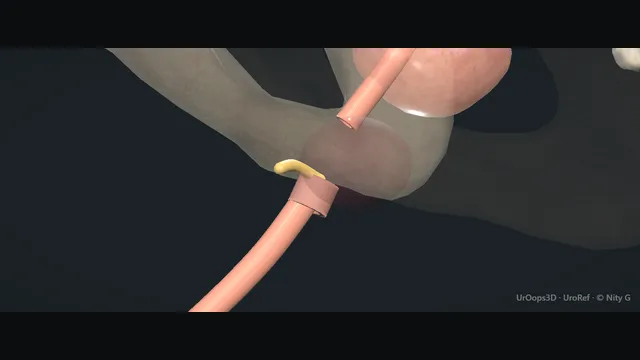
The membranous urethra is torn at the point where it is fixed at the pelvic floor: the prostate and its urethra shear off the bulbar urethra and ride high into the pelvic haematoma.
Blood at the meatus and a high-riding prostate are the signs at the bedside, a gentle retrograde urethrogram is the study, and nothing blind ever enters the urethra.
Advanced tierFRCS urological trauma, reconstructive urologyInteractive 3D
Midline slice from the patient's right, bladder down to the perineum. Schematic, not to scale; relationships only.
Look at a midline slice from the patient's right.
The bladder sits above, the prostate hangs from its neck, and the membranous urethra runs down through the pelvic floor, the one point where the urethra is tethered to bone and fascia, to join the bulbar urethra below and behind the pubic arch.
Pelvic fracture shears at exactly that tether: the prostate and prostatic urethra are torn free and ride high and forward into the pelvic haematoma, while the bulbar urethra stays anchored in the perineum.
The injury is the gap between those two healthy ends, and every decision that follows, the gentle study, the suprapubic drain, the months of maturation, the perineal anastomosis that mobilises the bulbar urethra back up to the settled prostate apex, is about spanning that gap without damaging the sphincter wrapped around its distal end.
›Membranous urethra - the short segment fixed at the pelvic floor between prostate apex and bulbar urethra; the level of the tear.
›Prostate apex - the avulsed proximal end, riding high and anterior in the haematoma; the target the reconstruction must reach.
›Bulbar urethra - the distal end, which stays with the perineum; mobilised cranially at delayed repair to bridge the gap.
›Distal sphincter - the continence mechanism downstream of the verumontanum; preserved at repair because the bladder neck may be scarred open.
›Suprapubic corridor - the diversion route into the bladder from above, placed under imaging once the urethra is out of bounds.
The shearing injuryPelvic fracture forces travel through the pubic arch and the pelvic floor, and the urethra absorbs them at its one fixed point. The membranous urethra is tethered where it pierces the floor between the prostate apex above and the bulbar urethra below, and that tether is where it tears. The prostate, with the prostatic urethra still attached, is avulsed and displaced superiorly and anteriorly into the expanding haematoma. The bulbar urethra stays anchored in the perineum, so both ends of the disrupted segment remain healthy tissue separated by a gap. Every decision that follows is about spanning that gap without adding to the injury. where it bites: the gap between the prostate apex and the bulbar urethra is the injury: both ends remain healthy away from it.
Blood at the meatus, prostate highBlood at the meatus, inability to void, and a high-riding or impalpable prostate on rectal examination make the clinical diagnosis before any imaging is taken. The findings can be subtle, so any pelvic fracture patient who cannot void is treated as having a urethral injury until proven otherwise. The meatus and perineum are inspected and the rectum examined gently, and what is found is recorded. From that moment the urethra is out of bounds: no catheter, no instrument, no dilatation, until a retrograde study has been seen and read. where it bites: one forceful blind catheter can convert a partial tear into a complete one, or carve a false passage alongside the lumen.
Gentle retrograde urethrogramContrast is instilled at the meatus with a soft tip or nozzle held in the fossa navicularis, gently and never against pressure, with oblique fluoroscopic imaging. Gentle technique keeps the study diagnostic: extravasation at the membranous level with contrast still entering the bladder defines a partial injury, and no contrast passing beyond the tear defines a complete one. The level and extent of extravasation are recorded because they plan any later reconstruction. A study that cannot be read cleanly is repeated properly rather than acted upon blindly. where it bites: forcing contrast under pressure extends the injury and adds a chemical peritonitis of the perineum.
Partial injury: one gentle attemptA partial tear may accept a single gentle catheter placement, by experienced hands and ideally under imaging control, with a soft catheter and generous lubrication. If the catheter meets genuine resistance the attempt stops and the plan converts to suprapubic drainage, because resistance at a fresh tear is the injury declaring itself. Once placed, the catheter drains the bladder and splints the healing urethra, and its position is confirmed before it is relied upon. One gentle attempt is the ceiling: the second forceful attempt does the damage the first was careful to avoid. where it bites: one attempt, gentle, by experienced hands: resistance means stop, not push.
Complete injury: suprapubicThe urethra is left completely alone: no catheter, no realignment attempt, no further instrumentation of any kind. The bladder is drained suprapubically, with the track placed under imaging control and mapped against any orthopaedic hardware first. The catheter buys complete drainage while the haematoma resolves and the injury declares its true length. The patient then enters a planned pathway: healing, elective imaging of both urethral ends, and delayed reconstruction in a mature field. where it bites: repeated urethral instrumentation now compromises the quality of the tissue available for later repair.
The expected strictureThe injured segment heals narrow whatever the initial strategy, because the injury itself has destroyed urethra and fibrosis replaces it. A stricture after realignment is an expected outcome rather than a failure, and it is assessed electively once the haematoma has matured and the field is quiet. Follow-up watches the stream objectively with flow rates and symptom review from the first clinic visit, because patients adapt to a slow decline and under-report it. Assessment defines the length and position of the obliteration and plans the repair, rather than reaching for an instrument to manage the symptom. where it bites: treating the expected stricture by repeated blind dilatation manages the symptom and destroys the reconstruction field.
Delayed anastomotic repairRepair is elective, once the haematoma has resolved, the fracture has been treated and the patient can be positioned for a perineal approach. The scarred segment is excised completely to healthy tissue at both ends, the bulbar urethra is mobilised to close the gap, and the ends are spatulated and joined as a tension-free mucosa-to-mucosa anastomosis. Length is bought by mobilisation first, and by the specialist manoeuvres around the pubis only where the gap defeats it. The distal sphincter is preserved throughout, because continence after repair rests on what remains of it. where it bites: the dissection stays close to the ends and clear of the sphincter: continence after repair depends on that structure.
The spatial relationships the animation is built to make automatic. Before you read each one, ask yourself: at this point in the operation, what is about to go wrong, and what is my first move? The save for each sits in the Complications tab.
RiskThe fixation point at the pelvic floor - the level of the shearing tear; also the level where a blind catheter leaves the lumen.
RiskThe meatus - blood there stops all blind instrumentation; the next instrument in the urethra is guided or none at all.
RiskThe distal sphincter - continence after injury and repair rests on it; excision and dissection at repair stay precise to it.
Trigger each of these live in the Animation tab, then read the save: the first move, the escalation trigger, and the line that goes in the op note.
False passage
a catheter forced past the tear burrows alongside the true lumen through the sphincter into the haematoma; every later attempt follows the false track.
The save
Stop. Leave the urethra completely alone: no repeat attempt, no dilatation, no guide passed blind.first move
Drain the bladder suprapubically, under imaging control, to break the cycle of instrumentation.
Document the event explicitly, including what was passed, how far, and what was felt.
Definitive urethral evaluation is deferred to the reconstructive setting, endoscopically guided, once inflammation settles.
A false passage that has become the dominant channel, or supplies a stricture that defeats endoscopic realignment → planned open reconstruction.escalation trigger
Op note: instrument, force used, findings, abandonment point, and the suprapubic plan.document
Post-traumatic stricture
the sheared segment heals as a fibrotic narrowing at the membranous level: weak stream, straining, spraying, and the presentation is expected rather than surprising.
The save
Recognise the symptom pattern at follow-up: poor stream, straining, spraying, incomplete emptying.first move
Assess with flow rate, endoscopy and contrast studies that show both the lumen and the length of the obliteration.
Avoid repeat blind dilatation: it buys hours of stream and builds scar that lengthens the defect.
Plan the repair electively: short obliterations are treated by excision and anastomosis, longer ones by substitution with graft.
Retention, sepsis or upper tract dilatation from the obstructed bladder → urgent diversion and decompression first.escalation trigger
Note: stricture length, calibre, position relative to the sphincter, and the operation planned.document
Incontinence after injury
the tear and the repair both sit at the distal sphincter; when the bladder neck is also scarred or open, continence rests entirely on the structure the injury threatened.
The save
Assess continence honestly after reconstruction, with a pad history and flow, before reassuring.first move
Distinguish sphincter weakness from overflow and from bladder overactivity: the pad looks the same, the treatments do not.
Preserve what sphincter remains: no further instrumentation or dilatation through the repaired segment.
Treat conservatively first: pelvic floor training and timed voiding have a genuine role.
Persistent significant leak → specialist continence assessment, urodynamics and discussion of options including implants.escalation trigger
Note: pad status pre and post repair, urodynamic findings, and the continence plan discussed.document
When the case turns: the problem, then the next move.
ActionCatheter will not pass gently at first attempt - stop and drain suprapubically under imaging; a partial injury declares itself on a later guided study rather than by force.
ActionExtravasation on urethrogram but the patient is stable - complete the imaging, drain suprapubically, and book the elective pathway; stability buys time, it does not buy instrumentation.
ActionSuprapubic placement needed with an unstable pelvis or fixator in place - image-guided insertion with the orthopaedic hardware mapped first; coordinate the track before anyone fixes anything.
ActionGap length cannot be defined on standard imaging - combine retrograde study with simultaneous cystoscopic or suprapubic contrast filling; the combined films show both ends and measure the defect for planning.
The examiner stems this scene prepares you for. Try to answer out loud first, then reveal, and learn the discriminator, the detail that turns a pass into a strong pass.
QPrimary realignment or suprapubic diversion with delayed repair: argue the case for both.reveal answer
The debate is real because each strategy trades something away.
Immediate primary realignment, most often endoscopic, passing a catheter across the gap in the early days after injury, offers the chance of healing without any stricture at all, and when it works the patient keeps a urethra and avoids a major reconstruction.
Against that, the majority who undergo realignment still develop a stricture, because the injury has removed or scarred a segment of urethra, and realignment does not restore it.
The realignment then has to be repeated, sometimes repeatedly, and each endoscopic manipulation is done through a recently injured, poorly defined field, with the attendant risks of false passage and channelling.
Suprapubic diversion with delayed anastomotic repair accepts an interval of catheter dependence and a definite operation later, but it buys three things: the haematoma resolves and declares the true gap, the tissue quality becomes predictable, and the repair is a single, planned, mucosa-to-mucosa anastomosis performed in a mature field, with the highest long-term patency of any option in experienced hands.
The practical position most units take: partial injuries may take one gentle guided catheter, complete injuries are diverted suprapubically, early endoscopic realignment is reasonable where expertise and imaging allow, and delayed anastomotic repair remains the durable answer for the strictures that follow either path.
Strong pass
Stating that most realigned patients still develop a stricture because realignment bridges but does not replace the destroyed segment, and that the delayed repair gains its results from operating in a mature, declared field.
QWhen and how do you reconstruct a membranous stricture after pelvic fracture?reveal answer
Timing first: reconstruction is elective, deferred until the pelvic haematoma has resolved, fracture treatment is complete and the patient can be positioned, at three months.
Earlier surgery enters an unreadable field of organised haematoma and freshly healing bone, and produces worse outcomes.
Assessment defines three things: the length of the defect, the position of the ends relative to the sphincter, and the state of the bladder neck and continence mechanism.
Combined retrograde and antegrade imaging, supplemented by endoscopy through the suprapubic tract, maps both ends.
The repair itself is a perineal anastomotic urethroplasty: through a perineal approach the bulbar urethra is mobilised generously, the scarred segment and the apex are identified, the fibrosis is excised to healthy tissue, both ends are spatulated, and a wide, tension-free anastomosis is created.
Length is bought by bulbar mobilisation, and where the gap defeats that, by inferior pubectomy, by rerouting the urethra over the crus, or by crural separation, manoeuvres that belong to specialist practice.
Longer defects that cannot be closed by approximation need substitution, with a buccal mucosa graft, accepting a lower single-stage success than anastomosis.
Bladder neck competence and the distal sphincter are respected throughout, and the suprapubic catheter stays until the anastomosis is proven.
Strong pass
Naming the specific length-buying manoeuvres of the perineal approach, mobilisation, inferior pubectomy and rerouting over the crus, and stating that long defects leave anastomosis for graft substitution.
QWhat is the role of endoscopic treatment in pelvic fracture urethral injury?reveal answer
Endoscopy has a defined but narrow role.
Early after injury, endoscopically assisted realignment can be used to pass a catheter across a complete tear in the first days, working from both ends with a flexible cystoscope through the suprapubic tract and a rigid scope from below, under fluoroscopy.
Where it succeeds and the urethra heals without stricture, the patient is spared a major operation, and even where a stricture forms, the ends have been kept in alignment, which commonly shortens the defect and makes the later perineal repair easier.
What endoscopy cannot do is reconstruct: direct visual internal urethrotomy and periodic dilatation have a poor durable record in post-traumatic strictures, because they cut through scar in a segment that is already obliterated rather than strictured, and the recurrent narrowing returns within months while each intervention adds fibrosis.
Serial dilatation is justified only for the patient who declines or cannot tolerate reconstruction, as a drainage strategy rather than a treatment.
So the honest structure is: endoscopy to align early where appropriate, endoscopy to evaluate and calibrate at any stage, reconstructive surgery for cure, and dilatation as a compromise chosen with the patient rather than defaulted into.
Strong pass
Say all four
Separating endoscopic alignment
Which preserves length for a later repair
From urethrotomy and dilatation
Which manage symptoms at the cost of the reconstruction field
Examiner note
Separating endoscopic alignment, which preserves length for a later repair, from urethrotomy and dilatation, which manage symptoms at the cost of the reconstruction field.
QDiscuss continence and sexual function after pelvic fracture urethral injury.reveal answer
Both functions are threatened by the same anatomy that the injury disrupts, and both deserve explicit discussion because the patient rarely volunteers them.
Continence depends on two structures acting together: the bladder neck proximally and the distal sphincter mechanism at the membranous urethra.
Pelvic fracture with urethral injury damages the distal sphincter directly, by shearing or by the haematoma, and in severe displacement the bladder neck can be injured as well.
Most patients remain continent because one mechanism suffices, but a proportion leak after injury or after reconstruction, and the risk climbs when both mechanisms are hurt.
Assessment is with pad history, endoscopy of the bladder neck and urodynamics before offering intervention.
Preservation at surgery is the strongest protection: the dissection at anastomotic repair stays precisely on the urethra and respects the sphincter fibres at the distal end.
Erectile dysfunction after pelvic fracture is common and is driven by injury to the cavernosal nerves and vessels as they pass the prostate apex and pelvic floor, the same corridor as the urethral tear.
It may recover late, and it is treatable in stages, first-line oral therapy, then local options.
The counselling point is that dysfunction after these injuries is a recognised part of the disease, treatable in most, and asked about routinely at follow-up.
Strong pass
Say all three
Explaining the two-mechanism continence model
Why most patients stay dry
That erectile dysfunction shares the injury corridor rather than being caused by the repair
Examiner note
Explaining the two-mechanism continence model, why most patients stay dry, and that erectile dysfunction shares the injury corridor rather than being caused by the repair.
Say it aloud, then reveal. Reading is not remembering.
01The relationshipSay the critical spatial relationship out loud, then reveal.reveal
Memory frame
Look at a midline slice from the patient's right.
Why?
The bladder sits above, the prostate hangs from its neck, and the membranous urethra runs down through the pelvic floor, the one point where the urethra is tethered to bone and fascia, to join the bulbar urethra below and behind the pubic arch. Pelvic fracture shears at exactly that tether: the prostate and prostatic urethra are torn free and ride high and forward into the pelvic haematoma, while the bulbar urethra stays anchored in the perineum. The injury is the gap between those two healthy ends, and every decision that follows, the gentle study, the suprapubic drain, the months of maturation, the perineal anastomosis that mobilises the bulbar urethra back up to the settled prostate apex, is about spanning that gap without damaging the sphincter wrapped around its distal end.
02The first moveFalse passage. Your first move?reveal
First move
Stop.
More detail
Leave the urethra completely alone: no repeat attempt, no dilatation, no guide passed blind.
03The escalation triggerFalse passage. What makes you call for help?reveal
Escalate when
A false passage that has become the dominant channel, or supplies a stricture that defeats endoscopic realignment → planned open reconstruction.
04The discriminatorWhat turns a pass into a strong pass?reveal
Strong pass
Stating that most realigned patients still develop a stricture because realignment bridges but does not replace the destroyed segment, and that the delayed repair gains its results from operating in a mature, declared field.
05The pitfallWhere does this operation bite first?reveal
Pitfall
The fixation point at the pelvic floor
Why?
the level of the shearing tear; also the level where a blind catheter leaves the lumen.
Revision practice with self-marked answers, not an assessment of clinical competence. Kept in
this browser only; it does not sync between devices.