
UrOops3D, danger-zone atlas
Operative animation, Urodynamics

Urodynamics: the filling and voiding study

Two pressure lines, one pump and a flowmeter, and a patient who has to leak or void in front of the team. The study reproduces the symptom while the trace records what the detrusor and the outlet did, so that the surgeon operates on a mechanism and not a story.

This page is the question, the set-up that makes the trace true, the filling and voiding phases, and the report written in ICS terms.

Core tier FRCS functional urology Interactive 3D

Operative visualisation

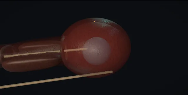
Schematic scene preview: Urodynamics
Preparing the 3D anatomy…

UrOops3D · UroRef · © Nity G

Click me next Section 2 of 8 Anatomy The structures this operation works between, and the one it must not cross.

The structures in the scene

Urodynamics: how the structures relateThe lower tract with both lines placed and zeroed at the symphysis. Detrusor pressure, calculated: vesical pressure; never forced past a stricture Urethra and the 6 French line (danger). Detrusor pressure, calculated: abdominal pressure, subtracted from vesical Rectum and the balloon line. Detrusor pressure, calculated: both lines zeroed here, or the trace is wrong Upper edge of the symphysis pubis (danger). Detrusor pressure, calculated: closed at rest in health Bladder neck. Detrusor pressure, calculated: site of dyssynergia and intrinsic deficiency External sphincter. Detrusor pressure, calculated: Qmax paired with detrusor pressure The flowmeter.Detrusor pressure,calculated!Urethra and the 6French lineRectum and theballoon lineUpper edge of thesymphysis pubisBladder neckExternal sphincterThe flowmetervesical pressure;never forced past astrictureabdominal pressure,subtracted fromvesicalboth lines zeroedhere, or the traceis wrongclosed at rest inhealthsite of dyssynergiaand intrinsicdeficiencyQmax paired withdetrusor pressureTargetDangerLandmarkContext
The lower tract with both lines placed and zeroed at the symphysis. Schematic, not to scale; relationships only.
  • The filling line runs through the urethra, past the sphincter and the prostate, to lie in the bladder;
  • the balloon line sits in the rectum behind the bladder base, and both are zeroed at the level of the top of the symphysis in front.
  • Filling raises the vesical line, a cough raises both, and only a detrusor contraction raises the difference.
  • On screening the neck, the prostatic urethra and the sphincter are seen as the outlet the contrast column has to pass.
  • ›The urethra and the 6 French line - the double-lumen catheter passes the same two curves as any catheter; it fills through one channel and measures vesical pressure through the other.
  • ›The rectum and the balloon line - ten centimetres in, half filled, it reads abdominal pressure; the vagina or a stoma serve when the rectum cannot.
  • ›Upper edge of the symphysis pubis - the reference height at which both transducers are zeroed to atmosphere; every pressure on the trace is measured from here.
  • ›Detrusor - not measured but calculated: vesical minus abdominal. A rise that is on both lines is not the detrusor.
  • ›Bladder neck - closed at rest in health; open at rest on screening in the neuropathic and after some prostate surgery.
  • ›External sphincter - the ring beyond the prostate; the site of dyssynergia and of the intrinsic deficiency that leaks at a low abdominal pressure.
  • ›The flowmeter - the last instrument in the chain; Qmax is read from it and paired with the detrusor pressure at that moment.
Click me next Section 3 of 8 Steps The operation in order, with the landmark that guards each move.

Operative steps

  1. The question before the catheter A urodynamic study is an answer, so the question is written on the request before anything is opened: what would the result change? The woman with mixed symptoms before a continence operation, the man whose flow rate and prostate size disagree, the neuropathic bladder whose upper tracts need protecting, the patient whose first operation failed. The bladder diary, flow rate and residual are read first and where they answer the question the study is not needed. The urine is dipped and the study postponed if it is infected. The patient is told they will be awake, catheterised, filled and asked to void with people in the room, because the study only works if it reproduces the symptom they came with.
  2. Set up: the lines and the zero A 6 French double-lumen catheter goes into the bladder after gel, one channel to fill and one to measure vesical pressure. A fluid-filled balloon catheter goes ten centimetres into the rectum for abdominal pressure, or into the vagina or a stoma if the rectum is not available. Both transducers are opened to air and zeroed to atmosphere, then fixed at the level of the upper edge of the symphysis pubis, the reference height. The machine subtracts abdominal from vesical to give detrusor pressure. The patient coughs: both lines jump together and the detrusor trace stays flat. The cough is repeated every fifty millilitres or every minute so a line that has slipped is caught at once.
    where it bites: a cough that shows on one line and not the other is a study that will be wrong from here; fix the line before the pump starts.
  3. Filling Room temperature saline, or dilute contrast if screening is planned, is pumped at 30 to 50 millilitres a minute in the adult, and at 10 to 20 in the neuropathic or the small painful bladder, where fast filling provokes contractions and a low compliance that is not real. The patient sits or stands, because leakage and detrusor overactivity are missed lying down. The resting pressures are checked before the pump starts: vesical and abdominal both in the range for the position, sitting roughly 15 to 40 centimetres of water, with the detrusor near zero. From here the trace is watched, not the machine; the pen writes what the bladder does and the operator writes what the patient says.
  4. Sensation and compliance The patient reports and the operator marks the trace: first sensation of filling, first desire to void, strong desire, and maximum cystometric capacity when they cannot hold more. In a normal bladder these arrive at roughly 150, 250, 400 and 500 millilitres and the detrusor pressure barely rises, because a compliant bladder stores at low pressure. Compliance is the change in volume divided by the change in detrusor pressure; a pressure that climbs steadily with the fill, or a compliance below about 20 millilitres per centimetre of water, is the low-compliance bladder that threatens the upper tracts. Pain, and the volume at which it comes, is written in the patient's own words.
    where it bites: a detrusor pressure that rises with every fifty millilitres is low compliance: slow the pump, and stop at 40 centimetres of water or when the patient asks, whichever is first.
  5. Provocation: detrusor overactivity Halfway through filling and again at capacity the bladder is provoked: coughing, heel bouncing, a running tap, a hand in cold water, standing from sitting. Detrusor overactivity is a phasic rise in detrusor pressure during filling, spontaneous or provoked, with or without the urgency the patient reports; it is neurogenic when there is a relevant neurological condition and idiopathic when there is not. A rise on the vesical line that is also on the rectal line is a cough or a strain, not the detrusor. A wave on the rectal line alone is a rectal contraction and appears as a false dip in detrusor pressure. The volume, the amplitude and whether it leaked are all marked.
  6. Leak: the stress test and the leak point pressures At a set volume, usually 200 to 300 millilitres, the patient coughs and then strains with the meatus watched. Urine appearing with the cough and no detrusor rise is urodynamic stress incontinence. The abdominal leak point pressure is the lowest abdominal pressure at which leakage occurs on straining: below about 60 centimetres of water suggests a sphincter that cannot close, above about 90 a hypermobile urethra with a sphincter that works, and the range between is mixed. The detrusor leak point pressure, sought in the neuropathic bladder, is the detrusor pressure at which leakage occurs without any strain; above 40 centimetres of water the kidneys are at risk.
    where it bites: a detrusor leak point pressure above 40 centimetres of water is the number that damages kidneys; it is the finding the neuropathic study exists to catch.
  7. Voiding: pressure and flow With permission to void the patient passes urine into the flowmeter with both lines in, as privately as the room allows. Detrusor pressure is plotted against flow, and the two numbers read are the maximum flow rate, Qmax, and the detrusor pressure at that flow, pdetQmax. The bladder outlet obstruction index is pdetQmax minus twice Qmax: above 40 is obstructed, below 20 is not, and between is equivocal. The bladder contractility index is pdetQmax plus five times Qmax, strong above 150 and weak below 100. The residual is measured at the end. High pressure with low flow is obstruction; low pressure with low flow is a detrusor that cannot, and no operation on the outlet will help it.
  8. Video: what the screening adds When contrast has been used the image runs beside the trace. During filling the operator looks for vesicoureteric reflux and the pressure at which it appears, for a bladder neck open at rest, for a trabeculated or diverticular bladder, and for descent of the bladder base on straining. During voiding the outlet is watched: the neck opens and the contrast column shows where the narrowing sits, at the neck, in the prostate, at the external sphincter or at a stricture. Detrusor sphincter dyssynergia is a detrusor contraction against a sphincter that will not open, seen on the screen and, on the trace, as a high pressure with a poor interrupted flow.
  9. The report answers the question The trace is read again after the patient has gone, artefacts marked and events annotated, and the report is written in ICS terms: the filling phase with sensation, compliance and any detrusor overactivity; leakage with its mechanism and leak point pressure; the voiding phase with Qmax, pdetQmax and the indices; the residual; the video findings. Then one sentence answers the question written before the catheter went in and says whether the symptoms were reproduced, because a study that did not reproduce them has answered less than it seems. The trace is kept with the record: a number without its trace cannot be checked by the surgeon who has to act on it.
Click me next Section 4 of 8 Danger zones Where this operation bites, and why it bites there.
Critical relationship

Danger zones

The spatial relationships the animation is built to make automatic. Before you read each one, ask yourself: at this point in the operation, what is about to go wrong, and what is my first move? The save for each sits in the Complications tab.

  • RiskThe urethra during placement - a stiff 6 French line in a stricture or a false passage; stop when it will not pass, do not force it.
  • RiskA line that has slipped or was never zeroed - the trace looks fine and is wrong; the cough test every fifty millilitres is the only defence.
  • RiskOverfilling the low-compliance bladder - pain, reflux and a pressure that goes to the kidneys; slow the pump and stop at 40 centimetres of water.
  • RiskRadiation and contrast in the video study - pregnancy excluded, the screening time kept short and the contrast allergy asked about.
Practise it Take me into the scene Opens the animation at a danger moment and asks you to predict the mechanism and your first move before it reveals.
Click me next Section 5 of 8 Complications The save for each one: the first move, the escalation trigger, the op-note line.

Complications and immediate saves

Trigger each of these live in the Animation tab, then read the save: the first move, the escalation trigger, and the line that goes in the op note.

Autonomic dysreflexia

  • the patient with a spinal lesion at or above T6: headache, flushing above the lesion, sweating and a blood pressure that climbs as the bladder fills.
The save
  1. Stop filling and drain the bladder through the catheter; sit the patient up. first move
  2. Check the blood pressure now and every two minutes; remove any other stimulus, a full rectum, a kinked line, tight clothing.
  3. A pressure that stays high after drainage: sublingual nifedipine or a GTN spray as the local protocol directs.
  4. A blood pressure that will not fall, chest pain or a neurological change: call for help and treat as a hypertensive emergency. escalation trigger
  5. Report: the volume and pressure at which it began, the peak blood pressure, the treatment, and a flag on the record for the next study. document

Infection after the study

  • dysuria, fever or rigors in the days after two catheters and a fill; sepsis in the patient who was already colonised.
The save
  1. Dip and culture before the study, and give antibiotic prophylaxis to the neuropathic patient, the patient with a residual, and anyone with a history of urosepsis. first move
  2. Tell every patient what fever after the study means and who to ring.
  3. Symptoms without sepsis: a culture and an oral antibiotic.
  4. Rigors, hypotension or a rising lactate: sepsis pathway, blood cultures and intravenous antibiotics within the hour. escalation trigger
  5. Report: the pre-study urine result, the prophylaxis given and the advice at discharge. document

Non-diagnostic study

  • artefact, a line that moved, a bladder filled too fast, or a patient who never leaked and never voided: a report that will mislead if it is written as if it were true.
The save
  1. Recognise it during the study, not afterwards: a cough that shows on one line, a detrusor trace that drifts, a resting pressure outside the range. first move
  2. Flush, reposition and re-zero, then repeat the cough test and refill if the trace was lost.
  3. Symptoms not reproduced: stand, provoke, refill slower or faster; consider ambulatory urodynamics.
  4. A neuropathic or pre-operative decision resting on a doubtful trace: repeat the study rather than operate on it. escalation trigger
  5. Report: state plainly what could not be answered and why; a study that says nothing is safer than one that says the wrong thing. document

Overfilling the low-compliance bladder

  • a pressure that climbs with every fifty millilitres in a bladder that was never going to hold the volume: pain, reflux on the screen, and a kidney taking the pressure.
The save
  1. Stop the pump when detrusor pressure reaches 40 centimetres of water, when reflux appears, or when the patient asks; drain the bladder. first move
  2. Refill at 10 millilitres a minute if a second fill is needed; the slow fill gives the true compliance.
  3. Reflux at low volume or pressure changes the management: the kidneys are imaged and the pressure treated.
  4. Pain, fever or loin pain after the study: image for upper tract obstruction or infection and admit if septic. escalation trigger
  5. Report: the volume and pressure at which filling was stopped, the compliance calculated from the slow fill, and any reflux with its pressure. document

Retention after the study

  • the patient, usually a man, who could not void with the lines in and cannot void after they come out.
The save
  1. Time, privacy, running water and a walk; a bladder scan before any catheter. first move
  2. A residual over about 400 millilitres with discomfort: an in-out catheter and a second attempt to void.
  3. Failure again: a catheter home with a trial without catheter in the week, and an alpha-blocker if the study showed obstruction.
  4. A neuropathic patient in retention with autonomic symptoms or pain, or a residual that will not clear: admit and drain. escalation trigger
  5. Report: that voiding with the lines in was not representative, the residual, the catheter and the plan. document
Click me next Section 6 of 8 Troubleshooting When the case turns: the problem, then the next move.

Intra-operative troubleshooting

When the case turns: the problem, then the next move.

  • ActionThe cough shows on the vesical line only - the rectal line is empty, blocked or out; flush it, reposition it, re-zero and cough again before filling.
  • ActionDetrusor pressure is negative from the start - the abdominal line reads higher than the vesical: the balloon is overfilled or the zero is wrong; empty the balloon to half, re-zero both lines at the symphysis.
  • ActionThe 6 French line will not pass - a stricture or a false passage: stop, do not force; the study waits for cystoscopy or goes suprapubic.
  • ActionPatient cannot void with the lines in - privacy, the tap on, the team out of the room, time; if still nothing, remove the lines, let them void into the flowmeter and report the pressure-flow as not obtained.
  • ActionSymptoms were not reproduced - stand, provoke, refill at a different rate; if still nothing, say so in the report and consider ambulatory urodynamics.
  • ActionBoth lines drift upward together - a kinked tubing or a full bag on the transducer; the detrusor trace is still valid but the resting pressures are not; find the kink and re-zero.
Click me next Section 7 of 8 FRCS viva Say it out loud, then mark yourself against the model answer.

FRCS viva questions

The examiner stems this scene prepares you for. Try to answer out loud first, then reveal, and learn the discriminator, the detail that turns a pass into a strong pass.

QWhat is urodynamics for, and who needs it before surgery?reveal answer
  • Urodynamics reproduces the patient's symptoms under measurement so that a mechanism, not a symptom, is treated.
  • I would request it when the answer would change management and the simpler tests have not given it: a bladder diary, flow rate and residual first.
  • In women with pure stress incontinence and a clear history and examination, NICE does not require urodynamics before a primary continence procedure;
  • I would perform it for mixed or urgency-predominant symptoms, a voiding dysfunction, previous continence surgery, or a prolapse that might be masking leakage.
  • In men I would use it when the flow rate, residual and prostate size do not agree, in the younger man, in the man with a neurological disease, and after a failed outlet operation.
  • In the neuropathic bladder the study is a safety test as much as a diagnostic one, looking for low compliance, a detrusor leak point pressure above 40 centimetres of water and dyssynergia, because those findings threaten the kidneys whatever the symptoms.
  • I would always write the question on the request and read the report against it.
Strong pass

Say both

  • Naming the NICE position that pure stress incontinence with a clear history does not need urodynamics before a first operation
  • Treating the neuropathic study as a kidney-protection test
Examiner note
Naming the NICE position that pure stress incontinence with a clear history does not need urodynamics before a first operation, and treating the neuropathic study as a kidney-protection test.
QDescribe how you set up a study and check that the trace can be trusted.reveal answer
  • I would follow ICS good urodynamic practice.
  • After a negative urine dip I would pass a 6 French double-lumen catheter into the bladder for filling and vesical pressure and a fluid-filled balloon catheter into the rectum for abdominal pressure.
  • I would zero both transducers to atmospheric pressure with the taps open to air and set them at the height of the upper edge of the symphysis pubis.
  • I would check the resting pressures are plausible for the position, roughly 5 to 20 centimetres of water lying, 15 to 40 sitting and 30 to 50 standing, with a detrusor pressure close to zero.
  • I would ask the patient to cough and confirm that both lines rise together and the subtracted detrusor trace stays flat, and I would repeat the cough every fifty millilitres or every minute.
  • I would fill at 30 to 50 millilitres a minute, slower in a neuropathic bladder, with the patient sitting or standing, annotating every sensation, provocation and leak as it happens.
  • I would read the trace live and repair a line the moment the cough test fails, and I would keep the trace with the report.
Strong pass

Say all three

  • The zero to atmosphere at the symphysis
  • The resting pressure ranges by position
  • The cough test repeated through the study rather than done once at the start
Examiner note
The zero to atmosphere at the symphysis, the resting pressure ranges by position, and the cough test repeated through the study rather than done once at the start.
QA man with LUTS has a pressure-flow study. How do you interpret it?reveal answer
  • I would read the filling phase first for detrusor overactivity, compliance and capacity, because storage findings alter what I would promise from an outlet operation.
  • In the voiding phase I would take the maximum flow rate and the detrusor pressure at maximum flow and calculate the bladder outlet obstruction index, pdetQmax minus twice Qmax: above 40 he is obstructed, below 20 he is not, and between 20 and 40 it is equivocal.
  • I would then calculate the bladder contractility index, pdetQmax plus five times Qmax: above 150 strong, 100 to 150 normal and below 100 weak.
  • A high-pressure low-flow void is obstruction and he is likely to benefit from surgery on the outlet.
  • A low-pressure low-flow void is detrusor underactivity, and I would warn him that outlet surgery may not improve his emptying and might leave him with a catheter or intermittent catheterisation.
  • I would check the residual and, on video, where the obstruction sits, at the bladder neck, the prostate or a stricture.
  • I would also check that voiding was representative; a man who could not void with the lines in has not had a pressure-flow study.
Strong pass

Say both

  • Quoting both indices with their thresholds
  • Drawing the clinical line between the obstructed man who benefits from surgery and the underactive detrusor that may not
Examiner note
Quoting both indices with their thresholds and drawing the clinical line between the obstructed man who benefits from surgery and the underactive detrusor that may not.
QWhat are the findings on a neuropathic urodynamic study that put the kidneys at risk?reveal answer
  • Three findings, and the study is designed to catch them.
  • First, low compliance: a detrusor pressure that rises steadily with filling, with a compliance below about 20 millilitres per centimetre of water, which means the bladder stores at a pressure that is transmitted to the upper tracts.
  • Second, a detrusor leak point pressure above 40 centimetres of water, the threshold from McGuire's work in myelodysplasia above which upper tract deterioration becomes likely.
  • Third, detrusor sphincter dyssynergia, the detrusor contracting against a closed external sphincter, producing high voiding pressures, incomplete emptying and reflux.
  • On video I would add vesicoureteric reflux and the pressure at which it appears, and a trabeculated bladder with diverticula as the marks of chronic high pressure.
  • I would fill slowly, at about 10 millilitres a minute, and stop at 40 centimetres of water, because fast filling exaggerates the compliance loss and a full-pressure fill can itself harm the kidney.
  • The management that follows is to lower the storage pressure with anticholinergics or botulinum toxin and intermittent catheterisation, or with augmentation, and the study is repeated to prove that it has worked.
Strong pass

Say all three

  • The 40 centimetres of water threshold with its origin
  • Dyssynergia named as a voiding-phase danger
  • The slow fill as part of the safety of the test itself
Examiner note
The 40 centimetres of water threshold with its origin, dyssynergia named as a voiding-phase danger, and the slow fill as part of the safety of the test itself.
Click me next Section 8 of 8 Evidence The guidance each of these answers is built on.

Evidence and guidance

Two minutes

Close the lap: recall it before you leave.

Say it aloud, then reveal. Reading is not remembering.

  1. 01 The relationship Say the critical spatial relationship out loud, then reveal. reveal
    Memory frame

    The filling line runs through the urethra, past the sphincter and the prostate, to lie in the bladder;

    Why?
    the balloon line sits in the rectum behind the bladder base, and both are zeroed at the level of the top of the symphysis in front. Filling raises the vesical line, a cough raises both, and only a detrusor contraction raises the difference. On screening the neck, the prostatic urethra and the sphincter are seen as the outlet the contrast column has to pass.
  2. 02 The first move Autonomic dysreflexia. Your first move? reveal
    First move

    Stop filling and drain the bladder through the catheter; sit the patient up.

  3. 03 The escalation trigger Autonomic dysreflexia. What makes you call for help? reveal
    Escalate when

    A blood pressure that will not fall, chest pain or a neurological change: call for help and treat as a hypertensive emergency.

  4. 04 The discriminator What turns a pass into a strong pass? reveal
    Strong pass

    Say both

    • Naming the NICE position that pure stress incontinence with a clear history does not need urodynamics before a first operation
    • Treating the neuropathic study as a kidney-protection test
    Examiner note
    Naming the NICE position that pure stress incontinence with a clear history does not need urodynamics before a first operation, and treating the neuropathic study as a kidney-protection test.
  5. 05 The pitfall Where does this operation bite first? reveal
    Pitfall

    The urethra during placement

    Why?
    a stiff 6 French line in a stricture or a false passage; stop when it will not pass, do not force it.

Revision practice with self-marked answers, not an assessment of clinical competence. Kept in this browser only; it does not sync between devices.