UrOops3D, danger-zone atlas

Schematic model.

Operative animation, Transvaginal VVF repair

Vesicovaginal fistula repair: the transvaginal layered repair

Repair through the vagina of a fistula between the bladder and the vaginal cuff after hysterectomy. Cystoscopy places the fistula against the trigone and the two ureteric orifices before anything is cut.

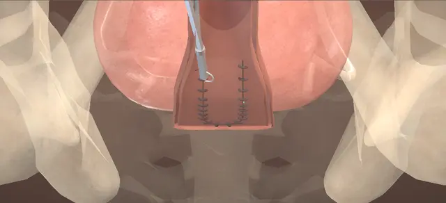
The fistula is circumscribed, an inverted-U flap of anterior vaginal wall is raised, the tract is excised down to the detrusor and the bladder is closed in two layers set across each other.

After a second cystoscopy and a dye fill, the flap is advanced over the repair as a third layer. The lesson is three suture lines that do not overlie one another, and two orifices kept out of every bite.

Advanced tier FRCS female urology, urinary fistula, reconstruction Interactive 3D

Operative visualisation

Schematic scene preview: Vesicovaginal fistula repair, transvaginal
Preparing the 3D anatomy…

UrOops3D · UroRef · © Nity G

How to use this module. Play through the eight steps, or use Back and Next. Four views take turns: the perineum, the bladder base from the front, the anterior vaginal wall from below, and the midline in section. The triggers show an orifice caught by a stitch and dye leaking through the repair.

Click me next Section 2 of 8 Anatomy The structures this operation works between, and the one it must not cross.

The structures in the scene

Transvaginal VVF repair: how the structures relateA fistula at the vaginal cuff after hysterectomy, and what lies round it. Fistula: bladder to vaginal cuff: lie a short way below and lateral; out of every bite Ureteric orifices, one each side (danger). Fistula: bladder to vaginal cuff: opens just above it, on the back wall Trigone and the ridge between the orifices. Fistula: bladder to vaginal cuff: tract dissected to it; edge closed with it Detrusor: the depth of the dissection. Fistula: bladder to vaginal cuff: flaps raised on it; imbricated over the first layer Perivesical fascia: the second layer. Fistula: bladder to vaginal cuff: advanced over the repair; its line lies beyond Anterior vaginal flap: the third layer. Fistula: bladder to vaginal cuff: the U runs from the fistula down to this level Bladder neck: base of the inverted U.Fistula: bladder tovaginal cuff!Ureteric orifices,one each sideTrigone and theridge between theorificesDetrusor: the depthof the dissectionPerivesical fascia:the second layerAnterior vaginalflap: the thirdlayerBladder neck: baseof the inverted Ulie a short waybelow and lateral;out of every biteopens just aboveit, on the backwalltract dissected toit; edge closedwith itflaps raised on it;imbricated over thefirst layeradvanced over therepair; its linelies beyondthe U runs from thefistula down tothis levelTargetDangerLandmarkContext
A fistula at the vaginal cuff after hysterectomy, and what lies round it. Schematic, not to scale; relationships only.
  • From the foot of the table in lithotomy the urethra is anterior and the vagina runs up and back to the cuff.
  • The bladder lies on the anterior vaginal wall with only the perivesical fascia between them.
  • A fistula at the cuff after hysterectomy opens through the back wall of the bladder just above the trigone, so a ureteric orifice lies a short distance below and to each side of it.
  • The flap is raised on the fascia from the fistula down to the level of the bladder neck.
  • ›The fistula - a short scarred tract between the back wall of the bladder and the anterior vaginal wall at the cuff, with an opening at each end.
  • ›Trigone - the fistula of this case lies just above it, on the back wall of the bladder.
  • ›Ureteric orifices - one each side, below and lateral to the bladder opening; seen at both cystoscopies.
  • ›Detrusor - the depth to which the tract is dissected, and what the deep stitches take with the edge of the opening.
  • ›Perivesical fascia - the plane the flaps are raised on, and the superficial layer of the bladder closure.
  • ›Anterior vaginal wall - carries the inverted U; the flap inside it becomes the cover over the repair.
  • ›Vaginal cuff - the apex after hysterectomy, where the fistula opens and the posterior flap lies.
  • ›Bladder neck - the level of the base of the inverted U.
  • ›Urethra - carries the catheter that keeps the bladder empty, and the cystoscope twice.
Click me next Section 3 of 8 Steps The operation in order, with the landmark that guards each move.

Operative steps

  1. Cystoscopy After antibiotics the patient is placed in dorsal lithotomy on well-padded stirrups, and the genitalia, the perineum and the lower abdomen are prepared. A rigid cystoscope is passed and the bladder is inspected thoroughly with angled lenses. Both ureteric orifices are seen in their normal position with clear efflux on each side. The fistula is found from inside the bladder and its position is recorded in relation to the trigone and to each orifice. The cystoscope is removed.
    where it bites: the orifices are a short distance from a vault fistula; where they are is known before the first incision.
  2. Exposure and the catheter through the fistula A urethral catheter is passed and the bladder drained. A weighted vaginal speculum is placed on the posterior wall and a self-retaining retractor is set to open the vagina. The anterior vaginal epithelium is infiltrated with local anaesthetic and adrenaline, which helps the dissection and the haemostasis. The fistula is identified anteriorly at the apex of the vagina and is intubated from the vagina with a small balloon catheter; the balloon is inflated.
    where it bites: a narrow tract is dilated carefully until it takes the catheter; forcing it tears the tract.
  3. The inverted U and the flaps The vaginal epithelium round the fistula is incised circumferentially. With the bladder completely decompressed, an inverted-U incision is made in the anterior vaginal epithelium, its apex contiguous with the circumscribed fistula and its base extending to the bladder neck. By sharp and blunt dissection the vaginal epithelium is dissected off the underlying perivesical fascia, creating vaginal flaps anterior and posterior to the fistula.
    where it bites: the bladder lies directly under the flap; it is kept completely empty while the plane is developed.
  4. The tract, down to the detrusor With gentle traction on the catheter, the fistulous tract is dissected proximally to the level of the detrusor muscle, until it has been mobilised round its whole circumference.
    where it bites: the traction is gentle, and the bladder end of the tract lies just above the trigone.
  5. Excision of the tract Once the mobilisation is adequate all the way round, the entire fistulous tract and the inflammatory tissue surrounding it are excised, and the catheter through the fistula is removed.
    where it bites: the edge that is left is what the first stitches hold in; it has to be healthy bladder wall.
  6. The bladder in two layers The fistula is closed in two opposing layers of interrupted absorbable sutures. The deep layer incorporates the edges of the fistula with the adjacent detrusor muscle. The superficial layer imbricates the perivesical fascia over the first. Opposing the two layers limits the overlap of their suture lines.
    where it bites: a deep lateral bite can take a ureteric orifice; each stitch is placed knowing where both orifices lie.
  7. Second cystoscopy and the dye fill The urethral catheter is removed and a thorough cystourethroscopy is done again: the proximal urethra and the bladder are intact, and there is clear efflux from both ureteric orifices. The cystoscope is replaced by the urethral catheter. The bladder is then filled with a dilute solution of dye in saline, which confirms an intact, watertight closure.
    where it bites: an absent efflux or a leak is put right at this point; once the flap is closed neither can be seen.
  8. The flap as a third layer The anterior vaginal wall flap is advanced over the two-layer repair and closed with a running interlocking absorbable suture. Haemostasis is by sparing use of diathermy throughout the operation. The wound is irrigated, the self-retaining retractor and the weighted speculum are removed, and the urethral catheter is connected to a drainage bag.
    where it bites: the flap covers the repair, so its suture line lies beyond the bladder closure and never on it.
Click me next Section 4 of 8 Danger zones Where this operation bites, and why it bites there.
Critical relationship

Danger zones

The spatial relationships the animation is built to make automatic. Before you read each one, ask yourself: at this point in the operation, what is about to go wrong, and what is my first move? The save for each sits in the Complications tab.

  • RiskThe two ureteric orifices - below and to each side of the bladder opening; seen before the dissection and again after the closure.
  • RiskThe bladder under the flap - the flaps are raised on the perivesical fascia with the bladder completely decompressed.
  • RiskThe tract on traction - gentle traction only; the dissection follows the tract to the detrusor and stops there.
  • RiskSuture lines on one another - two crossed layers in the bladder, then a flap advanced beyond them, so that no line overlies another.
  • RiskA closure that leaks - found by the dye fill, before the flap hides it.
Practise it Take me into the scene Opens the animation at a danger moment and asks you to predict the mechanism and your first move before it reveals.
Click me next Section 5 of 8 Complications The save for each one: the first move, the escalation trigger, the op-note line.

Complications and immediate saves

Trigger each of these live in the Animation tab, then read the save: the first move, the escalation trigger, and the line that goes in the op note.

Ureteric obstruction

  • a bite of the bladder closure catches a ureteric orifice or the ureter in the bladder wall; ureteric injury or obstruction is a listed complication of this operation, and the second cystoscopy is there to find it.
The save
  1. Watch both orifices for long enough to be sure: efflux on one side and none on the other, with the closure beside it. first move
  2. Take out the stitch nearest that orifice, the deep lateral bite of the first layer, and watch for efflux to return.
  3. Replace it with a smaller bite away from the orifice and repeat the cystoscopy before anything else is closed.
  4. No efflux once the stitch is out: senior help in theatre, and a ureteric catheter passed up that side to show the ureter is open. escalation trigger
  5. Op note: the side, the stitch, what was done, and efflux from each orifice at the end. document

Persistent fistula

  • dye comes through the repair at the fill; a recurrent or persistent fistula is a listed complication of this operation, and a closure that leaks on the table will leak afterwards.
The save
  1. Stop the fill and find the point the dye comes from, with the flap still held back. first move
  2. Take down that part of the closure, check that the edge is healthy bladder wall, and close it again in the same two layers.
  3. Fill again with dye against a fresh swab; the flap is not advanced until the swab stays dry.
  4. Tissue that will not hold a stitch, or a second leak: senior review in theatre, and a vascularised flap between the layers as the operative text describes for deficient tissue. escalation trigger
  5. Op note: the result of each fill, what was redone, and the plan for the catheter. document
Click me next Section 6 of 8 Troubleshooting When the case turns: the problem, then the next move.

Intra-operative troubleshooting

When the case turns: the problem, then the next move.

  • ActionThe introitus is too narrow to work through - posterolateral episiotomies, at five and seven o'clock, improve the exposure.
  • ActionThe fistula cannot be found from the vagina - dye instilled into the bladder shows the opening.
  • ActionThe tract is too narrow for the catheter - it is dilated carefully until it takes one; the alternative is a stay suture on each side of the fistula for traction.
  • ActionThe fistula lies near a ureteric orifice - that ureter is stented before the dissection, or both are, to reduce the chance of an inadvertent injury.
  • ActionThe vaginal tissue is deficient or compromised, as after radiotherapy - the repair is reinforced with a vascularised flap: labial fat (a Martius flap), peritoneum or gracilis muscle.
  • ActionDrainage is in doubt, or the wound oozes at the end - a suprapubic catheter can be placed as well as the urethral one, to drain well and to help the later trial of voiding; a rolled gauze pack in the vagina helps haemostasis.
  • ActionThe fistula is high, fixed at the vault and out of reach - an abdominal approach. The European guideline indicates the abdominal route for a high fistula fixed at the vault that cannot be reached through the vagina.
Click me next Section 7 of 8 FRCS viva Say it out loud, then mark yourself against the model answer.

FRCS viva questions

The examiner stems this scene prepares you for. Try to answer out loud first, then reveal, and learn the discriminator, the detail that turns a pass into a strong pass.

QA woman leaks urine continuously from the vagina some weeks after an abdominal hysterectomy. How do you assess her?reveal answer
  • I would take a history and examine her with a speculum, looking for the leak at the vault; the European guideline on female lower urinary tract symptoms calls history and examination with direct inspection essential.
  • To confirm a vesicovaginal fistula I would do a cystoscopy and fill the bladder with coloured fluid with a tampon in the vagina, which the guideline recommends, as a weak recommendation.
  • I would send any fluid I could collect for creatinine to show it is urine.
  • I would not stop at the bladder, because a ureter can be injured at the same operation: a double-dye test can help, and the guideline recommends contrast CT with a late excretory phase when the diagnosis is difficult or a ureterovaginal fistula is suspected.
  • At cystoscopy I would record the size and site of the fistula and its relation to the trigone and to each ureteric orifice, because that decides the approach and whether a ureter needs protecting.
  • On how common this is, I would quote Hilton and Cromwell: among 343,771 women who had a hysterectomy in English NHS hospitals, the rate of urogenital fistula within a year was 1 in 788 overall, and 1 in 87 after radical hysterectomy for cervical cancer.
  • I would drain the bladder with a catheter meanwhile.
  • The guideline gives a spontaneous closure rate of 13 plus or minus 23 per cent, largely in fistulae under a centimetre, so I would not promise her that drainage alone will close it.
Strong pass

Say all three

  • Looking for a ureteric injury as well as the bladder fistula
  • Describing the fistula by its size
  • Its site and its relation to the orifices
Examiner note
Looking for a ureteric injury as well as the bladder fistula, and describing the fistula by its size, its site and its relation to the orifices.
QTalk me through a transvaginal repair of a fistula at the vaginal cuff.reveal answer
  • Antibiotics, dorsal lithotomy on padded stirrups.
  • I start with a cystoscopy: I find the fistula, see both ureteric orifices with clear efflux and note how far each is from it.
  • I pass a urethral catheter to empty the bladder, place a weighted speculum and a self-retaining retractor, and infiltrate the anterior vaginal epithelium.
  • I pass a small balloon catheter through the fistula from the vagina and inflate the balloon.
  • I incise the epithelium round the fistula, then, with the bladder empty, make an inverted-U incision with its apex on that incision and its base at the bladder neck.
  • I dissect the vaginal wall off the perivesical fascia to make flaps anterior and posterior to the fistula.
  • With gentle traction on the catheter I dissect the tract down to the detrusor all the way round, excise the tract with the inflamed tissue round it and take the catheter out.
  • I close the bladder in two opposing layers of interrupted absorbable sutures: the edges of the opening with the adjacent detrusor, then the perivesical fascia imbricated over that.
  • I take the urethral catheter out and repeat the cystoscopy to see an intact bladder and efflux on each side, replace the catheter and fill the bladder with dilute dye to prove the closure is watertight.
  • I then advance the vaginal flap over the repair as a third layer and close it with a running interlocking suture.
  • The catheter goes on free drainage.
Strong pass

Say both

  • Three layers whose suture lines do not overlie one another
  • With both ureteric orifices seen before the dissection and again after the closure
Examiner note
Three layers whose suture lines do not overlie one another, with both ureteric orifices seen before the dissection and again after the closure.
QWhen would you repair it, by which route, and would you add an interposition flap?reveal answer
  • I would be honest that the evidence is thin on all three.
  • On timing, the European guideline finds only small uncontrolled series, which suggest no difference in success between repair within four weeks and repair after three months, and it recommends tailoring the timing to the patient once oedema, inflammation, necrosis and infection have resolved.
  • On route, it says there are no randomised trials of abdominal against vaginal repair.
  • It describes dissection and repair in layers, the flap-splitting technique, as the more commonly used vaginal method, and it indicates the abdominal route for a high fistula fixed at the vault that cannot be reached vaginally, and for a fistula that persists after repair.
  • This woman has a first, non-irradiated fistula that I can reach, so I would offer the vaginal repair.
  • On interposition, the operative text I follow adds a vascularised flap, labial fat, peritoneum or gracilis, when the vaginal tissue is deficient or compromised, as after radiotherapy.
  • The guideline says flaps are mostly used for recurrence, radiation, large fistulae and poor tissue, that no high-level evidence shows they improve outcome, and, as a weak recommendation, that an interposition graft is used when a radiation fistula is repaired.
  • I would not add one to a first repair in healthy tissue.
  • On trimming, the guideline cites a single randomised trial, by Shaker and colleagues in 64 women with obstetric fistulae: cure was 75 per cent without trimming and 67.6 per cent with it, not a significant difference, though fistulae that recurred after trimming tended to be larger.
  • The operative text excises the tract, and that is what I have described.
Strong pass

Say all three

  • Saying how weak the evidence is: no trial of route
  • No trial showing that a flap helps
  • Timing left to the state of the tissue
Examiner note
Saying how weak the evidence is: no trial of route, no trial showing that a flap helps, and timing left to the state of the tissue.
QWhat do you tell her about success, and how do you look after the bladder afterwards?reveal answer
  • I would tell her that most of these repairs succeed and that the first attempt is the best chance.
  • The European guideline gives overall closure rates after surgical repair of 58 to 100 per cent and says that second and later repairs do less well than the first.
  • The UK figures I would quote are from Hilton, a personal series of 348 women referred to one tertiary unit over 25 years: anatomical closure at the first operation in 95.7 per cent, and closure more often in women who had had no attempt at repair before referral, 98.2 against 88.2 per cent.
  • That is one experienced unit, so I would present it as what a specialist service achieves, and it is my reason for sending her to one.
  • I would tell her the complications the operative text lists: bleeding, infection, ureteric injury or obstruction, a recurrent or persistent fistula, vaginal shortening and painful intercourse.
  • Afterwards the bladder must stay empty while the repair heals.
  • The guideline recommends continuous drainage until healing is confirmed, with expert opinion of ten to fourteen days for a simple fistula after surgery, and notes that most series drained for at least ten days;
  • the operative text allows a suprapubic catheter as well as the urethral one.
  • A blocked catheter in that time is an emergency for the repair, so I would make sure the ward knows it.
Strong pass

Say both

  • Knowing that a failed first repair lowers the chance of the next
  • That drainage continues until healing is confirmed
Examiner note
Knowing that a failed first repair lowers the chance of the next, and that drainage continues until healing is confirmed.
Click me next Section 8 of 8 Evidence The guidance each of these answers is built on.

Evidence and guidance

Operative Dictations in Urologic Surgery
Armenakas NA, Fracchia JA, Golan R. Vesicovaginal fistula repair. Chapter 97. Wiley, 2019
The indication, the eight essential steps and their order, the template dictation followed step by step here, the variations (episiotomies for a narrow introitus, dye in the bladder to show the fistula, dilating a narrow tract or stay sutures for traction, stenting a ureter when the fistula is near an orifice, a vascularised flap for deficient tissue, a suprapubic catheter, a vaginal pack) and the complication list: bleeding, infection, ureteric injury or obstruction, recurrent or persistent fistula, vaginal shortening, dyspareunia. The chapter gives catheter sizes, a suture material and a fill volume; they stay off the page. It lists complications without their management: the first, escalate and document steps, the danger lines and the remark about a blocked catheter are general surgical practice. The scene is schematic. Where the orifices lie on the bladder base, the width of the cuff and of the flap, the direction of each bladder suture line and the size of every bite are drawing choices; the dictation places no ureteric catheters and none are drawn.
Open guideline
EAU Guidelines
Non-neurogenic Female LUTS, 2026: Disease management, section 4.8 Urinary fistula, read 13 October 2026
Diagnosis: history and examination with direct inspection (strong); cystoscopy and retrograde filling with coloured fluid and a vaginal tampon (weak); a double-dye test, mentioned as sometimes useful; creatinine in the fluid; contrast CT with a late excretory phase when the diagnosis is difficult or a ureterovaginal fistula is suspected (weak). Spontaneous closure 13 plus or minus 23%, largely fistulae under 1 cm. Closure after surgical repair 58 to 100%; secondary repairs less successful. Timing: small uncontrolled series, no difference between repair within four weeks and after three months; timing tailored once oedema, inflammation, necrosis and infection have resolved (weak). Vaginal techniques: partial colpocleisis, and the more commonly used dissection and repair in layers; no comparison. No randomised trial of abdominal against vaginal repair; abdominal route for high fistulae fixed at the vault and for persistent fistulae (weak). Tissue interposition: no high-level evidence of benefit; an interposition graft for radiation fistulae (weak). Drainage until healing is confirmed, expert opinion ten to fourteen days for simple or post-surgical fistulae (weak); most series ten days or more. Its statement on trimming rests on the Shaker trial listed below.
Open guideline
BJOG
Hilton P, Cromwell DA. The risk of vesicovaginal and urethrovaginal fistula after hysterectomy performed in the English National Health Service: a retrospective cohort study examining patterns of care between 2000 and 2008. BJOG 2012;119:1447-1454
Abstract read 13 October 2026. Hospital Episode Statistics, 343,771 women: urogenital fistula within one year of hysterectomy in 1 in 788 overall; highest after radical hysterectomy for cervical cancer (1 in 87), lowest after vaginal hysterectomy for prolapse (1 in 3,861). The rate counts vesicovaginal and urethrovaginal fistulae together. This is the study the guideline cites for a rate about nine times higher after radical hysterectomy; only the figures of the study itself are quoted here.
Open guideline
BJU International
Hilton P. Urogenital fistula in the UK: a personal case series managed over 25 years. BJU Int 2012;110:102-110
Abstract read 13 October 2026. A prospective series from one tertiary unit, 348 women referred between 1986 and 2010: two-thirds of surgical aetiology, almost half after hysterectomy; spontaneous closure in 6.9%; 291 had surgery; anatomical closure at the first operation in 95.7%; closure more likely with no attempt at repair before referral (98.2% against 88.2%). One surgeon and one unit: not a figure for every service.
Open guideline
Neurourology and Urodynamics
Shaker H, Saafan A, Yassin M, Idrissa A, Mourad MS. Obstetric vesico-vaginal fistula repair: should we trim the fistula edges? A randomized prospective study. Neurourol Urodyn 2011;30:302-305
Abstract read 13 October 2026. Sixty-four women with obstetric fistulae, all repaired vaginally in three layers with a Martius flap, randomised to trimming of the bladder edges or none: cure 67.6% with trimming and 75% without, not significantly different; fistulae that recurred after trimming tended to be larger than the original. Obstetric fistulae, so it bears only indirectly on a fistula after hysterectomy. This is the trial behind the guideline statement on trimming.
Open guideline

Curriculum. Supports learning relevant to the ISCP Urology Curriculum syllabus topic Female urology. Not a workplace-based assessment and not evidence of competence.

Two minutes

Close the lap: recall it before you leave.

Say it aloud, then reveal. Reading is not remembering.

  1. 01 The relationship Say the critical spatial relationship out loud, then reveal. reveal
    Memory frame

    From the foot of the table in lithotomy the urethra is anterior and the vagina runs up and back to the cuff.

    Why?
    The bladder lies on the anterior vaginal wall with only the perivesical fascia between them. A fistula at the cuff after hysterectomy opens through the back wall of the bladder just above the trigone, so a ureteric orifice lies a short distance below and to each side of it. The flap is raised on the fascia from the fistula down to the level of the bladder neck.
  2. 02 The first move Ureteric obstruction. Your first move? reveal
    First move

    Watch both orifices for long enough to be sure: efflux on one side and none on the other, with the closure beside it.

  3. 03 The escalation trigger Ureteric obstruction. What makes you call for help? reveal
    Escalate when

    No efflux once the stitch is out: senior help in theatre, and a ureteric catheter passed up that side to show the ureter is open.

  4. 04 The discriminator What turns a pass into a strong pass? reveal
    Strong pass

    Say all three

    • Looking for a ureteric injury as well as the bladder fistula
    • Describing the fistula by its size
    • Its site and its relation to the orifices
    Examiner note
    Looking for a ureteric injury as well as the bladder fistula, and describing the fistula by its size, its site and its relation to the orifices.
  5. 05 The pitfall Where does this operation bite first? reveal
    Pitfall

    The two ureteric orifices

    Why?
    below and to each side of the bladder opening; seen before the dissection and again after the closure.

Revision practice with self-marked answers, not an assessment of clinical competence. Kept in this browser only; it does not sync between devices.