
Varicocele treatment
Occlusion of refluxing gonadal veins by microsurgical, laparoscopic or radiological routes, with artery, lymphatic and recurrence risks compared.
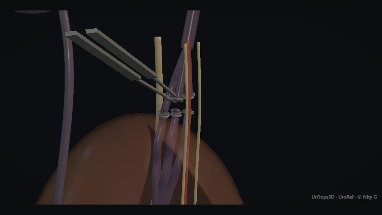 Interactive 3D Interactive operative model Open the 3D animation
Teaching catalogue only. This coverage record is not a patient pathway or operative instruction.
Typical place in practice
Selected infertility, testicular-growth concern or persistent discomfort associated with a clinically relevant varicocele.
Alternatives and neighbouring procedures
Observation, fertility treatment without repair, analgesic/supportive care, or radiological versus surgical treatment according to anatomy and preference.
Same procedure family, explicit technique branches
These are variations of this canonical operation. Complication modules below identify whether they apply to all branches or only to named approaches.
Microsurgical subinguinal or inguinal repair
Divides refluxing veins while preserving the testicular artery, lymphatics and vas.
Laparoscopic ligation
Controls gonadal vessels through an abdominal minimally invasive route.
Open high ligation
Uses a more proximal non-laparoscopic route.
Radiological embolisation
Treats reflux endovascularly without scrotal or inguinal incision.
High-level sequence
Coverage outline only; not a step-by-step technical guide.
- Confirm laterality, indication and baseline testicular/fertility status
- Identify the venous anatomy through the selected route
- Preserve artery, lymphatics and vas where intended
- Occlude refluxing veins while maintaining testicular perfusion
- Review wound, hydrocele, recurrence and relevant fertility outcomes
Operative notes
Concise operative notes covering the key steps, landmarks and watch points, in the order you encounter them.
Fertility surgery
What it does: operations to find and fix blocked pipes, or to fetch sperm.
Varicocele (open routes; coils via radiology)
- Palomo (high, back of tummy): whole pedicle tied. Artery may go with it. Collaterals save the testis. Fewer veins. More hydroceles. ~15% back if the artery is spared.
- Ivanissevich (groin canal): cord lifted. Separate artery (papaverine, Doppler) and lymphatics from the ~3 veins. Artery NOT tied here. Cut the cremaster vein too, or it recurs.
- Marmar (below the canal, at the ring): no fascia opened. Many veins, branched artery. The confident microsurgeon's route.
Vasography (blocked vas, before repair)
- Transverse nick in the vas. Do not divide it.
- Fluid to a slide. No sperm = block is on the testis side.
- Saline flush runs free = open beyond. Or blue dye + catheter in bladder: blue urine.
- 0.5 mL X-ray contrast down the vas maps the level.
Vasovasostomy (rejoin the vas)
- Trim to healthy ends. Both bleed.
- 2-layer micro join: 3 x 10/0 stitches in the lining, turn 180, 3 more. 9/0 in the muscle. Then sheath layers.
Vaso-epididymostomy (join vas to epididymis)
- Fat-to-thin tube change marks the block.
- Open 1 tube. Sperm confirmed on a slide.
- Vas fixed to the sheath, 6/0. Lining joined 10/0.
- Sperm back in ~70%. Later narrowing common.
TURED (ejaculatory duct)
- Scan rectally first. Dye + contrast into ducts.
- Resect the veru. Spare neck and sphincter.
- Dye in the bed = in the duct.
- Sperm back ~50%. Risks: reflux, dry ejaculation.
MESA / PESA (sperm retrieval)
- MESA: open an epididymal tube. Pipette the fluid. Blocked system: best sperm sit HIGH (unusual).
- PESA: 27G butterfly into the head of the epididymis, held between fingers. Gentle pull. Press firmly 5 min after.
- 2026 update: micro-TESE is the standard for non-blocked azoospermia. Name it in vivas.
From the UroRef Operative Quick Guide. Open the note page →
Every failure mode is its own module.
Timing and variation coverage are recorded for each module.
Testicular artery injury and atrophy
Loss of arterial supply can cause ischaemic pain and later testicular volume loss.
Applies to: All variantsHydrocele or lymphatic swelling
Lymphatic interruption can lead to fluid accumulation around the testis.
Applies to: All variantsPersistent or recurrent varicocele
Missed collaterals, recanalisation or alternative venous drainage can preserve reflux.
Applies to: All variantsBleeding, infection or chronic pain
Access-site morbidity varies across inguinal, abdominal and venous routes.
Applies to: All variantsCoil migration, venous thrombosis or contrast-related event
The endovascular route adds embolic-device, access-vessel and contrast-specific complications.
Applies to: Radiological embolisationIntra-abdominal organ or vessel injury
Port and retroperitoneal dissection add abdominal access risks.
Applies to: Laparoscopic ligationSources for this module
Penile prosthesis, Peyronie disease, priapism and reproductive surgery.
Accessed 2026-08-03 · opens source European Association of Urology EAU Guidelines on Paediatric UrologyCommon congenital, acute scrotal and paediatric reconstructive procedures.
Accessed 2026-08-03 · opens source British Association of Urological Surgeons Patient information leaflets by procedureUK patient-facing procedure, alternatives, recovery and complication framing.
Accessed 2026-08-03 · opens source