
Revision sheet
Varicocele: embolisation or ligation
6 steps, each one the authored title and caption from the interactive animation, with the frame captured from that step. Read it before the case, or print it and work through the steps in order.
Teaching only, not for diagnosis or treatment decisions. The text on this sheet is the animation's own step copy, reproduced verbatim.
UrOops3D · UroRef · © Nity G
- 01
The reflux tract
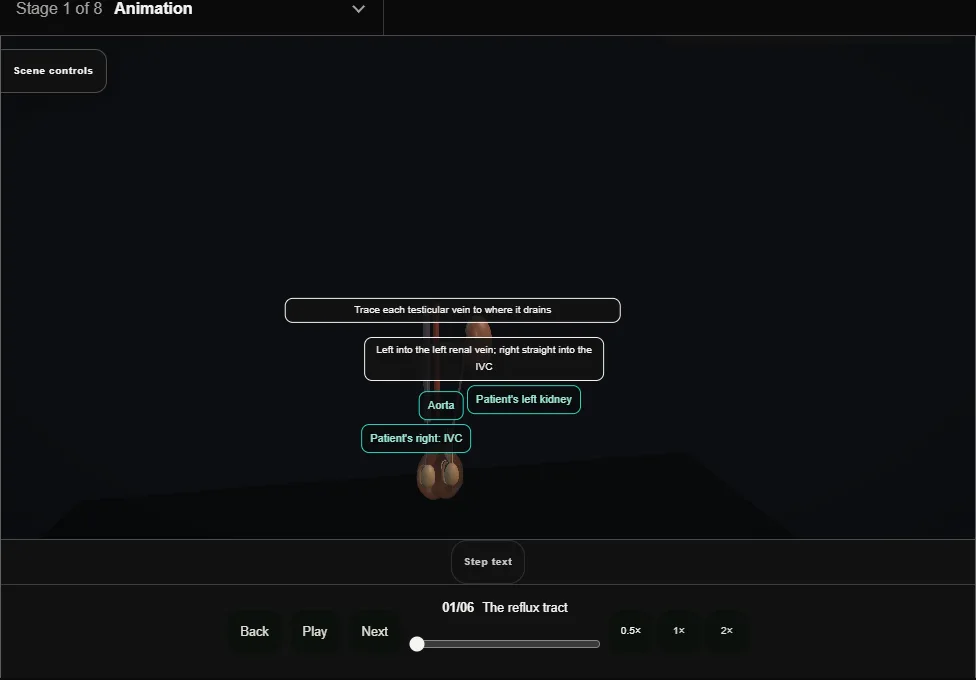
- The left spermatic vein drains into the left renal vein; the right drains directly into the IVC.
- The left's longer course and right-angled entry favour reflux, which fills the pampiniform plexus around the cord.
- 02
Assessment + indications
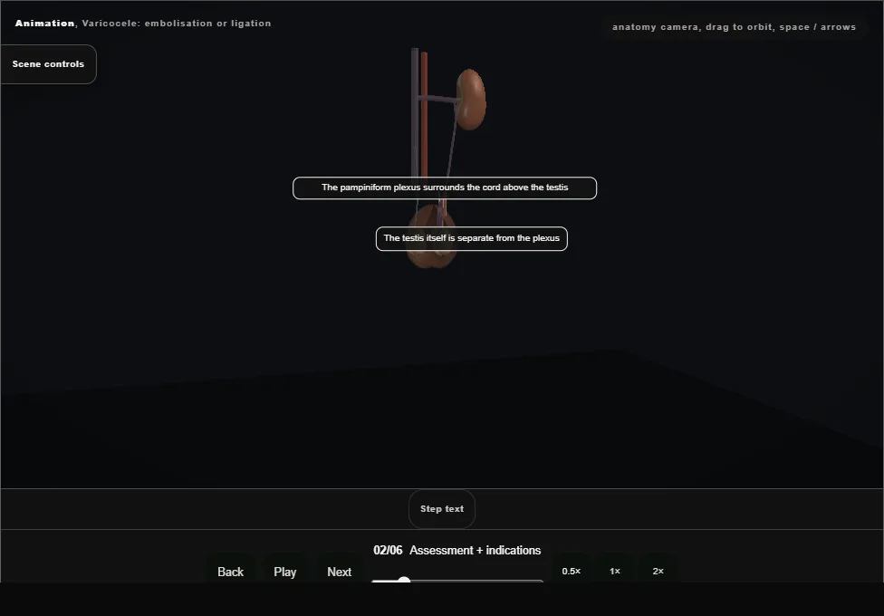
- Examine standing: a bag of worms that fills with Valsalva and empties lying flat.
- One that does not empty lying flat, or a new right-sided one, needs the testis and kidney imaged, because a varicocele can be a symptom, not a diagnosis.
- 03
Embolisation: access + venography
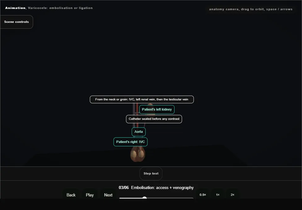
- Via jugular or femoral access a catheter is seated in the left spermatic vein and venography maps the channel, its collaterals and its size, before anything is deployed.
- 04
Coil + sclerosant deployment
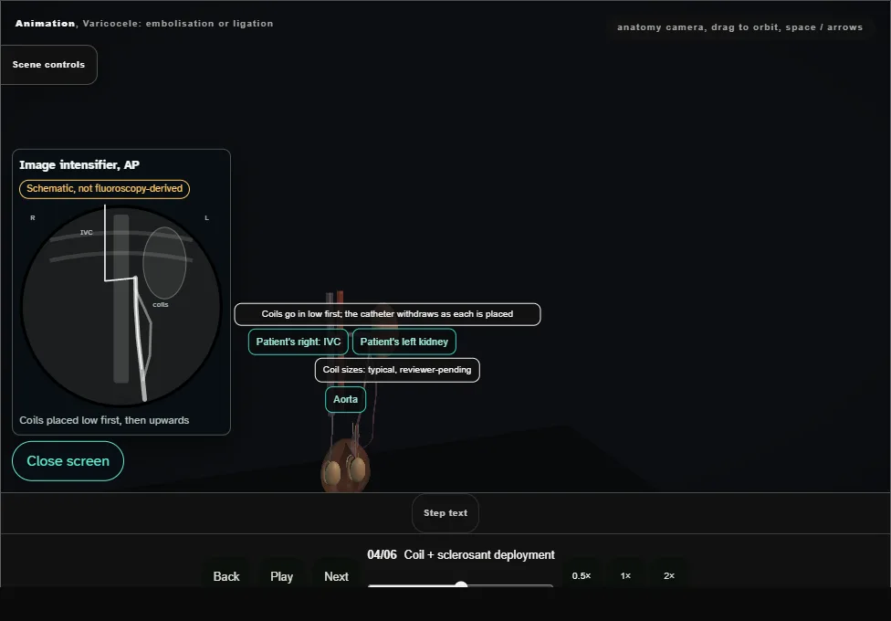
- Coils are packed into the spermatic vein, often with sclerosant, from low in its course up to just below the renal vein junction, the catheter withdrawing as each is placed.
- The retrograde flow that filled the plexus stops.
- 05
The surgical geometry: subinguinal ligation
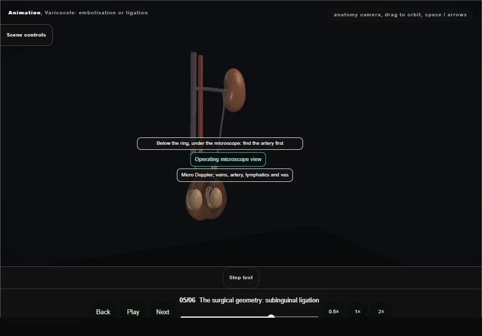
- The alternative: ligation below the ring, under magnification, taking the veins while sparing the testicular artery and the lymphatics.
- Division of lymphatics is the mechanical cause of the post-ligation hydrocele.
- 06
Aftercare + the recurrence watch
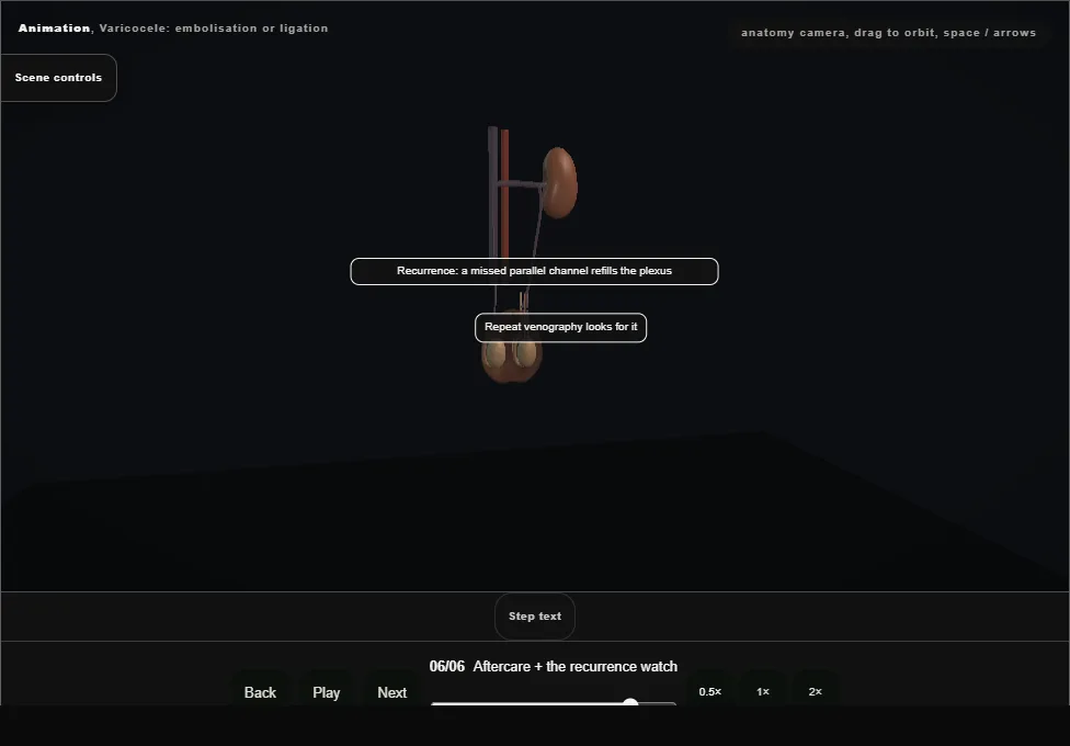
- Persistence or recurrence means flow has found a way around the occlusion, through a missed collateral or a parallel channel.
- Examine for recurrence, hydrocele and testicular volume on follow-up.
Derived from VaricoceleScene.tsx at build time: every step title and caption above is the authored
ProcedureDef text, reproduced verbatim, and this page shares its readable-points renderer with the animation
itself. 6 of 6 steps have a captured frame.
Text fingerprint e67b1f8b51a6bbef. A caption edit without a re-capture fails the production
check, so the sheet and the animation cannot drift apart silently.