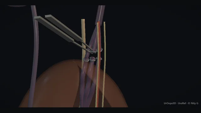
A varicocele is a geometry problem: reflux down the left spermatic vein from its right-angled entry into the left renal vein, filling a pampiniform plexus that hangs around the cord onto the testis, while the right side drains directly to the IVC.
Two treatments attack the same channel from opposite ends, coils deployed from within at the renal junction, or the veins taken at subinguinal ligation with the testicular artery and lymphatics spared.
Core tierFRCS andrology, infertility, endourologyInteractive 3D
Standing, the left venous column from renal vein to testis. Schematic, not to scale; relationships only.
Stand the patient and follow the column downwards: left renal vein at the midline, spermatic vein leaving its underside and descending lateral to the midline to the internal ring, then the pampiniform plexus fanning around the cord onto the posterosuperior testis.
Valsalva fills the column from above, which is why the cough impulse is felt at the upper scrotum and why the hand should rest there during examination.
The two treatments are the two ends of the same column: embolisation occludes it at the top, below the renal junction, while subinguinal ligation interrupts it at the bottom, below the ring, where the plexus has separated into individual veins and the artery and lymphatics can be picked out and spared.
›Left spermatic (testicular) vein - the refluxing channel; it leaves the underside of the left renal vein and runs to the internal ring.
›Left renal vein - crosses the midline to the IVC under the superior mesenteric artery; the junction where the reflux begins.
›Pampiniform plexus - the bag of worms around the cord from ring to testis; it fills with Valsalva and is the examination finding.
›Testicular artery - runs in the cord among the veins; the structure whose sparing defines proper subinguinal ligation.
›Cord lymphatics - fine channels draining the scrotal coverings; divided with the veins, they are the mechanical cause of the post-ligation hydrocele.
The reflux tractThe left spermatic vein drains into the left renal vein, while the right drains directly into the inferior vena cava. The geometry explains the asymmetry: the left column is longer and enters at a right angle, and both features favour reflux. Reflux descends that column under standing pressure and fills the pampiniform plexus around the cord, which is the bag of worms the examination finds. Valsalva fills the column from above, which is why the impulse is felt at the upper scrotum and why the veins empty when the patient lies flat. A unilateral right varicocele of sudden onset breaks the pattern and demands imaging of the kidney and the retroperitoneum before anything is treated. where it bites: the anatomy explains the side: a unilateral right varicocele of sudden onset demands the kidney and the retroperitoneum be imaged.
Assessment + indicationsThe examination is performed standing, with the cord palpated at the upper scrotum during Valsalva: the plexus fills like a bag of worms and empties when the patient lies down, and testicular volumes are measured and compared side to side. Indications to treat are discomfort the patient actually feels, the fertility association with abnormal semen parameters, and persistent asymmetry of testicular growth in a young man. Every new varicocele has the testis examined as well, and imaged when the story is atypical, because a varicocele can be a symptom of retroperitoneal disease rather than a diagnosis in itself. Treatment decided without a standing examination is treatment decided on the wrong evidence. where it bites: every new varicocele needs the testis examined and, when the story is atypical, imaged: a varicocele can be a symptom, not a diagnosis.
Embolisation: access + venographyVia jugular or femoral venous access a catheter is advanced to the renal vein and selectively seated in the spermatic vein at its junction. Venography is then performed to map the channel before anything is deployed: the calibre of the vein, the position of the junction, and above all the collateral and parallel channels running alongside it. The venogram is the operation plan and is read like a map rather than glanced at, because a collateral missed at this stage is the recurrence presented later. Access that cannot be negotiated from one route is attempted from the other, since the angle at the renal junction decides which approach reads better. where it bites: the venogram is the operation plan; collateral channels missed here are the recurrence of later.
Coil + sclerosant deploymentCoils are deployed into the upper spermatic vein, often with sclerosant added to reach the smaller parallel channels, working methodically from the renal vein junction downwards. The column is covered from the top because a patent channel left above the coils keeps doing exactly what it did before. Deployment continues until the reflux column is occluded and the check venogram no longer fills it, including every collateral the mapping identified. A coil undersized for the vein can migrate, and a column occluded only in its middle leaves channels open above and below it: these are the two classic technical failures of the procedure. where it bites: a coil too small migrates and a column too short under-covered leaves parallel channels open above and below.
The surgical geometry: subinguinal ligationThe surgical alternative interrupts the same column from below: a small subinguinal incision, the cord delivered into the wound, and the individual veins ligated separately under magnification. At this level the plexus has separated into identifiable channels, which is what allows the testicular artery to be recognised and the lymphatics to be spared alongside it. Magnification is not optional in this operation, because the artery and the lymphatics are its fine print: taken with the veins, they produce ischaemic atrophy and the post-ligation hydrocele respectively. The veins are ligated at two levels on the delivered cord so that no channel within the field escapes. where it bites: the artery and the lymphatics are the operation's fine print; taking them produces ischaemic atrophy and the post-ligation hydrocele.
Aftercare + the recurrence watchFollow-up examines the patient standing for recurrence, feels for a hydrocele around the testis, and compares testicular volumes against the baseline recorded before treatment. Persistence or return means flow has found another way, through a collateral missed at the renal junction or a parallel channel beyond the coils or the clips. The recurrent varicocele is re-imaged and re-mapped before it is re-treated, because the second attempt needs the anatomy of the first failure rather than a repeat of it. Surveillance is part of the plan from the outset, and recurrence is described to the patient as a recognised outcome of both routes rather than a surprise. where it bites: a recurrent varicocele is re-imaged and re-mapped, not simply re-operated; the second attempt needs the first attempt's anatomy.
The spatial relationships the animation is built to make automatic. Before you read each one, ask yourself: at this point in the operation, what is about to go wrong, and what is my first move? The save for each sits in the Complications tab.
RiskThe renal junction and its collaterals - embolisation must cover the proximal channel and any parallel collateral; anything missed is the recurrence.
RiskTesticular artery in the cord - accompanies the veins below the ring; the non-sparing operation risks ischaemic atrophy of the testis.
RiskCord lymphatics - invisible without magnification; divided en masse with the veins, they leave the scrotum unable to drain and a hydrocele follows.
Trigger each of these live in the Animation tab, then read the save: the first move, the escalation trigger, and the line that goes in the op note.
Post-ligation hydrocele
lymphatics divided with the veins leave the tunica unable to drain; fluid collects around the testis in the weeks after ligation.
The save
Examine the swelling as a hydrocele: transilluminate, get above it, confirm the testis is normal.first move
Most post-ligation hydroceles are observed first; a proportion settle without intervention.
A tense or persistent hydrocele is aspirated for symptomatic relief, accepting that aspiration alone recurs.
Definitive management when symptomatic and persistent → hydrocele repair, with the lymphatic-sparing principle now non-negotiable.escalation trigger
Note: side, size, examination findings and the plan with intervals, so the follow-up is structured.document
Persistence / recurrence
the veins refill because flow found another way: a collateral missed on venography, or parallel channels beyond the clips or coils.
The save
Re-examine standing with Valsalva and compare with the pre-treatment examination and imaging.first move
Re-image the tract: venography after embolisation, ultrasound with reflux assessment after ligation.
Map the escape route before choosing the second attempt; a collateral at the renal junction and a parallel cord channel are re-treated by different routes.
Symptomatic recurrence with a defined channel → re-embolisation or re-operation according to what the mapping shows.escalation trigger
Note: findings before and after each treatment, and the anatomical reason identified for the failure.document
Testicular atrophy (arterial injury)
the rare disaster of non-sparing surgery: the testicular artery taken with the veins leaves the testis ischaemic and it shrinks.
The save
Detect it on follow-up: a smaller, softer testis compared with the contralateral side, measured consistently.first move
Confirm with ultrasound, including intra-testicular flow assessment, and document baseline volume.
There is no restoration once the artery is lost; management is surveillance of volume and of the hormonal axis in the young.
Suspected arterial injury at operation → immediate vascular surgical input, though prevention by identification is the real strategy.escalation trigger
Note: pre-operative testicular volume, the sparing technique used, and the follow-up measurements.document
When the case turns: the problem, then the next move.
ActionSpermatic vein cannot be catheterised from the IVC - try the alternative access, jugular or femoral; the renal junction angle decides which approach reads better.
ActionVenography shows multiple parallel channels - coil and sclerose each identified channel from the junction down; under-coverage of one is the recurrence at six months.
ActionArtery not identifiable at subinguinal dissection - use the Doppler probe, deliver the cord fully and separate the planes; if still in doubt, take a wider patch of veins rather than the artery.
ActionBilateral varicocele with predominantly right-sided symptoms - image the retroperitoneum first; a right-sided varicocele of sudden appearance has a defined differential that must be excluded before treatment.
The examiner stems this scene prepares you for. Try to answer out loud first, then reveal, and learn the discriminator, the detail that turns a pass into a strong pass.
QShould adolescents with varicocele be treated? Take me through the controversy.reveal answer
The adolescent controversy exists because the indication is preventive rather than symptomatic, and the thing being prevented is imperfectly measurable.
On one side is the observation that varicocele is associated with impaired testicular growth and, in the long term, with subfertility, and that catch-up growth of the affected testis after treatment is demonstrable in a proportion of boys, which is used as the proxy for future fertility.
On the other side, most adolescents with varicocele will not need assisted conception, the follow-up into paternity is long and loss-prone, and both treatments carry real complications, hydrocele after ligation, recurrence after either, and the rare arterial injury.
The settled middle ground is watchful surveillance for the asymptomatic boy with symmetrical testes, serial measurement of testicular volume, and treatment for defined indications: failing growth of the affected testis relative to its partner, significant asymmetry that persists, pain, or a varicocele in a solitary testis where the margin for error disappears.
Counselling the family is part of the treatment: no one can promise which boy is protected, so the decision is made together, with the volume trend as the objective anchor.
The discriminator in practice is the growth trend, not the size of the veins at a single visit.
Strong pass
Say both
Anchoring the decision on serial testicular volume trends rather than varicocele grade
Naming catch-up growth as the contested surrogate
Examiner note
Anchoring the decision on serial testicular volume trends rather than varicocele grade, and naming catch-up growth as the contested surrogate.
QCompare embolisation with surgical ligation for varicocele.reveal answer
Both occlude the same refluxing column; they differ in where, in what they spare, and in their failure modes.
Embolisation approaches from the renal junction downwards, through jugular or femoral access: it is minimally invasive, needs no scrotal or inguinal dissection, and venography during the procedure maps collateral channels in real time, which suits the redo case after failed surgery.
Its limitations are anatomical and technical: it depends on catheterisable veins, it exposes nothing, so a channel it cannot see or reach persists, and coil migration and access complications are its own risks.
Surgical ligation interrupts the column from below: subinguinal ligation under magnification is the operation most sympathetic to the anatomy, because the plexus has separated into individual veins that can be taken while the testicular artery and lymphatics are identified and spared;
higher ligation at the inguinal or laparoscopic plane takes the cord as a bundle, which is quicker but less sparing.
Ligation's price is the wound, a slightly deeper dissection, and a hydrocele rate that tracks with lymphatic division.
Recurrence differs by mechanism: parallel channels and missed collaterals for both, but embolisation failures tend to be at the top and surgical failures at the bottom of the column.
Selection follows the patient: the body-conscious young man, the redo and the needle-averse diverge in opposite directions.
Strong pass
Say all three
Contrasting the failure mechanisms anatomically
Recurrence from the top after embolisation and from the bottom after ligation
Rather than reciting complication lists
Examiner note
Contrasting the failure mechanisms anatomically, recurrence from the top after embolisation and from the bottom after ligation, rather than reciting complication lists.
QWhy do varicoceles recur after apparently successful treatment?reveal answer
Recurrence is the same word for several different anatomies, which is why I would map before re-treating.
The first mechanism is the missed collateral: the spermatic vein rarely travels alone, and parallel channels at the renal junction can be dormant on the first venogram or simply outside the field of the first ligation;
when the main column is closed, they recruit.
The second is under-coverage: coils packed too low leave the proximal channel patent above them, and a ligation that takes the visible veins at one level leaves channels that join the cord above or below it.
The third is the testicular arterial supply's travelling companions: small veins adherent to the artery, deliberately or unavoidably spared at an artery-sparing operation, can dilate and reflux later;
this is the specific price of the operation done properly.
The fourth is genuinely new disease, rare and slow.
The clinical implication is that the second treatment is planned from images, not from memory of the first: venography for the post-embolisation recurrence, ultrasound with reflux mapping for the post-ligation one, and the re-treatment route chosen by where the escape route runs.
The honest counsel before any first treatment is that recurrence is a recognised outcome of both routes, and surveillance for it is part of the plan.
Strong pass
Say both
Separating the recurrence mechanisms by anatomy
Noting that the veins travelling with the spared artery are the specific cost of artery-sparing surgery
Examiner note
Separating the recurrence mechanisms by anatomy, and noting that the veins travelling with the spared artery are the specific cost of artery-sparing surgery.
QA thirty-year-old with a left varicocele and a year of primary infertility is referred to you. How do you proceed?reveal answer
I begin by establishing that the varicocele is the whole story, because it is the commonest finding and the least provable cause.
The history covers duration, pain, and prior groin or scrotal surgery;
the examination is standing, with the cord palpated at the upper scrotum during Valsalva, testicular volumes assessed with an orchidometer, and the right side examined too, because a new right varicocele changes the differential to retroperitoneal pathology and mandates imaging.
Semen analysis, at least two samples properly taken, documents the impairment; the partner's assessment happens in parallel, because treating the man for a couple's problem wastes a year if her factors dominate.
Imaging with ultrasound confirms reflux, measures testicular volume and excludes a renal cause for the left column.
The treatment conversation then follows evidence and preference: discomfort with demonstrable reflux, testicular asymmetry and abnormal semen parameters in a man trying to conceive are the standard indications, and he is offered embolisation or magnification-guided ligation with their different failure modes explained.
If semen parameters are normal and the varicocele is asymptomatic, treatment is not automatic and surveillance is defensible.
I set expectations honestly: semen parameters improve in a proportion of treated men, and the pathway to assisted conception stays open either way.
Strong pass
Sequencing the assessment, two semen samples, partner assessment, standing examination with volumes, before any treatment decision, and stating when treatment is not indicated.
Say it aloud, then reveal. Reading is not remembering.
01The relationshipSay the critical spatial relationship out loud, then reveal.reveal
Memory frame
Stand the patient and follow the column downwards
Why?
Stand the patient and follow the column downwards: left renal vein at the midline, spermatic vein leaving its underside and descending lateral to the midline to the internal ring, then the pampiniform plexus fanning around the cord onto the posterosuperior testis. Valsalva fills the column from above, which is why the cough impulse is felt at the upper scrotum and why the hand should rest there during examination. The two treatments are the two ends of the same column: embolisation occludes it at the top, below the renal junction, while subinguinal ligation interrupts it at the bottom, below the ring, where the plexus has separated into individual veins and the artery and lymphatics can be picked out and spared.
02The first movePost-ligation hydrocele. Your first move?reveal
First move
Examine the swelling as a hydrocele: transilluminate, get above it, confirm the testis is normal.
03The escalation triggerPost-ligation hydrocele. What makes you call for help?reveal
Escalate when
Definitive management when symptomatic and persistent → hydrocele repair, with the lymphatic-sparing principle now non-negotiable.
04The discriminatorWhat turns a pass into a strong pass?reveal
Strong pass
Say both
Anchoring the decision on serial testicular volume trends rather than varicocele grade
Naming catch-up growth as the contested surrogate
Examiner note
Anchoring the decision on serial testicular volume trends rather than varicocele grade, and naming catch-up growth as the contested surrogate.
05The pitfallWhere does this operation bite first?reveal
Pitfall
The renal junction and its collaterals
Why?
embolisation must cover the proximal channel and any parallel collateral; anything missed is the recurrence.
Revision practice with self-marked answers, not an assessment of clinical competence. Kept in
this browser only; it does not sync between devices.