
Revision sheet
Anterior vaginal prolapse repair
9 steps, each one the authored title and caption from the interactive animation, with the frame captured from that step. Read it before the case, or print it and work through the steps in order.
Teaching only, not for diagnosis or treatment decisions. The text on this sheet is the animation's own step copy, reproduced verbatim.
UrOops3D · UroRef · © Nity G
- 01
The prolapse
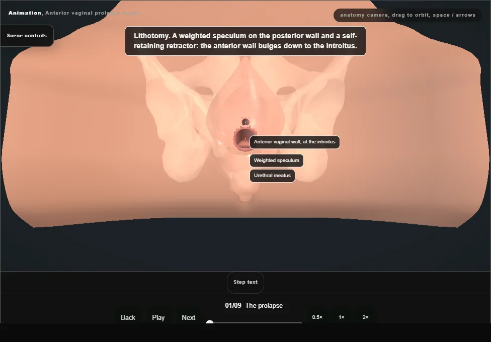
- Lithotomy, with a weighted speculum on the posterior wall and a self-retaining retractor.
- The anterior vaginal wall bulges down to the introitus, and the bulge is the bladder base.
- A urethral catheter drains the bladder and it is kept empty throughout.
- The fascia has given way centrally and, on each side, at the arcus tendineus.
- 02
Midline incision
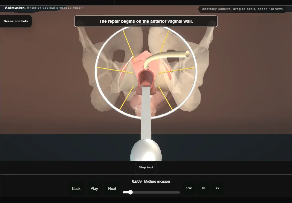
- The anterior vaginal epithelium is infiltrated to lift it off the fascia and to help haemostasis.
- With the bladder decompressed, a midline incision is made in the anterior wall from just below the urethral meatus to the apex of the defect.
- 03
Vaginal flaps
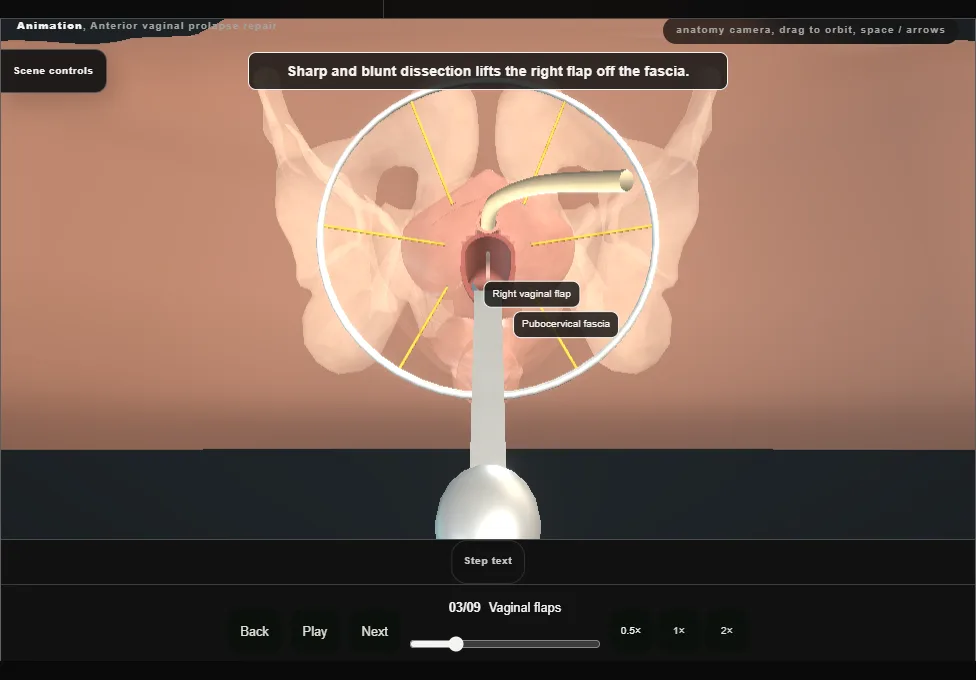
- By sharp and blunt dissection each vaginal flap is lifted off the periurethral and pubocervical fascia, which exposes the central defect.
- The bladder lies directly behind the fascia, so the dissection keeps to the flap.
- 04
Plication stitches
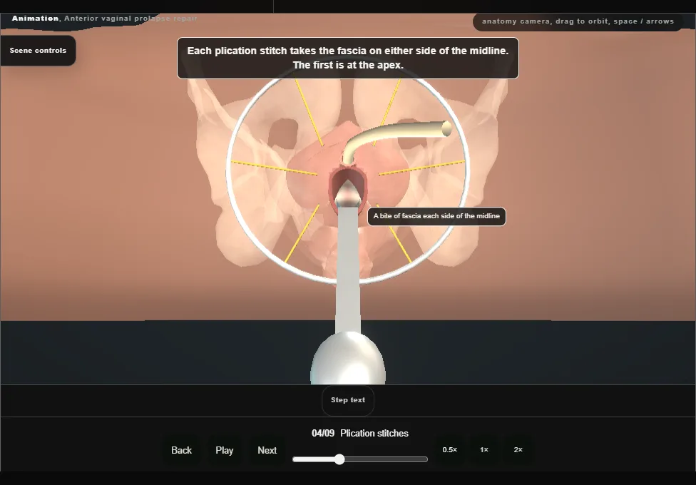
- The bladder is reduced and interrupted stitches are placed across the midline.
- Each takes the vaginal muscularis and adventitia, the layer called the pubocervical fascia, on either side of the defect.
- The bladder wall and the ureters lie above that layer and are left out of every bite.
- 05
Tie the plication
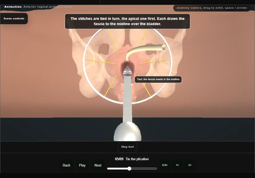
- The stitches are tied in turn.
- Each brings the fascia together in the midline over the bladder, and with each the bladder base is carried back into the pelvis.
- This is the anterior colporrhaphy: it closes the central defect.
- 06
Lateral defect, first side
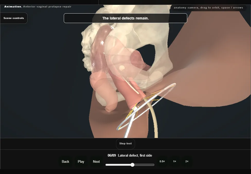
- The bladder is retracted medially and the dissection continues laterally to the arcus tendineus fasciae pelvis, which exposes the lateral defect.
- Five sutures are placed through the arcus, the lateral edge of the pubocervical fascia and the inner surface of the vaginal flap.
- Once all are in they are tied one at a time.
- 07
Lateral defect, other side
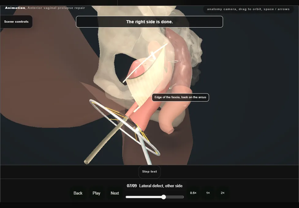
- The identical repair is made at the opposite arcus tendineus.
- Throughout the dissection the bladder and the urethra are not entered, and bleeding is controlled with diathermy.
- 08
Cystourethroscopy
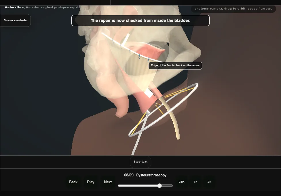
- The catheter is removed and the bladder and urethra are inspected.
- Three findings are wanted: the bladder and proximal urethra intact, clear efflux from both ureteric orifices, and no suture in the bladder.
- The catheter is then replaced.
- 09
Trim and close
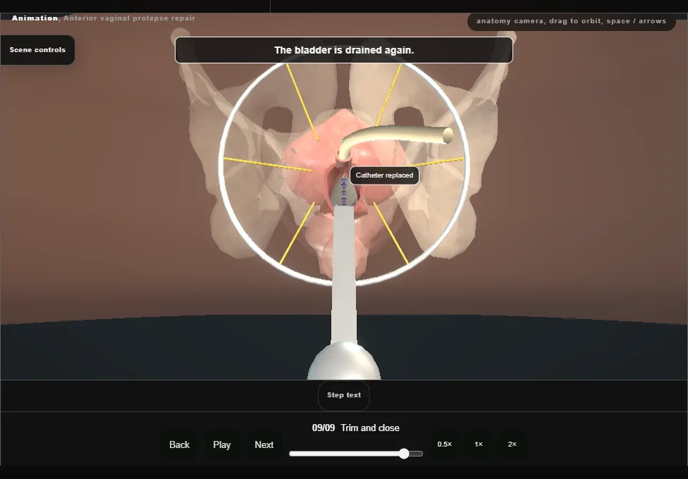
- The wound is irrigated and haemostasis confirmed again.
- The edges of the vaginal epithelium are trimmed only where they are redundant, and the wall is closed in the midline with interrupted stitches.
- The retractor and the speculum come out and the catheter is left on drainage.
Derived from female/AnteriorColporrhaphyScene.tsx at build time: every step title and caption above is the authored
ProcedureDef text, reproduced verbatim, and this page shares its readable-points renderer with the animation
itself. 9 of 9 steps have a captured frame.
Text fingerprint 60d685b0b71c6e92. A caption edit without a re-capture fails the production
check, so the sheet and the animation cannot drift apart silently.