
Revision sheet
Bladder neck and anastomotic stenosis
8 steps, each one the authored title and caption from the interactive animation, with the frame captured from that step. Read it before the case, or print it and work through the steps in order.
Teaching only, not for diagnosis or treatment decisions. The text on this sheet is the animation's own step copy, reproduced verbatim.
UrOops3D · UroRef · © Nity G
- 01
Who
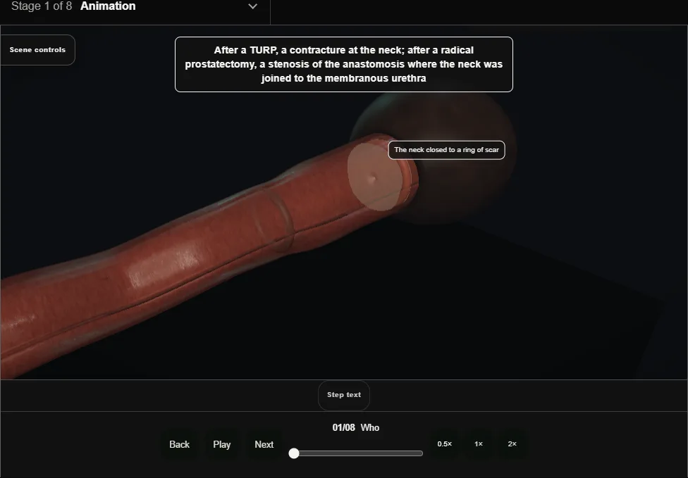
- A contracture of the neck after TURP, or a stenosis of the vesicourethral anastomosis after radical prostatectomy.
- What differs is what lies beyond: after a prostatectomy the sphincter is a centimetre away and is the only thing holding the urine.
- 02
Assessment
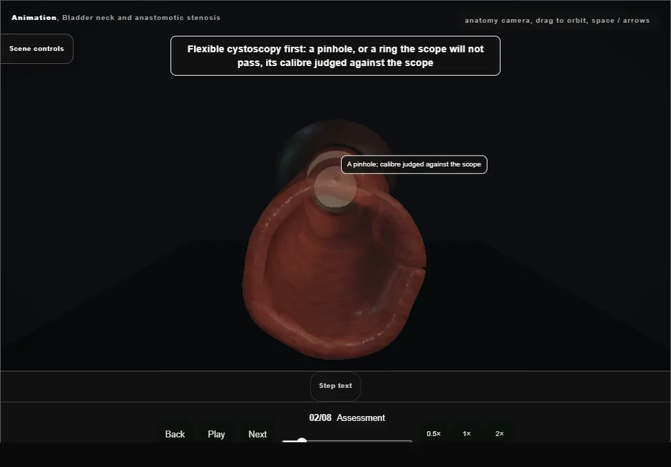
- Flexible cystoscopy of the pinhole and its calibre; the continence status that decides how aggressive to be.
- A dry man is treated gently; an incontinent one can have the neck opened wide and a sphincter later.
- 03
The sheath and the wire
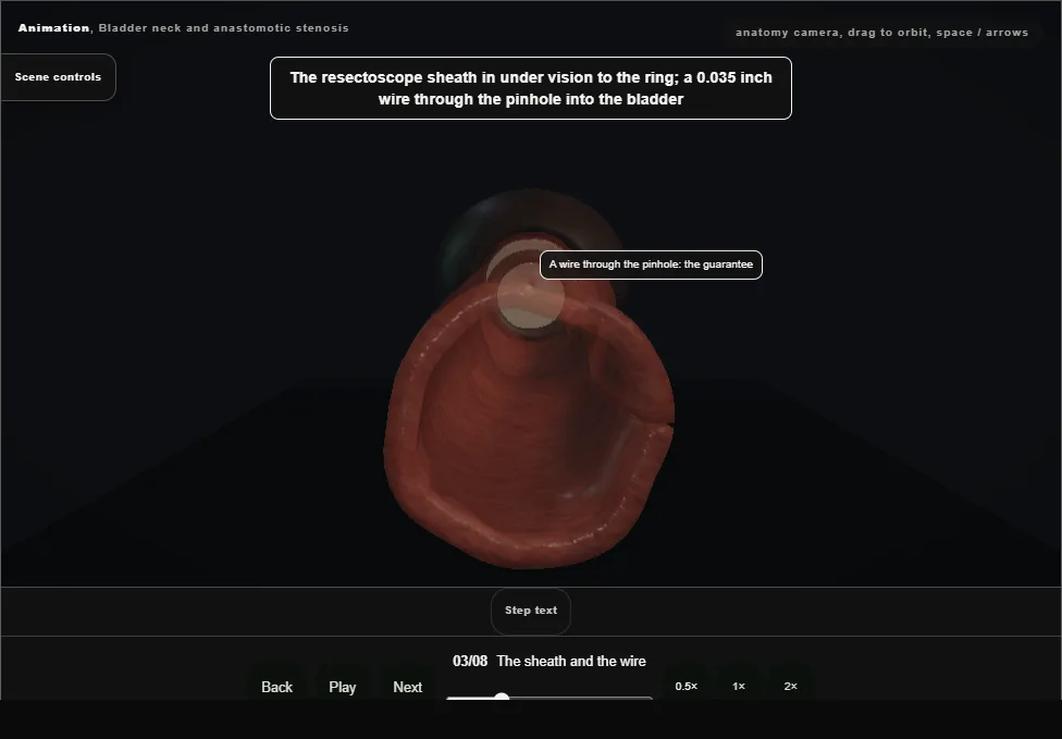
- The resectoscope sheath in under vision to the ring and a 0.035 inch wire through the pinhole into the bladder: the guarantee that every cut stays on the true lumen.
- 04
Incisions at three and nine
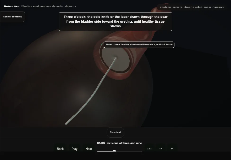
- The cold knife or the laser drawn through the scar at three o'clock from the bladder side toward the urethra until soft tissue shows, then nine.
- Never six, where the rectum lies, never twelve, where the pubis and the dorsal veins are.
- 05
Dilatation
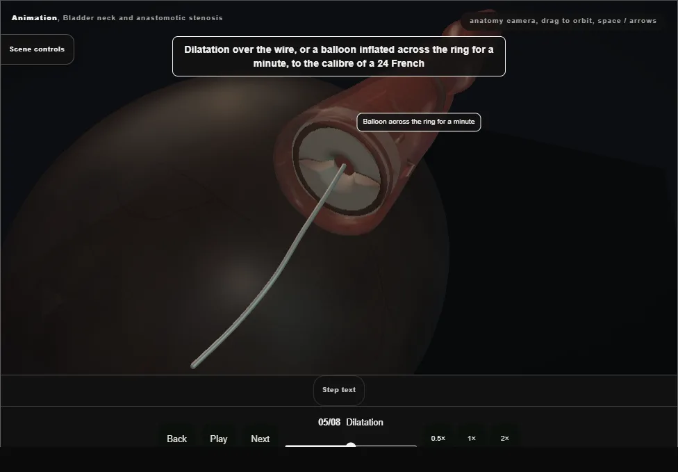
- Over the wire, or a balloon inflated across the ring for a minute, to the calibre of a 24 French.
- The scope then passes into the bladder for the orifices and a survey.
- 06
Adjuncts
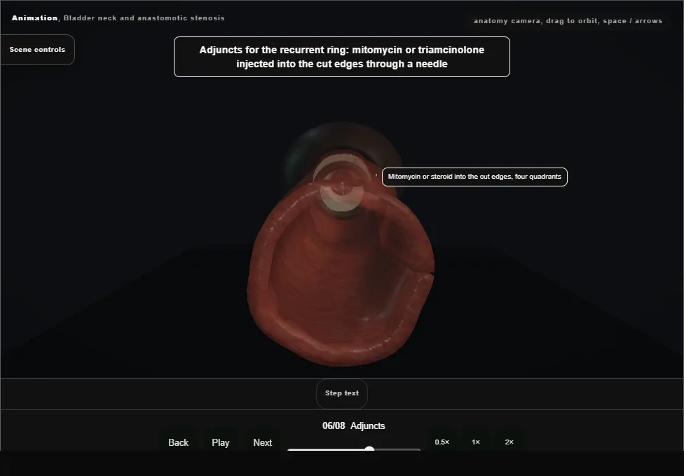
- For the recurrent ring: mitomycin or triamcinolone injected into the cut edges in four quadrants, a small volume each, away from the sphincter.
- 07
The catheter and the trial
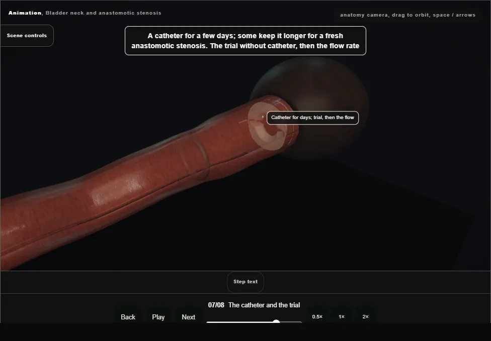
- A catheter for a few days, longer for a fresh anastomosis; the trial without catheter, then a flow rate.
- Some men self-dilate for months to keep it open.
- 08
Recurrence and the ladder
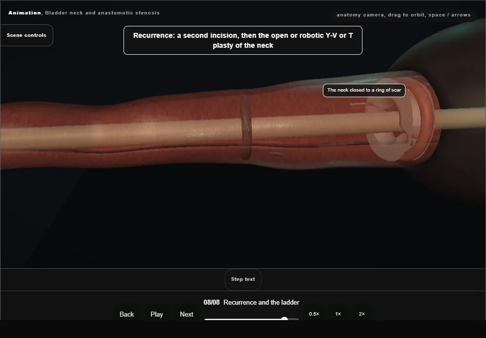
- A second incision, then a Y-V or T plasty of the neck, open or robotic.
- The man made incontinent by the treatment gets an artificial sphincter once the neck is stable: patency first, continence rebuilt after.
Derived from endoscopy/BladderNeckStenosisScene.tsx at build time: every step title and caption above is the authored
ProcedureDef text, reproduced verbatim, and this page shares its readable-points renderer with the animation
itself. 8 of 8 steps have a captured frame.
Text fingerprint 8331e0a1391bd36b. A caption edit without a re-capture fails the production
check, so the sheet and the animation cannot drift apart silently.