
Revision sheet
ECIRS (Galdakao-modified supine)
6 steps, each one the authored title and caption from the interactive animation, with the frame captured from that step. Read it before the case, or print it and work through the steps in order.
Teaching only, not for diagnosis or treatment decisions. The text on this sheet is the animation's own step copy, reproduced verbatim.
UrOops3D · UroRef · © Nity G
- 01
Supine position + retrograde access
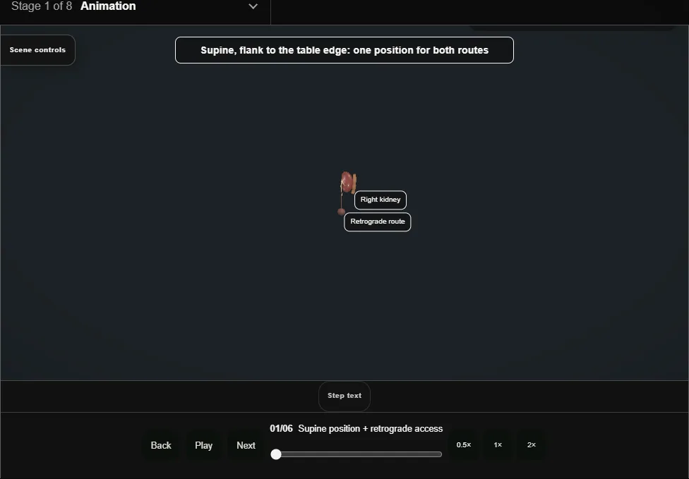
- The patient lies supine, flank flush with the table edge, one position for both approaches.
- Cystoscopically a safety wire (teal) goes up to the kidney, a ureteral access sheath to the PUJ beside it, and through it the flexible ureteroscope into the target calyx, before any puncture.
- 02
Puncture onto the waiting scope
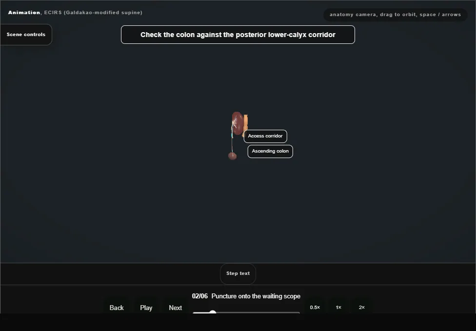
- The needle walks from the flank to a posterior lower-pole calyx.
- In the supine position the ascending colon can lie on this corridor: ultrasound sees bowel, fluoroscopy sees the calyx, and both must agree.
- The ureteroscope in the calyx watches the needle tent the papilla and come through its tip.
- 03
Two antegrade wires
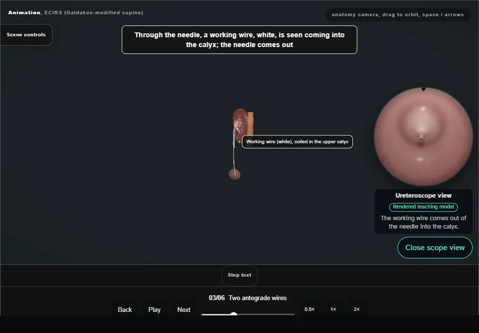
- Through the needle the working wire (white) is seen entering the calyx and coils in the upper calyx; an 8/10 Fr coaxial adds the antegrade safety wire (teal).
- Both stay for the whole case and every exchange happens over them.
- 04
Tract dilation + sheath
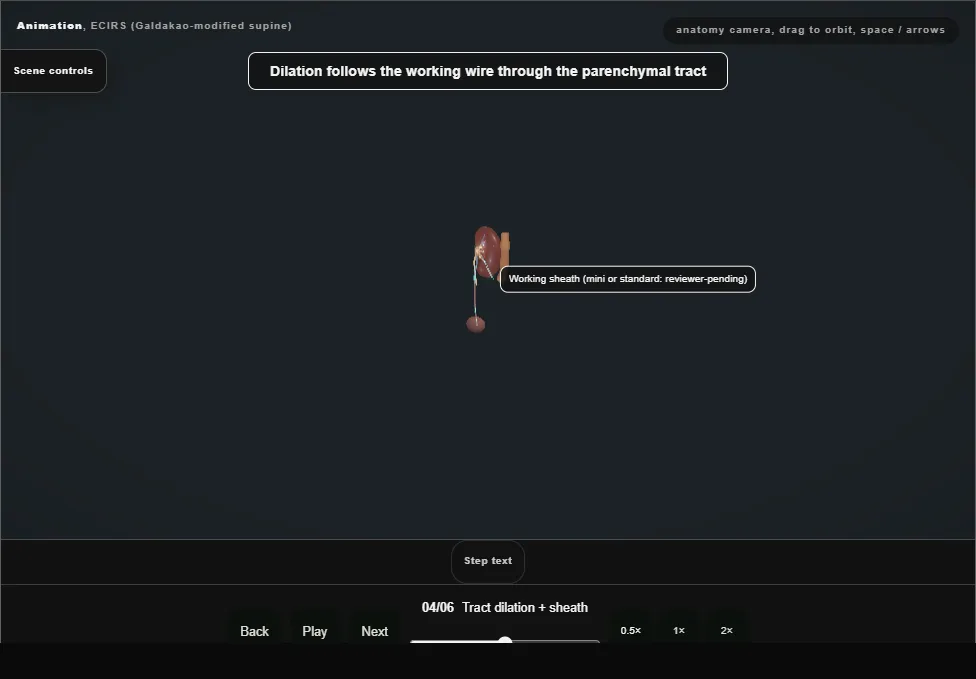
- Dilators pass over the working wire and the sheath is seated in the calyx under the flexible scope's eye.
- The tract crosses parenchyma that the segmental arteries bracket: dilation stays on the wire, and bleeding means tamponade with the sheath, not deeper dilation.
- 05
Dual-scope fragmentation
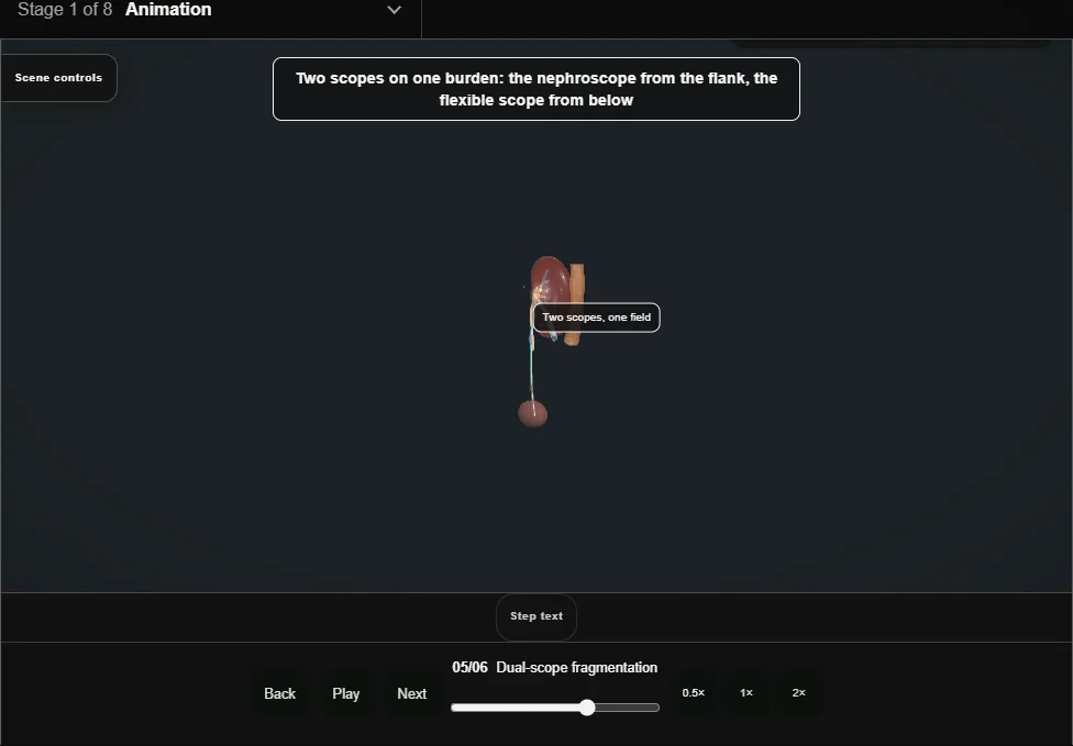
- The nephroscope works down the sheath while the flexible ureteroscope works from below: two angles on one burden.
- Each piece is broken on its own; the flexible scope baskets fragments to the nephroscope and they leave through the sheath one at a time.
- One fragment out is not stone-free.
- 06
Exit strategy
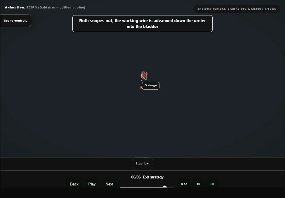
- When drainage is adequate and the field quiet the case ends tubeless with a JJ stent placed antegradely over the working wire, its coils confirmed on screening.
- A nephrostomy is kept when bleeding or obstruction makes a second look plausible.
Derived from EcirsScene.tsx at build time: every step title and caption above is the authored
ProcedureDef text, reproduced verbatim, and this page shares its readable-points renderer with the animation
itself. 6 of 6 steps have a captured frame.
Text fingerprint 94e26e7c8d70edcc. A caption edit without a re-capture fails the production
check, so the sheet and the animation cannot drift apart silently.