
Revision sheet
Flexible cystoscopy: the outpatient look
8 steps, each one the authored title and caption from the interactive animation, with the frame captured from that step. Read it before the case, or print it and work through the steps in order.
Teaching only, not for diagnosis or treatment decisions. The text on this sheet is the animation's own step copy, reproduced verbatim.
UrOops3D · UroRef · © Nity G
- 01
Before the scope
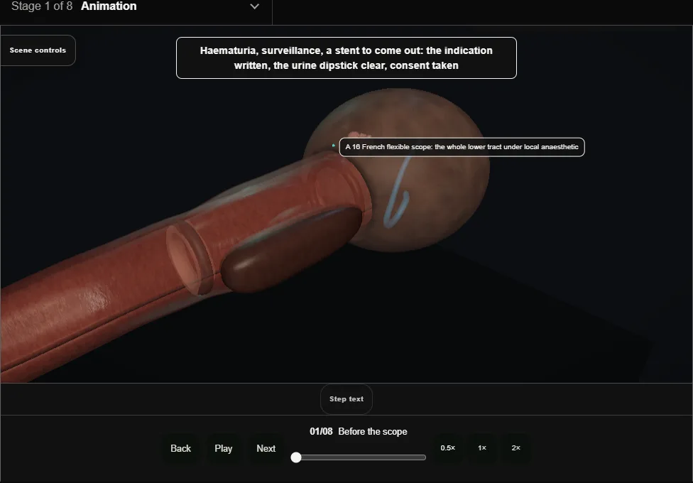
- The indication written, the dipstick clear, consent for the look and for anything planned through the working channel; a chaperone offered, and allergies to lidocaine, chlorhexidine or latex checked.
- Antibiotic cover for a stent removal by local policy; the scope's decontamination record logged to the patient.
- Gel in the urethra and held for a few minutes; the scope white-balanced with irrigation running.
- 02
The urethra under vision
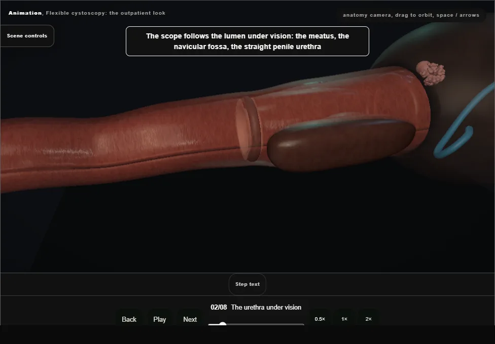
- Meatus, navicular fossa, the straight penile urethra, the bulb turning upward.
- The external sphincter is closed and opens as the patient breathes out slowly and the irrigation runs; the scope never pushes it.
- 03
The prostatic urethra and the neck
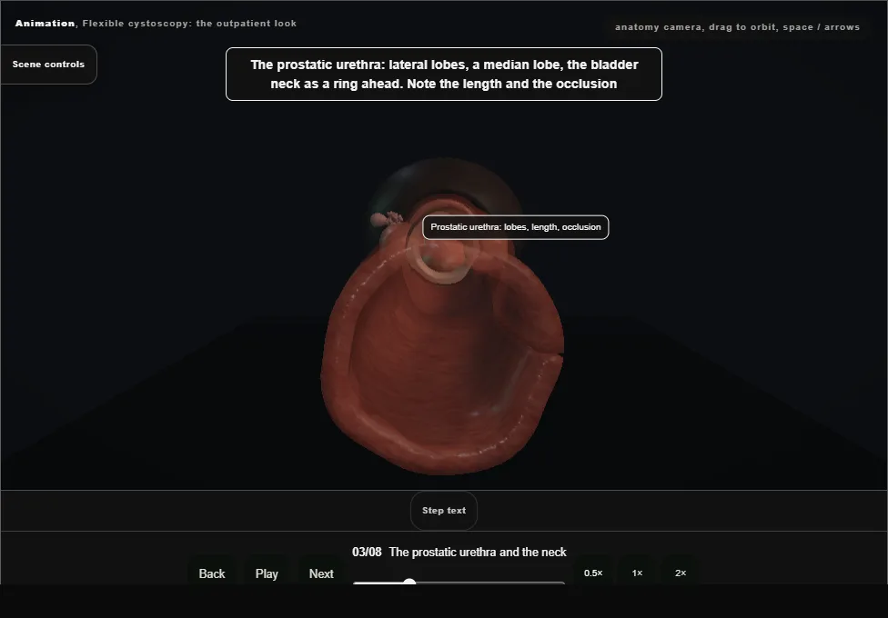
- Lateral lobes, a median lobe, the length and the degree of occlusion noted; then the bladder neck as a ring and into the bladder, where the air bubble at twelve o'clock is the compass.
- 04
The systematic survey
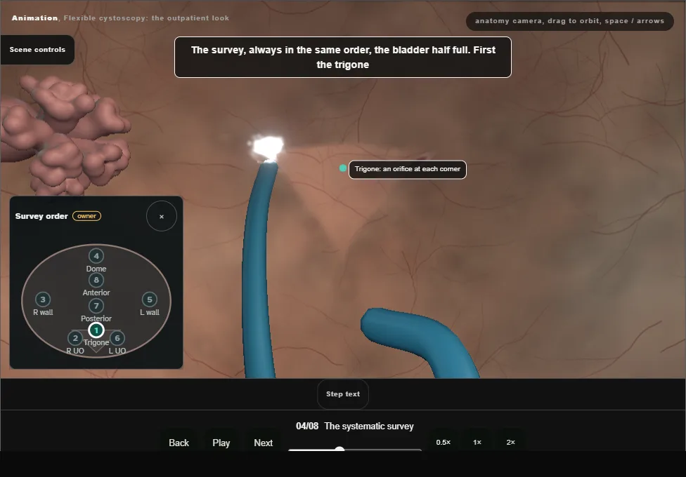
- Always the same order, the bladder half full: trigone, right orifice, right lateral wall, dome, left lateral wall, left orifice, posterior wall, anterior wall.
- The trigone with an orifice at each corner, each watched for a jet of urine.
- Tumours, red velvety patches, stones and trabeculation looked for on every wall; a diverticulum is looked into.
- 05
Retroflexion
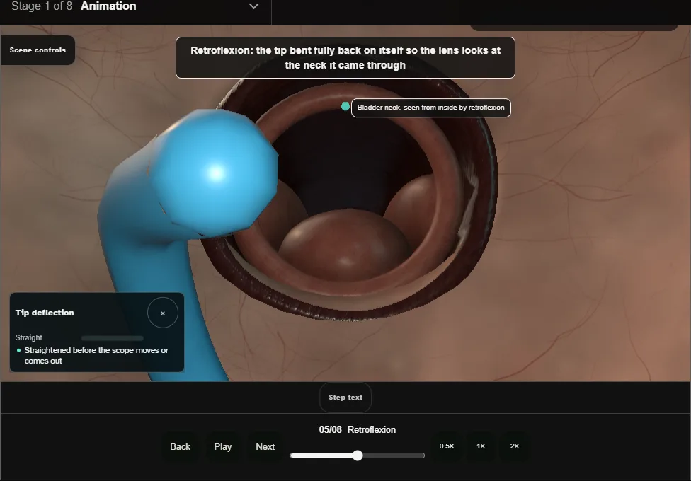
- The tip bent fully back so the lens looks at the neck it came through: the anterior bladder neck and an intravesical median lobe are seen from nowhere else.
- The tip is straightened before the scope moves on or comes out; a retroflexed scope pulled back injures the neck.
- 06
The finding
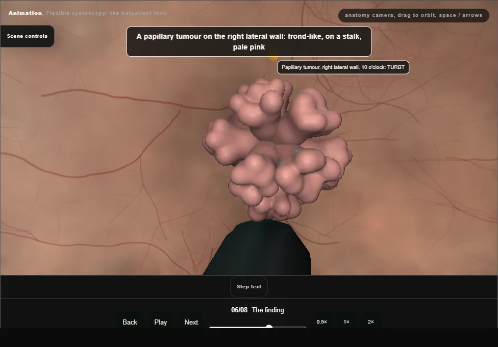
- A papillary tumour on the right lateral wall: clock position, wall, size against the scope, number, stalk or broad base, relation to the orifice.
- The rest of the bladder still surveyed; photographed and booked for TURBT; a flexible scope does not biopsy this.
- The patient told today, placed on the cancer pathway, and the upper tracts imaged if not already done.
- 07
The working channel: a stent out
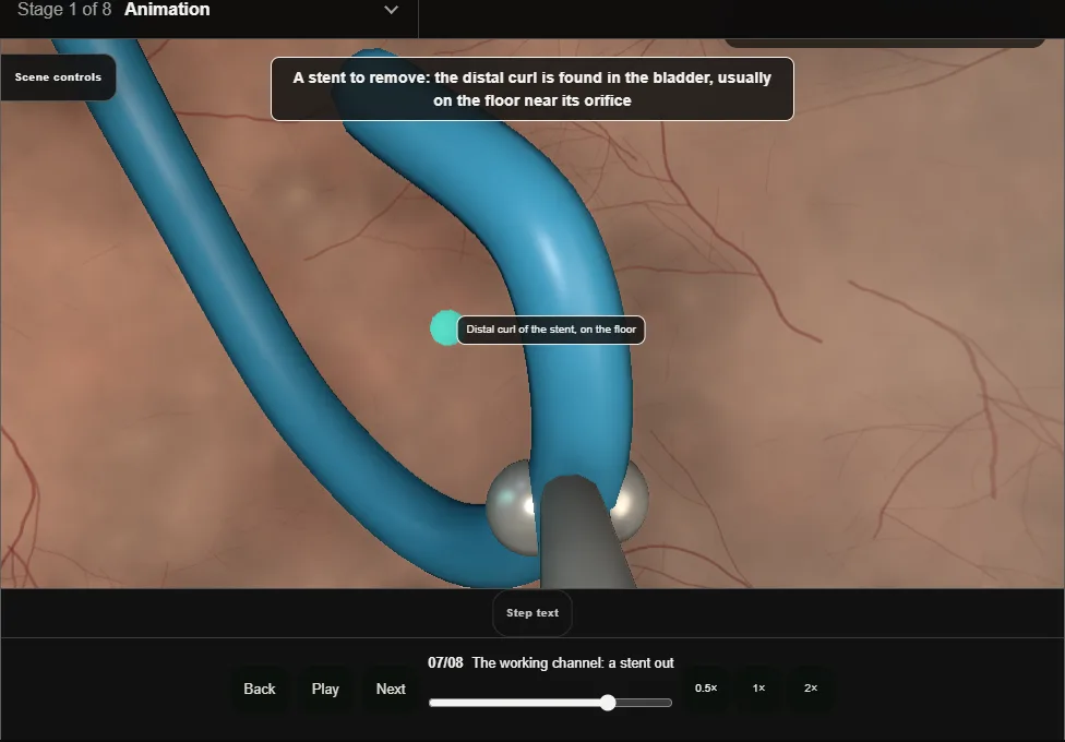
- The stent checked against the register first: side, date inserted and why.
- The distal curl found on the floor near its orifice, graspers through the channel, closed on the curl, and the whole scope withdrawn with the stent held.
- Encrusted or resisting: stop, do not pull.
- Checked intact; the register updated.
- 08
Withdraw and write
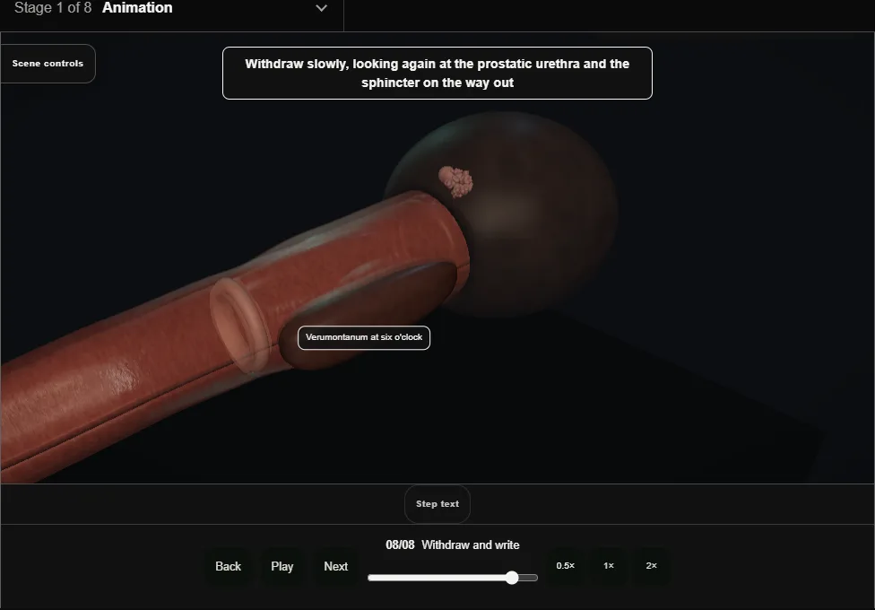
- A second look at the prostatic urethra and the sphincter on the way out.
- The note is a map: urethra, prostate, each wall, the orifices and their efflux, the finding and the plan.
- Before leaving: stinging and pink urine for a day or two are expected; fever, clots or being unable to pass urine mean come back.
Derived from endoscopy/FlexibleCystoscopyScene.tsx at build time: every step title and caption above is the authored
ProcedureDef text, reproduced verbatim, and this page shares its readable-points renderer with the animation
itself. 8 of 8 steps have a captured frame.
Text fingerprint af9f475dd898eb86. A caption edit without a re-capture fails the production
check, so the sheet and the animation cannot drift apart silently.