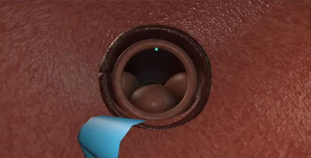
The commonest procedure in urology and the one that finds most of the bladder cancers: a flexible scope under local anaesthetic through a urethra that opens for a breathing patient, into a bladder that is surveyed the same way every time.
This page is the look, the map, and the small things done through the working channel.
Core tierFRCS core urology, haematuria and bladder cancer surveillanceInteractive 3D
From inside, meatus to bladder with the flexible scope. Schematic, not to scale; relationships only.
From inside, the urethra is a tunnel that turns upward at the bulb, closes to a ring at the sphincter and opens again over the veru into the prostatic urethra.
The bladder beyond the neck is a chamber with the air bubble at its roof, the trigone on its floor and an orifice at each back corner;
the anterior neck is behind the scope and is only seen by bending the tip back on itself.
›Meatus and penile urethra - straight under vision; a stricture here is a ring the scope will not pass.
›External sphincter - a closed ring that opens for a breathing patient; the false passage is made by pushing it.
›Prostatic urethra - lateral lobes, median lobe, the length and the occlusion noted on the way in and on the way out.
›Air bubble - at twelve o'clock, the compass for the whole survey.
›Trigone and ureteric orifices - the raised triangle on the floor with an orifice at each corner, each watched for a jet.
›Anterior bladder neck - seen only by retroflexion; the intravesical median lobe lives here.
›The scope - 16 French, deflecting tip, working channel for graspers, a ureteric catheter or a small basket.
Before the scopeThe indication is written: visible or non-visible haematuria, surveillance after a bladder tumour, symptoms that need the bladder looked at, or a stent to come out. The dipstick is checked; a positive one with symptoms defers the scope or covers it with an antibiotic. The consent conversation names dysuria and pink urine for a day, infection in a few per cent, the small chance the scope will not pass, and any intervention planned through the channel. The patient is told they will feel the scope and that the sphincter is the uncomfortable moment.
The scope and the roomA 16 French flexible cystoscope with a deflecting tip, a working channel, and a light lead; single-use scopes are now common. The stack: light source, camera, monitor, irrigation of saline or water by gravity, and a specimen pot. Lidocaine gel, 11 ml, is put into the urethra and held for a few minutes. The scope is white-balanced and focused on a swab before it goes anywhere near the patient, and the irrigation is checked to be running through it.
The urethra under visionThe scope follows the lumen: the meatus, the navicular fossa, the straight penile urethra, and the bulbar urethra opening out and turning upward. A stricture shows here as a ring the scope will not pass and is noted, not forced. The external sphincter is a closed ring; the patient is asked to breathe slowly or to try to pass urine and it opens. The scope never pushes it. Beyond it the verumontanum is on the floor and the prostatic urethra follows, with its lateral lobes and median lobe, its length and how much it occludes noted. where it bites: a scope pushed against a closed sphincter or a stricture leaves the lumen and makes a false passage.
Into the bladderThe bladder neck is a ring ahead, and beyond it the scope enters the bladder. The air bubble sits at twelve o'clock, at the top of the bladder, and is the compass for everything that follows: whichever way the tip is bent, the bubble tells the surgeon where the dome is. The bladder is filled to half; a full bladder flattens the mucosa and hides small lesions, and hurts.
The surveyAlways the same order so that nothing is missed: the dome, the anterior wall, the right lateral wall, the posterior wall, the left lateral wall, and the floor with the trigone. The trigone is a raised triangle with a ureteric orifice at each corner, and each orifice is watched for a jet of urine, which means that kidney is draining. A diverticulum is looked into, not past. Anything abnormal is described by its wall, its clock position, its size against the scope, its number and its shape.
RetroflexionThe tip is bent fully back on itself so the lens looks at the bladder neck it came through, with the shaft of the scope crossing the view. The anterior bladder neck and a median lobe that hangs into the bladder are seen from nowhere else, and a tumour sitting just inside the neck is missed without this manoeuvre. The scope is straightened again before it is withdrawn. where it bites: the anterior neck and an intravesical median lobe are only seen retroflexed; a survey without it is incomplete.
The findingA papillary tumour is frond-like, pale pink, often on a stalk. Its clock position and wall, its size against the tip of the scope, its number, whether the base is broad or stalked, and its relation to the orifices are recorded and it is photographed. A flexible scope does not biopsy a tumour of this kind: cold-cup forceps through a flexible channel give a small burnt sample and bleeding. The tumour is booked for a TURBT, and the map is drawn so the resecting surgeon can find it.
The working channelA ureteric stent is removed through the channel: the distal curl is found in the bladder, usually on the floor near its orifice, graspers are passed, opened, closed on the curl, and the whole scope is withdrawn with the stent held in the graspers. It comes out in one movement and is checked to be complete. The stent register is updated before the patient leaves. Small stones, a foreign body, a retrograde study through a ureteric catheter and a bladder wash for cytology are the other things the channel does.
Withdrawal and the noteThe scope is withdrawn slowly with a second look at the prostatic urethra and the sphincter. The note is a map: the urethra and any stricture, the prostate and its occlusion, the bladder wall by wall, the trigone and both orifices with their efflux, the finding with its clock position and size, what was done through the channel, and the plan. The patient is told what was seen and warned about pink urine and burning for a day, and to come back with fever.
The spatial relationships the animation is built to make automatic. Before you read each one, ask yourself: at this point in the operation, what is about to go wrong, and what is my first move? The save for each sits in the Complications tab.
RiskThe closed sphincter and the stricture - the two places the scope stops; pushing either makes a false passage.
RiskThe anterior bladder neck - the blind spot of a survey without retroflexion.
RiskThe ureteric orifices - seen and watched, never instrumented without a plan.
RiskThe infected urine - instrumented into the bloodstream: sepsis within a day.
When the case turns: the problem, then the next move.
ActionCannot see anything after the meatus - the irrigation is off or the lens is against the wall: check the flow, withdraw a little.
ActionThe scope stops in the bulb - the curve, or a stricture: straighten the penis, let the patient breathe, and do not force.
ActionLost orientation in the bladder - find the air bubble at twelve o'clock and start the survey again from the dome.
ActionCannot see the neck from inside - retroflex: bend the tip fully back and look at the shaft coming through.
ActionThe stent curl cannot be found - look on the floor by its orifice and in the prostatic urethra; a stent that has migrated up needs a rigid scope or a ureteroscope.
ActionThe patient cannot tolerate it - stop; more gel and time, or list for a rigid look under anaesthetic.
The examiner stems this scene prepares you for. Try to answer out loud first, then reveal, and learn the discriminator, the detail that turns a pass into a strong pass.
QDescribe your technique for a diagnostic flexible cystoscopy.reveal answer
I would confirm the indication, check the dipstick and take consent that names dysuria, haematuria, infection and the chance the scope will not pass.
After instilling lidocaine gel and holding it, I would white-balance and focus the scope with irrigation running and pass it under vision, following the lumen through the penile and bulbar urethra.
At the external sphincter I would ask the patient to breathe slowly and wait for it to open rather than push.
I would note the prostatic urethra, its lobes and occlusion, then enter the bladder, use the air bubble at twelve o'clock for orientation and fill to half.
I would survey in a fixed order: dome, anterior, right, posterior, left, floor and trigone, watching each orifice for efflux.
I would retroflex to see the anterior neck and any median lobe, describe any lesion by wall, clock position, size, number and morphology, photograph it, withdraw with a second look at the urethra, and document a complete map with the plan.
Strong pass
Say all four
The fixed survey order with retroflexion
The bubble as the compass
Breathing at the sphincter
Rather than "pass the scope and look around"
Examiner note
The fixed survey order with retroflexion, the bubble as the compass, and breathing at the sphincter, rather than "pass the scope and look around".
QYou find a 2 cm papillary tumour on the right lateral wall. What do you do and what do you tell the patient?reveal answer
I would complete the survey, because a second tumour changes the plan, and I would look at the tumour's relation to the right ureteric orifice.
I would record its wall, clock position, size against the scope, number, stalk or broad base, and photograph it.
I would not biopsy it with the flexible scope: a cold-cup bite through a flexible channel gives a small sample and bleeding, and the diagnosis and staging come from a complete transurethral resection.
I would tell the patient there is a growth in the bladder that is very likely a bladder cancer, that most such growths are confined to the lining, and that the next step is a resection under anaesthetic which both removes it and tells us the stage.
I would arrange CT urography for the upper tracts, an urgent TURBT with a single dose of intravesical chemotherapy after it, and the cancer nurse specialist would be involved from that day.
I would document the map for the resecting surgeon.
Strong pass
Say all four
Completing the survey
Checking the orifice relation
No flexible biopsy
Naming the TURBT with single-dose intravesical chemotherapy and upper-tract imaging
Examiner note
Completing the survey and checking the orifice relation, no flexible biopsy, and naming the TURBT with single-dose intravesical chemotherapy and upper-tract imaging.
QHow do you remove a ureteric stent with a flexible cystoscope?reveal answer
I would confirm from the record which side and which stent, and that removal is planned and not an exchange over a wire.
After the usual preparation I would pass the scope into the bladder and find the distal curl, usually lying on the floor near its orifice.
I would pass graspers through the working channel, open them, close them on the curl and then withdraw the whole scope with the stent held in the graspers, in one movement, rather than trying to pull the stent through the channel.
I would check that the stent is complete and intact, because a fractured or encrusted stent leaves a fragment above, and I would record the removal on the stent register.
If the curl cannot be found, or the stent will not come with gentle traction because it is encrusted, I would stop and arrange a rigid cystoscopy or ureteroscopy under anaesthetic rather than pull hard and avulse the ureter.
Strong pass
Say all four
Withdrawing scope and stent together
Checking the stent is intact
Updating the register
Stopping if it is encrusted rather than pulling
Examiner note
Withdrawing scope and stent together, checking the stent is intact, updating the register, and stopping if it is encrusted rather than pulling.
QWhat are the complications of flexible cystoscopy and how do you reduce them?reveal answer
Infection is the commonest, in a few per cent, reduced by a dipstick before the procedure, deferring or covering a positive one, and antibiotic cover for higher-risk patients such as those with a catheter or a stent.
Urethral trauma and false passage follow forcing the scope at the sphincter or a stricture, and are prevented by following the lumen, waiting for the sphincter with the patient breathing, and stopping at a stricture.
Haematuria and dysuria are usual for a day and the patient is warned.
Failure to pass is planned for rather than fought.
A missed lesion is a complication of technique: a fixed survey order, a half-full bladder and retroflexion every time.
Retention is rare and affects the man with a big prostate.
I would document the completeness of the examination so that a negative result can be trusted.
Strong pass
Say both
Naming the missed lesion as a complication of technique alongside infection and trauma
With the specific prevention for each
Examiner note
Naming the missed lesion as a complication of technique alongside infection and trauma, with the specific prevention for each.
Say it aloud, then reveal. Reading is not remembering.
01The relationshipSay the critical spatial relationship out loud, then reveal.reveal
Memory frame
From inside, the urethra is a tunnel that turns upward at the bulb, closes to a ring at the sphincter and opens again over the veru into the prostatic urethra.
Why?
The bladder beyond the neck is a chamber with the air bubble at its roof, the trigone on its floor and an orifice at each back corner; the anterior neck is behind the scope and is only seen by bending the tip back on itself.
02The first moveThe scope will not pass. Your first move?reveal
First move
Stop.
More detail
Do not force the scope; let the patient breathe and try once more with the tip in the centre of the lumen.
03The escalation triggerThe scope will not pass. What makes you call for help?reveal
Escalate when
A patient in retention who cannot be passed needs a suprapubic catheter, not repeated attempts.
04The discriminatorWhat turns a pass into a strong pass?reveal
Strong pass
Say all four
The fixed survey order with retroflexion
The bubble as the compass
Breathing at the sphincter
Rather than "pass the scope and look around"
Examiner note
The fixed survey order with retroflexion, the bubble as the compass, and breathing at the sphincter, rather than "pass the scope and look around".
05The pitfallWhere does this operation bite first?reveal
Pitfall
The closed sphincter and the stricture
Why?
the two places the scope stops; pushing either makes a false passage.
Revision practice with self-marked answers, not an assessment of clinical competence. Kept in
this browser only; it does not sync between devices.