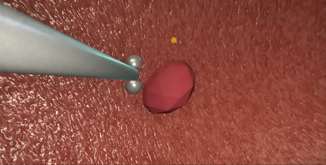
Operative animation, Rigid cystoscopy, biopsy and fulguration
Rigid cystoscopy, cold-cup biopsy and fulguration
The rigid look under anaesthetic: a stable sheath, two telescopes, and a working channel wide enough to take a real bite of the bladder wall and to burn what bleeds.
This page is the stack, the survey, the biopsy taken cold so the histology survives, the fulguration that stays on the point, and the mapping biopsies that answer a positive cytology.
Core tierFRCS core urology, bladder cancer diagnosisInteractive 3D
The bladder through the rigid sheath, 30 and 70 degree lenses. Schematic, not to scale; relationships only.
The rigid sheath enters the bladder along the axis of the urethra, so the 30 degree lens sees the trigone and the posterior and lateral walls easily and the 70 degree lens is needed to look back at the dome and the anterior wall.
The working channel places the cup or the electrode just ahead of the lens, in the centre of the view, so what is biopsied or burnt is what is being looked at.
›Bulbar urethra and sphincter - the curve the sheath follows and the ring it passes without push; the false passage is made here.
›The two telescopes - 30 degrees for the survey and the working channel, 70 degrees for the dome and the anterior wall.
›Trigone and orifices - the triangle on the floor with an orifice at each corner; seen before and after, never biopsied at the orifice.
›The dome - the thinnest wall and the one a distended bladder thins further; biopsied half full, pressed not driven.
›The lateral walls - obturator territory: current here can jerk the leg.
›Cold-cup forceps - a mucosal bite with lamina propria, taken without current so the histology survives.
›Button and Bugbee electrodes - coagulation at the point, short bursts, never painted across the wall.
Before the sheathThe indication: a tissue diagnosis that a flexible scope cannot make, a red patch or a flat lesion to biopsy, a positive cytology with nothing seen, a bleeding point to burn, or an examination the patient cannot tolerate awake. General or spinal anaesthetic, lithotomy in supports with the legs padded at the peroneal nerve and the buttocks at the end of the table. Antibiotics at induction where the urine or the history asks for them. The consent names infection, bleeding, urethral injury and the small chance of a perforation.
The stackA 21 French sheath with its obturator, a bridge with a working channel, a 30 degree telescope for the survey and a 70 degree for the dome and anterior wall, the camera and light lead, irrigation of saline by gravity from a bag no higher than sixty centimetres. On the trolley: cold-cup biopsy forceps, the button or Bugbee electrode with the generator on coagulation, a ureteric catheter, specimen pots already labelled by site, and formalin. The plate goes on the thigh if monopolar diathermy is used.
The sheath in under visionThe sheath goes in on the visual obturator with the 30 degree lens looking through it, following the lumen: never blind, never forced. The bulbar urethra turns upward and the sheath is dropped between the legs to follow the curve. The sphincter is a closed ring that the anaesthetic relaxes; the sheath passes it without push. The veru, the prostatic urethra with its lobes, and the bladder neck are noted on the way through, and the obturator comes out for the bridge and the telescope. where it bites: a rigid sheath forced through a stricture or a closed sphincter makes a false passage; the fix is to withdraw and find the lumen with the lens.
The surveyThe bladder is emptied and refilled to half. The air bubble at twelve o'clock orients the view; the trigone is on the floor with an orifice at each corner, each watched for a jet. The survey is in a fixed order: dome, anterior wall, right wall, posterior wall, left wall, floor. The 70 degree lens looks round the corner at the dome and the anterior wall that the 30 cannot reach, and suprapubic pressure from the assistant brings the dome down into view. A red velvety patch, a flat lesion, a diverticulum or a papillary tumour is described by wall, clock position and size before it is touched.
The cold-cup biopsyThe cold-cup forceps go through the bridge and are opened and pressed against the lesion so that the mucosa domes into the cup, then closed and pulled sharply away. The bite is mucosa with some lamina propria, taken without current so the histology is not burnt; a second bite from the same site if the first was small. Each bite goes straight into the pot labelled with its site. The site bleeds a little, which is expected and is dealt with once the specimen is safe. where it bites: a cold-cup bite at the thin dome of a distended bladder can go through the wall; the dome is biopsied with the bladder half full and the cup pressed, not driven.
FulgurationThe button or Bugbee electrode on coagulation is touched to the bleeding point, not painted across the wall, and a short burst is given until the point blanches. The current goes to where the electrode touches, and a distended bladder makes the wall thinner under it, so the bladder is kept half full. A small flat lesion can be fulgurated in full once its biopsy is taken; a papillary tumour is resected, not burnt, because burning destroys the stage. Fulguration near the lateral wall can stimulate the obturator nerve and jerk the leg: shorter bursts and lower power.
Mapping biopsiesWhen the cytology is positive and the bladder looks normal, or when carcinoma in situ is suspected, cold-cup bites are taken from named sites: the trigone, both lateral walls, the posterior wall and the dome, each in its own labelled pot, and the prostatic urethra with a loop or a cup. Nothing is biopsied at the orifice itself. The sites are drawn on a bladder map in the note so that a positive result can be placed. Random biopsies of a normal-looking bladder without a reason are not taken.
Haemostasis and the checkThe irrigation is slowed and every biopsy site inspected; a spurting point is fulgurated, an ooze is left. Both orifices are seen again with their jets, because a bite or a burn near one can obstruct it. The bladder is emptied through the sheath and the sheath is withdrawn under vision with a last look at the prostatic urethra and the sphincter. A catheter is left only if the bleeding or the number of biopsies asks for it.
The note and the specimensThe note draws the bladder and marks every site with what was done there: biopsy, fulguration, both. The pots and the request forms match the map, site by site, and the number of pots is counted against the number of sites before the patient leaves theatre. The patient is warned about pink urine and burning for a few days and told when and how the histology will be given. The team that requested the biopsies chases the result.
The spatial relationships the animation is built to make automatic. Before you read each one, ask yourself: at this point in the operation, what is about to go wrong, and what is my first move? The save for each sits in the Complications tab.
RiskThe dome of a full bladder - the thinnest wall, thinned further by distension; a cold cup or a burn here can perforate.
RiskThe ureteric orifices - a bite or a burn at the orifice obstructs the kidney; the site is chosen away from them.
RiskThe lateral wall - obturator territory; current here jerks the leg and, with a loop, perforates.
RiskThe sphincter and a stricture - a rigid sheath forced through either makes a false passage.
When the case turns: the problem, then the next move.
ActionThe sheath will not pass the bulb - drop the sheath between the legs to follow the curve; the obturator and the lens, never force.
ActionCannot see the dome - change to the 70 degree lens and ask for suprapubic pressure.
ActionThe cup slides off a flat lesion - a less full bladder so the mucosa is loose, press the open cup so the mucosa domes into it, then close and pull.
ActionThe burn does nothing - check the plate and the generator setting; a coated electrode is cleaned or changed.
ActionThe leg jerks - obturator stimulation: stop, shorter bursts, lower power, less bladder filling.
ActionMore pots than sites, or fewer - stop and reconcile before the patient leaves theatre; an unlabelled biopsy is worthless.
The examiner stems this scene prepares you for. Try to answer out loud first, then reveal, and learn the discriminator, the detail that turns a pass into a strong pass.
QHow would you investigate and manage a patient with positive urine cytology and a normal flexible cystoscopy?reveal answer
Positive cytology with a normal-looking bladder means carcinoma in situ, a lesion missed at flexible cystoscopy, or upper-tract urothelial cancer until proved otherwise.
I would image the upper tracts with CT urography, and I would arrange a rigid cystoscopy under anaesthetic with mapping biopsies: cold-cup bites from the trigone, both lateral walls, the posterior wall and the dome, each in a separate labelled pot, and a biopsy of the prostatic urethra with a loop or cup, because carcinoma in situ involves the prostatic urethra in a proportion of men.
I would take the bites cold so the histology is interpretable, avoid the orifices, and draw a map in the note.
If enhanced cystoscopy such as blue light or narrow band imaging is available I would use it to target biopsies.
If the bladder biopsies and the prostatic urethra are negative and the CT is normal, I would proceed to bilateral retrograde studies with selective upper-tract cytology and ureteroscopy as indicated.
Carcinoma in situ found in the bladder is treated with induction and maintenance BCG.
Strong pass
Say all three
Naming the prostatic urethral biopsy and the upper-tract work-up
With separate labelled pots and a map
Rather than a vague "random biopsies"
Examiner note
Naming the prostatic urethral biopsy and the upper-tract work-up, with separate labelled pots and a map, rather than a vague "random biopsies".
QWhy is a bladder biopsy taken with a cold cup rather than a resectoscope loop?reveal answer
The cold cup takes a small mucosal sample with some lamina propria without electrical current, so there is no thermal artefact and the pathologist can assess the urothelium, the lamina propria and any invasion at that level.
That is what is wanted for a flat red patch, a suspected carcinoma in situ or a mapping biopsy, where the question is the presence and grade of flat disease rather than depth of invasion.
A loop biopsy with current chars the specimen and the small sample is often uninterpretable.
For a papillary or solid tumour the question is different: depth of invasion into detrusor decides the stage and the treatment, and only a resection with a loop that reaches muscle can answer it, with the base sent separately.
So the rule is cold cup for flat lesions and mapping, loop resection with detrusor for tumours, and a lesion that bleeds after a cold-cup bite is fulgurated afterwards so that the histology is safe before any current is used.
Strong pass
Say all three
Linking the instrument to the pathological question: flat disease and grade for the cup
Depth and detrusor for the loop
With current only after the specimen is taken
Examiner note
Linking the instrument to the pathological question: flat disease and grade for the cup, depth and detrusor for the loop, with current only after the specimen is taken.
QA patient develops abdominal pain and distension after a cystoscopy with dome biopsies. Manage this.reveal answer
This is a bladder perforation until proved otherwise, and at the dome an intraperitoneal one is likely because the peritoneum lies over it.
I would resuscitate, examine for peritonism, stop any irrigation, and confirm a catheter is draining; if it is not in, I would place one.
I would obtain a CT cystogram, which distinguishes an extraperitoneal leak, with contrast confined to the perivesical space, from an intraperitoneal one with contrast outlining bowel loops.
An extraperitoneal perforation is managed with catheter drainage for about a week and a cystogram before removal.
An intraperitoneal perforation with peritonism needs a laparotomy or laparoscopy, repair of the bladder in two layers with absorbable suture, washout and a catheter, with senior help.
I would treat with antibiotics, monitor for ileus and sepsis, and document the site, the type, the imaging and the plan.
Prevention is biopsying the dome with the bladder half full and the cup pressed rather than driven.
Strong pass
Say both
The CT cystogram to separate extra- from intraperitoneal
Knowing that the dome perforation is the intraperitoneal one that needs repair
Examiner note
The CT cystogram to separate extra- from intraperitoneal, and knowing that the dome perforation is the intraperitoneal one that needs repair.
QWhat is the obturator reflex and how do you prevent and manage it?reveal answer
The obturator nerve runs along the lateral pelvic wall, close to the lateral bladder wall near the ureteric orifice, and current passed through the wall there stimulates it directly, causing a sudden adduction of the thigh.
Under general anaesthesia without muscle relaxation, or under spinal anaesthesia, the reflex is not abolished.
The danger is with a resection loop: the jerk drives the loop through the wall and perforates the bladder, often intraperitoneally.
With a button electrode during fulguration the risk is smaller but the movement still disturbs the field.
I would prevent it by warning the anaesthetist when working on a lateral wall so that muscle relaxation is given, or an obturator nerve block is placed, by keeping the bladder less full, using short bursts and lower power, and by using bipolar energy where available.
If a kick occurs I would stop the current immediately and inspect the wall for a perforation before continuing.
Strong pass
Say both
Muscle relaxation or an obturator block arranged with the anaesthetist before lateral wall work
Inspecting for perforation after a kick
Examiner note
Muscle relaxation or an obturator block arranged with the anaesthetist before lateral wall work, and inspecting for perforation after a kick.
Say it aloud, then reveal. Reading is not remembering.
01The relationshipSay the critical spatial relationship out loud, then reveal.reveal
Memory frame
The rigid sheath enters the bladder along the axis of the urethra, so the 30 degree lens sees the trigone and the posterior and lateral walls easily and the 70 degree lens is needed to look back at the dome and the anterior wall.
Why?
The working channel places the cup or the electrode just ahead of the lens, in the centre of the view, so what is biopsied or burnt is what is being looked at.
02The first movePerforation. Your first move?reveal
First move
Stop.
More detail
Empty the bladder through the sheath and do not refill it.
03The escalation triggerPerforation. What makes you call for help?reveal
Escalate when
Peritonism, abdominal distension or a large irrigant deficit: the intraperitoneal one, and a laparotomy with senior help.
04The discriminatorWhat turns a pass into a strong pass?reveal
Strong pass
Say all three
Naming the prostatic urethral biopsy and the upper-tract work-up
With separate labelled pots and a map
Rather than a vague "random biopsies"
Examiner note
Naming the prostatic urethral biopsy and the upper-tract work-up, with separate labelled pots and a map, rather than a vague "random biopsies".
05The pitfallWhere does this operation bite first?reveal
Pitfall
The dome of a full bladder
Why?
the thinnest wall, thinned further by distension; a cold cup or a burn here can perforate.
Revision practice with self-marked answers, not an assessment of clinical competence. Kept in
this browser only; it does not sync between devices.