
Rigid cystoscopy, bladder biopsy and fulguration
Rigid transurethral inspection with stable working access for targeted or systematic biopsy and limited coagulation of selected mucosal lesions or bleeding points.
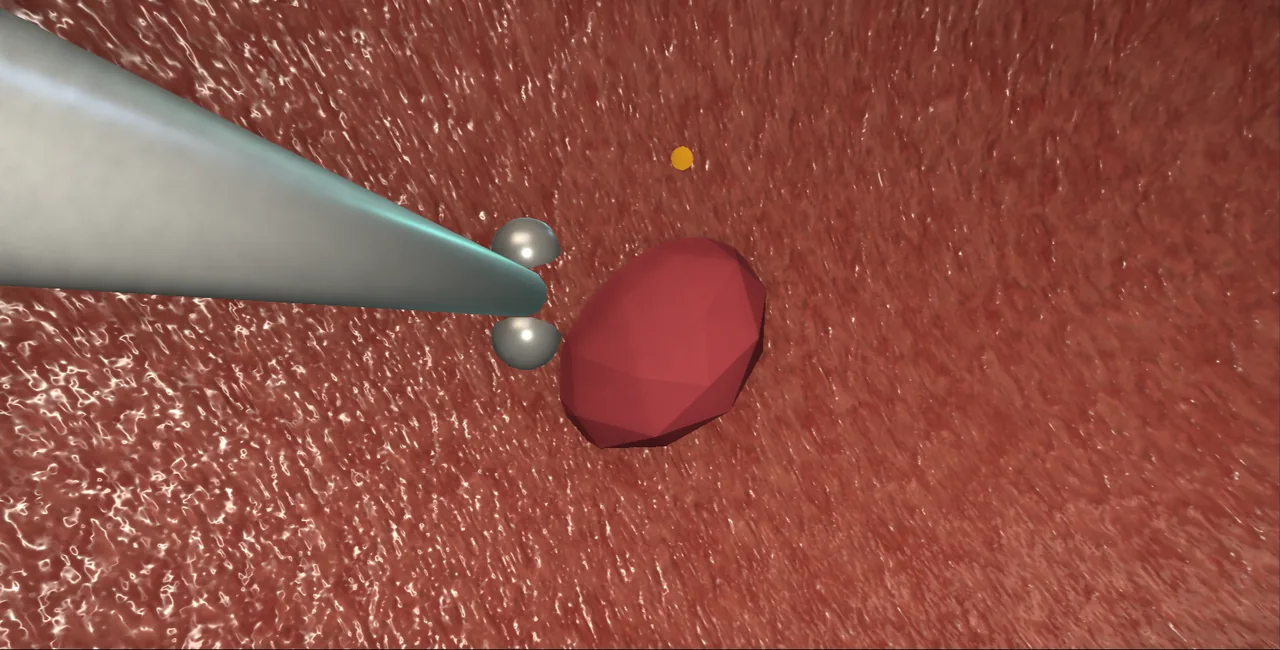 Interactive 3D Interactive rigid cystoscopy, biopsy and fulguration Open the 3D animation
Teaching catalogue only. This coverage record is not a patient pathway or operative instruction.
Typical place in practice
Tissue diagnosis of bladder abnormalities, mapping in selected urothelial pathways, assessment requiring anaesthesia and controlled treatment of a small mucosal target.
Alternatives and neighbouring procedures
Flexible cystoscopy, imaging and urine cytology may complement assessment; larger or suspicious tumours belong in the TURBT pathway.
Same procedure family, explicit technique branches
These are variations of this canonical operation. Complication modules below identify whether they apply to all branches or only to named approaches.
Rigid diagnostic cystoscopy
A rigid sheath provides stable optics and permits a complete examination under anaesthesia.
Targeted cold-cup biopsy
Visible abnormalities are sampled with a cold-cup instrument while preserving site orientation.
Mapping biopsy
Defined bladder and, when indicated, prostatic-urethral sites are sampled for occult disease assessment.
Limited bladder fulguration
A selected small lesion or bleeding surface is coagulated without a formal deep resection.
High-level sequence
Coverage outline only; not a step-by-step technical guide.
- Confirm indication, infection status and consented interventions
- Enter the urethra under vision and inspect systematically
- Survey bladder landmarks with controlled distension
- Biopsy or fulgurate only the planned sites
- Achieve haemostasis, label specimens precisely and document a bladder map
Operative notes
Concise operative notes covering the key steps, landmarks and watch points, in the order you encounter them.
Rigid cystoscopy
What it does: look inside the bladder with a straight scope.
- Assemble the scope. White-balance the camera. Check the focus.
- Never force. Always follow the lumen. Lose the view? Pull back till you find it.
- Too tight? Go smaller, dilate, or do an Otis cut. (Otis = a blade that slits a tight urethra at 12.)
- Left hand: penis vertical, gentle stretch. 10 mL gel. Right hand: scope vertical. Water on.
- Pass under vision. Penile urethra, then bulbar. The sphincter looks like a fold-narrowing.
- At the sphincter, drop your hand as you go in. The scope rides over the veru. (Veru = verumontanum, the mound at the prostate tip.)
- Note the prostate lobes and the bladder neck.
- In the bladder: drain and measure what comes out.
- Find the trigone and both ureteric orifices first.
- Survey all of the bladder. Rotate and slide. Order does not matter. Completeness does.
- Press the lower abdomen to see the front wall. Use the 70 degree scope near the bladder neck and for the dome. Hold the lens still: the view turns up.
- Do not over-fill. The front wall drifts away.
- Pull out the way you went in. Lift hands first.
- Watch: The light cable tip gets hot. Standby the light.
- Bladder stone: Stone found: survey first, then break it. Laser: the larger fibre, 365, 500 or 550 micron. 1.5 to 2.0 J at 20 to 50 Hz. Work one edge, wall in view behind every shot. Stone punch: jaws open under vision, take an edge. Look up. Lift the stone to the middle of the bladder, clear of the wall, lens clear. Then crunch. Never close on a stone resting on the wall. Wash the pieces out. Treat the cause the same day.
From the UroRef Operative Quick Guide. Open the note page →
Bladder biopsy
What it does: take a small sample of bladder lining.
- Biopsy with the bladder half full, not full. A full bladder has a thin wall. It perforates.
- Open the forceps. Push on the mucosa. Close firmly. Tug sharply.
- Hot forceps: touch the site. Coagulate till dry.
- Cold forceps: swap in a wire electrode. Coagulate.
- Watch: Remember the biopsy spot. Bleeding hides it.
- Watch: Biopsy last. Never distend a fresh biopsied bladder.
- Watch: Scars have thin walls. Be gentle there.
From the UroRef Operative Quick Guide. Open the note page →
Every failure mode is its own module.
Timing and variation coverage are recorded for each module.
Urinary infection or sepsis
Lower-tract instrumentation can introduce or disseminate infection, particularly when drainage is impaired.
Applies to: Rigid diagnostic cystoscopy · Targeted cold-cup biopsy · Mapping biopsy · Limited bladder fulgurationUrethral trauma or later stricture
A rigid sheath can injure a narrow or angulated urethra, with acute bleeding or later narrowing.
Applies to: Rigid diagnostic cystoscopy · Targeted cold-cup biopsy · Mapping biopsy · Limited bladder fulgurationHaematuria or clot retention
Biopsy sites or incomplete haemostasis can bleed and clot may obstruct bladder drainage.
Applies to: Targeted cold-cup biopsy · Mapping biopsy · Limited bladder fulgurationBladder perforation
Deep sampling, instrument movement, coagulation or work in a diverticulum can breach the bladder wall.
Applies to: Rigid diagnostic cystoscopy · Targeted cold-cup biopsy · Mapping biopsy · Limited bladder fulgurationUreteric-orifice injury or obstruction
Sampling or coagulation close to an orifice can impair drainage or cause reflux-related consequences.
Applies to: Targeted cold-cup biopsy · Mapping biopsy · Limited bladder fulgurationInadequate, damaged or mislabelled tissue
Small, crushed, cauterised or poorly mapped specimens can limit diagnosis and undermine subsequent treatment decisions.
Applies to: Targeted cold-cup biopsy · Mapping biopsySources for this module
Infection prevention, recognition and procedure-associated infection context.
Accessed 2026-08-03 · opens source British Association of Urological Surgeons Patient information leaflets by procedureUK patient-facing procedure, alternatives, recovery and complication framing.
Accessed 2026-08-03 · opens source