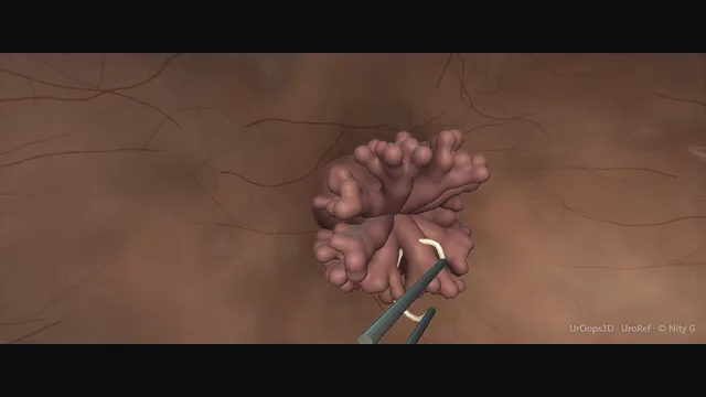
A papillary tumour on the right lateral wall beside the ureteric orifice, with the obturator nerve running just beyond that wall. Map first, take the tumour, then the base down to detrusor, then the ring of normal mucosa. The muscle is the message.
Core tierFRCS uro-oncology, non-muscle-invasive bladder cancerInteractive 3D
Through the right lateral bladder wall, lumen outwards. Schematic, not to scale; relationships only.
The loop, the bladder wall, the obturator nerve and the ureteric orifice sit in a line on the lateral wall, in that order, with only millimetres of detrusor between the resection plane and the nerve.
›Right lateral wall - the tumour bed; thin wall outside, obturator nerve just beyond it.
›Ureteric orifices - the reference points for everything; the right one is beside this tumour.
›Detrusor muscle - the depth that stages the disease; the endpoint of the deep pass.
›Obturator nerve - runs on the pelvic side wall lateral to the bladder; monopolar current finds it.
Cystoscopy + mappingA bimanual examination under anaesthesia before the scope goes in records whether any mass is palpable or fixed. Run a full survey of the bladder before any cutting, wall by wall, so no second lesion is discovered after the resection has begun. Then fix the anatomy: tumour site by clock face, distance from the ureteric orifices and the prostate, size, number and configuration, recorded as the map the histology and the MDT will be read against. Identify both ureteric orifices before cutting and state which lies in the field, so direct involvement is recognised and followed rather than discovered after resection. where it bites: map both orifices before cutting and document any direct involvement.
Resect the exophytic tumourBecause this tumour sits on the lateral wall, the anaesthetist is warned and relaxation agreed where the anaesthetic allows before the first stroke; then loop resection takes the exophytic tumour piecemeal from its top down towards its base, working in orderly strokes so the bulk disappears in layers rather than at random. The bladder is kept only half filled throughout: a full bladder thins and splints the wall, while a half-filled one stays thick and mobile and rides with the loop rather than against it. Chips are cleared when they gather and hide the working edge, because resecting through a carpet of chips is resecting blind. The endpoint of this pass is a visible base, not yet the staging depth itself. where it bites: an over-filled bladder thins the wall; the perforation is mechanical, not just electrical.
Base + marginal ringA deeper pass is then taken across the base with the deliberate aim of including detrusor muscle, in clean strokes so the pathologist receives muscle and architecture rather than char. Around the base a ring of normal-looking mucosa is resected as a separate margin and sent in its own pot, mapped to its site. Muscle in the specimen is what stages the cancer: no muscle means an unstageable tumour and a planned repeat resection. Pot by pot labelling is part of the operation, because an unlabelled sample is one the MDT cannot place. where it bites: no muscle in the specimen means an unstageable cancer and a repeat resection.
The lateral-wall momentOn the lateral wall the operation changes character: monopolar current spread through the thin wall can fire the obturator nerve, and one adductor jerk drives the hip and the loop together. The anaesthetist was warned before the first lateral-wall stroke, so the jerk is an expected event and relaxation has been agreed where the anaesthetic allows. Use considered current: short strokes, the loop energised only in contact with tissue, and cutting rather than coagulation current as the loop crosses the region nearest the nerve. If the view is lost or the wall moves with every stimulus, stop and reassess rather than cut blind into the jerk. where it bites: the kick is expected on the lateral wall: relaxant where possible, considered current, and never cutting blind.
Protect the ureteric orificeTumour involving or overlying a ureteric orifice is resected deliberately and completely, because oncological completeness still matters at this site. Avoid indiscriminate coagulation across the intramural ureter. Re-identify the orifice after resection and assess its patency and drainage. A wire, ureteric catheter or JJ stent is used selectively when the orifice has been directly resected, is oedematous or obstructed, or cannot be identified; proximity alone is not presented as an automatic indication. State the involvement, drainage assessment and follow-up plan in the op note. where it bites: direct orifice resection needs documented patency assessment and upper-tract follow-up; stenting is selective.
Haemostasis + instillationHaemostasis is completed systematically over the whole field rather than spot by spot, the rollerball resting on each point bleeder until it stops, and the final view confirms a dry base, margins, both ureteric orifices and an intact wall before the instruments leave. Chips are evacuated with the Ellik into their labelled pots, tumour and base apart, and the collected tissue is checked against the map taken at the start. Where indicated, a single immediate post-operative instillation of intravesical chemotherapy is given through the catheter. The instillation is omitted where perforation is suspected, because chemotherapy instilled across an uncontained breach soaks the perivesical tissues. A bimanual examination is repeated at the end and both findings are written in the note.
The spatial relationships the animation is built to make automatic. Before you read each one, ask yourself: at this point in the operation, what is about to go wrong, and what is my first move? The save for each sits in the Complications tab.
RiskTumour over the obturator nerve - the classic kick zone: the thin lateral bladder wall separates the active loop from the extravesical obturator nerve; exact distance varies with anatomy and bladder filling.
RiskThe tumour base - depth here stages the cancer; too deep perforates, too shallow under-stages.
RiskUreteric orifice in the field - resection remains oncologically complete; re-identify and assess drainage, then use a wire, catheter or stent selectively rather than automatically for proximity alone.
Trigger each of these live in the Animation tab, then read the save: the first move, the escalation trigger, and the line that goes in the op note.
Obturator kick
monopolar current stimulates the obturator nerve; the adductors fire and the loop is driven through the wall.
The save
Expect it: on the lateral wall, pause cutting as the loop approaches the tumour edge nearest the nerve.first move
Prevention: muscle relaxation where the anaesthetist agrees, and consideration of bipolar or laser resection.
If it fires: stop immediately, inspect the wall where the loop landed, and complete a full survey for perforation.
Signs of perforation alongside → manage as perforation (below), not as bleeding.
Uncontrolled jerks with a monopolar loop → change strategy, not persistence: bipolar, laser, or complete and stage.escalation trigger
Op note: laterality, relaxant used, kick observed, wall inspection result.document
Bladder perforation
loop or kick through the wall; irrigant extravasates, usually retroperitoneally laterally, intraperitoneally at the dome.
The save
Stop resecting. Keep the scope in the bladder; do not pull out and lose the view.first move
Drop irrigation pressure and stop inflow; a perforation under pressure fills the retroperitoneum fast.
Inspect: retroperitoneal fat or bowel means perforation; the return of irrigant stops or goes extravesical.
Extraperitoneal: catheter drainage, antibiotics consideration, and observation - most seal without surgery.
Intraperitoneal, rising abdomen, bowel in the field or instability → open repair and washout.escalation trigger
Op note: site and suspected type of perforation, drainage plan, instillation omitted, follow-up imaging.document
Ureteric orifice injury
direct resection can cause transient oedema and, uncommonly, a later ureterovesical-junction stricture; obstruction may be clinically silent.
The save
Recognise on the table: identify the orifice after resection of any tumour near it.first move
Assess efflux and patency; pass a wire or catheter and place a JJ stent selectively if the directly resected orifice is oedematous, obstructed or cannot be identified.
Arrange upper-tract imaging after direct orifice resection and before any stent removal; use functional imaging when anatomical imaging leaves drainage uncertain.
Late presentation with hydronephrosis → re-stent or dilate; a tight fibrotic stenosis needs formal re-implantation.escalation trigger
Op note: orifice involved, stent placed, upper-tract imaging plan before removal.document
When the case turns: the problem, then the next move.
ActionTumour bleeds and hides itself - resect the base systematically rather than chasing bleeders; the view returns with the tumour gone and irrigation steady.
ActionLoop will not cut cleanly - check current settings and loop contact; a dragging loop tears more than it cuts.
ActionChips obscure the dependent bladder - evacuate early with the Ellik and come back; operating through a carpet of chips hides a fresh tumour edge.
ActionTumour at the dome under air - reduce irrigation and use manual pressure; never chase a dome tumour with the bladder over-filled.
The examiner stems this scene prepares you for. Try to answer out loud first, then reveal, and learn the discriminator, the detail that turns a pass into a strong pass.
QWhat makes a TURBT adequate rather than merely complete?reveal answer
An adequate TURBT delivers complete visible resection where safely achievable, correctly orientated specimens with detrusor muscle when required for staging, and a documented map of site, size, number and configuration.
Absence of detrusor muscle increases residual-disease and under-staging risk and commonly triggers repeat resection, with guideline exceptions such as Ta low-grade disease and primary carcinoma in situ considered explicitly rather than ignored.
Completeness also means the ureteric orifices and prostatic urethra were assessed and that a single immediate post-operative instillation was given only where indicated and not contraindicated.
Technique serves pathology: the operation is a biopsy with therapeutic intent, staged by its depth.
Strong pass
Muscle in the specimen is the quality metric
Muscle→In the specimen: the operative quality metric
Map→Part of the operation, not paperwork
Examiner note
Framing muscle in the specimen as the operative quality metric, and the map as part of the operation rather than paperwork.
QHow do you prevent the obturator kick during lateral-wall TURBT?reveal answer
The kick is a predictable physics problem: monopolar current spreading to the obturator nerve on the pelvic side wall fires the adductor mass and moves the table.
Prevention is layered.
First, pharmacological: muscle relaxation agreed with the anaesthetist removes the motor end of the reflex for lateral-wall work.
Second, technical: cutting current rather than coagulation near the nerve, the loop energised only in contact with tumour, short strokes, and a half-filled bladder so the wall is mobile rather than splinted.
Third, alternative energy: bipolar resection or laser vaporisation largely sidesteps the reflex.
I also warn the anaesthetist before starting the lateral wall, so the jerk is an expected event and not a call for help.
Strong pass
Say both
Naming the layered strategy (relaxant, current discipline, alternative energy) and the pre-emptive anaesthetic conversation
Not just "be careful"
Examiner note
Naming the layered strategy (relaxant, current discipline, alternative energy) and the pre-emptive anaesthetic conversation, not just "be careful".
QYou suspect an extraperitoneal perforation mid-TURBT. Walk me through the next ten minutes.reveal answer
I stop resecting immediately and keep the scope in the bladder.
Irrigation inflow stops, so the extravasation is not pressurised.
I inspect the wall where the loop was working: retroperitoneal fat, an irregular wall defect or loss of return localises the perforation, and a quick survey tells me whether bowel is in the field, which would redefine it as intraperitoneal.
I complete haemostasis only where it is safe, abandon any residual resection, evacuate the chips I have, and catheterise with a large-bore catheter for continuous drainage.
I omit the post-operative instillation, start antibiotics where indicated by contamination or prophylaxis policy, monitor urine output and the abdomen, and image if the patient fails to progress.
Most extraperitoneal perforations seal on catheter drainage within days; a follow-up cystogram or a flexible check before or after catheter removal confirms healing.
An intraperitoneal pattern, a distending abdomen or instability means open repair and washout.
Strong pass
Say all three
Stopping inflow to depressurise the leak
Omitting the instillation
The clear extraperitoneal-versus-intraperitoneal fork
Examiner note
Stopping inflow to depressurise the leak, omitting the instillation, and the clear extraperitoneal-versus-intraperitoneal fork.
QA 3 cm solid-looking tumour covers the right ureteric orifice. How does that change your operation?reveal answer
The orifice changes the plan, not the oncological goal: complete visible resection with appropriate staging tissue remains the objective.
I would map the tumour carefully against the orifice and may pass a ureteric catheter or guidewire early when it will help identify the lumen.
I would resect deliberately at the orifice, avoid indiscriminate coagulation across the intramural ureter, then re-identify the opening and assess drainage.
A JJ stent is selective rather than automatic: I would use one if the orifice had been directly resected and was oedematous, obstructed, of uncertain patency or no longer identifiable.
I would document involvement and arrange upper-tract imaging, including before removal if a stent was placed.
I would also anticipate the obturator nerve if the tumour extends posterolaterally and counsel the patient about possible stenting and delayed obstruction.
Strong pass
Say all three
Preserving oncological completeness while making patency assessment
Selective drainage
Imaging follow-up explicit rather than treating every nearby resection as an automatic stent
Examiner note
Preserving oncological completeness while making patency assessment, selective drainage and imaging follow-up explicit rather than treating every nearby resection as an automatic stent.
Say it aloud, then reveal. Reading is not remembering.
01The relationshipSay the critical spatial relationship out loud, then reveal.reveal
Memory frame
Lateral wall, in a line: loop, wall, nerve, orifice
1→Loop
2→Bladder wall
3→Obturator nerve
4→Ureteric orifice
Only millimetres of detrusor between the resection plane and the nerve.
Why?
The loop, the bladder wall, the obturator nerve and the ureteric orifice sit in a line on the lateral wall, in that order, with only millimetres of detrusor between the resection plane and the nerve.
02The first moveObturator kick. Your first move?reveal
First move
Expect the kick: pause at the edge nearest the nerve
On the lateral wall, as the loop approaches the tumour edge.
More detail
Expect it: on the lateral wall, pause cutting as the loop approaches the tumour edge nearest the nerve.
03The escalation triggerObturator kick. What makes you call for help?reveal
Escalate when
Uncontrolled jerks on monopolar: change strategy
Bipolar
Laser
Complete and stage
Change strategy, not persistence.
More detail
Uncontrolled jerks with a monopolar loop → change strategy, not persistence: bipolar, laser, or complete and stage.
04The discriminatorWhat turns a pass into a strong pass?reveal
Strong pass
Muscle in the specimen is the quality metric
Muscle→In the specimen: the operative quality metric
Map→Part of the operation, not paperwork
Examiner note
Framing muscle in the specimen as the operative quality metric, and the map as part of the operation rather than paperwork.
05The pitfallWhere does this operation bite first?reveal
Pitfall
Tumour over the obturator nerve: the kick zone
A thin lateral wall separates the active loop from the extravesical nerve; the distance varies with anatomy and bladder filling.
Why?
Tumour over the obturator nerve: the classic kick zone: the thin lateral bladder wall separates the active loop from the extravesical obturator nerve; exact distance varies with anatomy and bladder filling.
Revision practice with self-marked answers, not an assessment of clinical competence. Kept in
this browser only; it does not sync between devices.