
Revision sheet
TURBT: lateral-wall bladder tumour
6 steps, each one the authored title and caption from the interactive animation, with the frame captured from that step. Read it before the case, or print it and work through the steps in order.
Teaching only, not for diagnosis or treatment decisions. The text on this sheet is the animation's own step copy, reproduced verbatim.
UrOops3D · UroRef · © Nity G
- 01
Cystoscopy + mapping
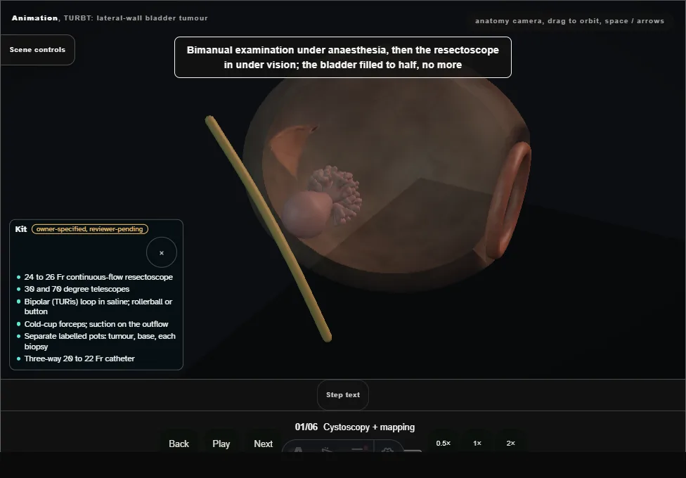
- Survey the whole bladder, half full, in a fixed order, then fix the anatomy: tumour on the right lateral wall, both ureteric orifices identified, relationship of tumour to the right UO declared.
- Avoid over-distension and use only enough fill for orientation and control.
- 02
Resect the exophytic tumour
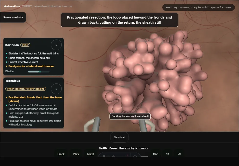
- The loop is placed beyond the fronds and drawn back, cutting on the return stroke with the sheath still: short swipes, piecemeal from the top down.
- Chips fall to the dependent bladder base and are collected at the end.
- 03
Base + marginal ring
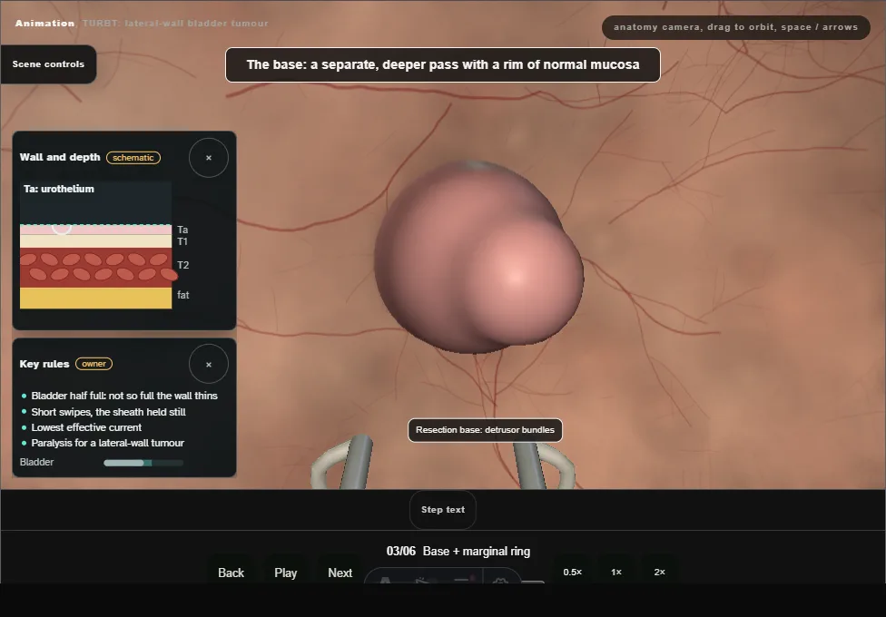
- A second, deeper pass takes the base with detrusor muscle and a ring of normal-looking mucosa around it.
- No muscle in the specimen is an incomplete TURBT and a biased staging.
- 04
The lateral-wall moment
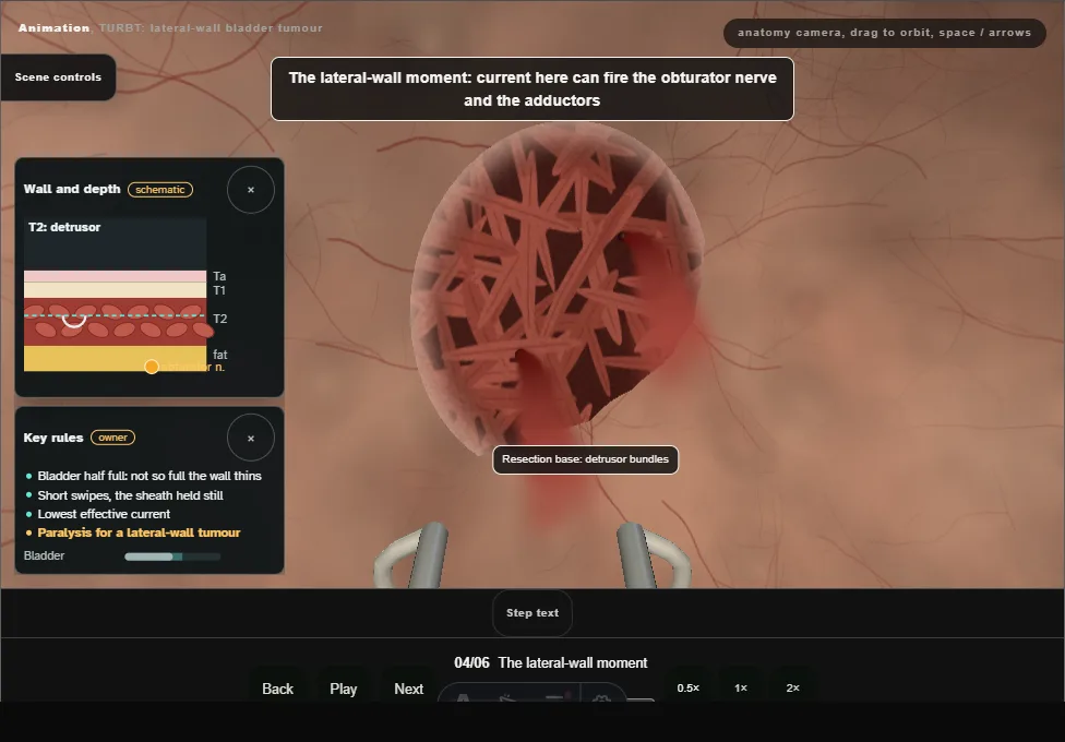
- Current near the obturator nerve can fire the adductors: paralysis requested for a lateral-wall tumour, bipolar in saline at the lowest effective current, short swipes and a half-full bladder.
- Bipolar reduces the kick but does not abolish it; this is the kick that perforates.
- 05
Protect the ureteric orifice
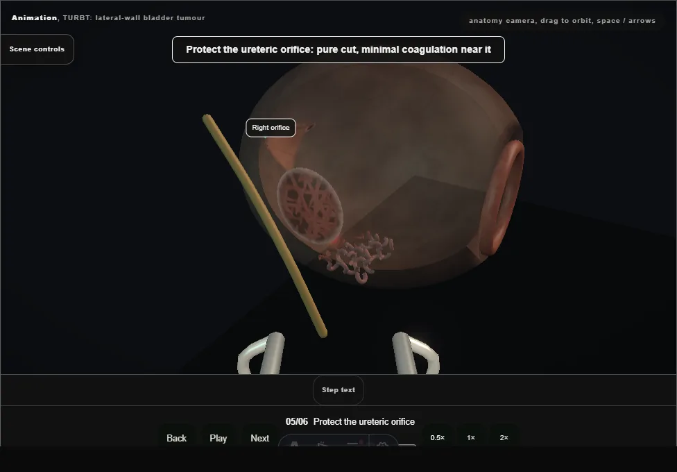
- Tumour involving the orifice is resected deliberately and completely.
- Re-identify the orifice afterwards and assess patency; wire, catheter and JJ stent use is selective when the orifice is directly resected, oedematous, obstructed or cannot be identified.
- A true late stricture is uncommon but earns imaging follow-up.
- 06
Haemostasis + instillation
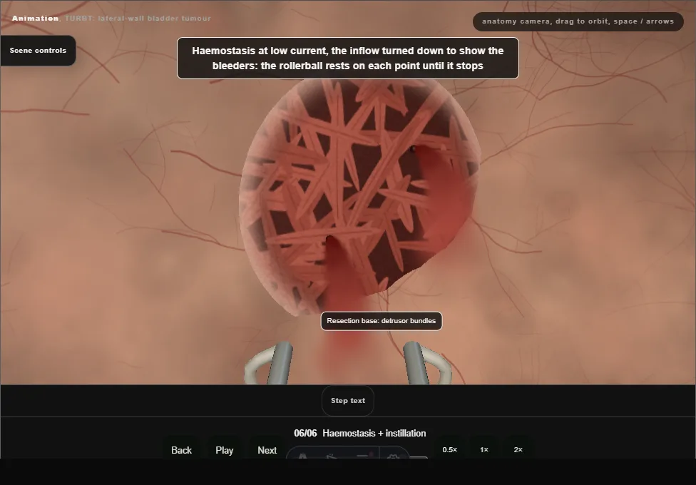
- Systematic haemostasis, Ellik evacuation of chips, then a single immediate post-operative instillation where indicated and not contraindicated by perforation or heavy resection.
Derived from TurbtScene.tsx at build time: every step title and caption above is the authored
ProcedureDef text, reproduced verbatim, and this page shares its readable-points renderer with the animation
itself. 6 of 6 steps have a captured frame.
Text fingerprint 731572ea95073279. A caption edit without a re-capture fails the production
check, so the sheet and the animation cannot drift apart silently.