
Revision sheet
HoLEP: laser enucleation of the prostate
10 steps, each one the authored title and caption from the interactive animation, with the frame captured from that step. Read it before the case, or print it and work through the steps in order.
Teaching only, not for diagnosis or treatment decisions. The text on this sheet is the animation's own step copy, reproduced verbatim.
UrOops3D · UroRef · © Nity G
- 01
The adenoma inside its capsule
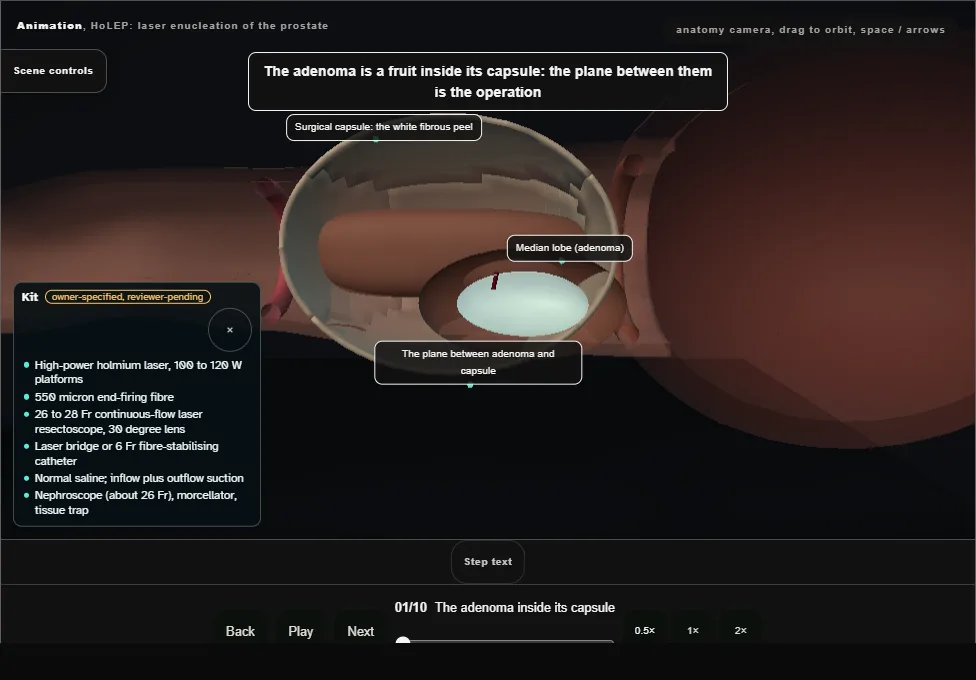
- The adenoma sits inside the surgical capsule like fruit in its peel, with a thin plane between them.
- The plane is the operation.
- Small capsular vessels cross it; distal to the veru lies the external sphincter.
- 02
Cystoscopic survey
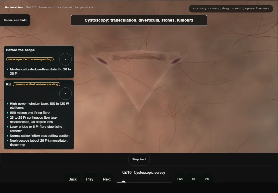
- Trabeculation, diverticula, stones and tumours; both orifices found against the bladder neck, because a median lobe can hide them; the veru and sphincter fix the distal limit.
- 03
Bladder neck incisions at 5 and 7 o'clock
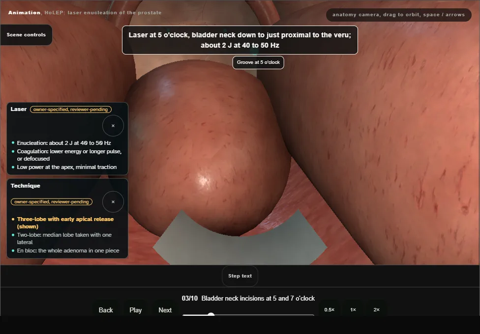
- Each groove runs from the bladder neck to just proximal to the veru and is deepened through adenoma until the white circular fibres of the capsule show at its floor.
- 04
Median lobe
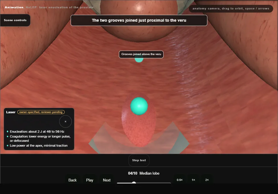
- The grooves are joined above the veru; the plane is developed distal to proximal, the beak lifting and the laser cutting, capsular vessels coagulated as they appear.
- Freed at the neck and pushed into the bladder, the trigone never undermined.
- 05
Early apical release
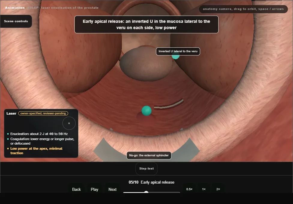
- An inverted U in the mucosa lateral to the veru on each side, at low power with minimal traction, and the 12 o'clock apical strip divided early, so the sphincter mucosa is never stretched or torn.
- 06
Left lateral lobe
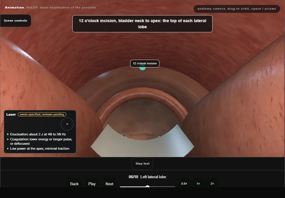
- A 12 o'clock incision defines the top of each lateral lobe.
- The left lobe is entered at the apex, swept laterally and upward, retrograde, and peeled round until it hangs only at the bladder neck.
- 07
Right lateral lobe
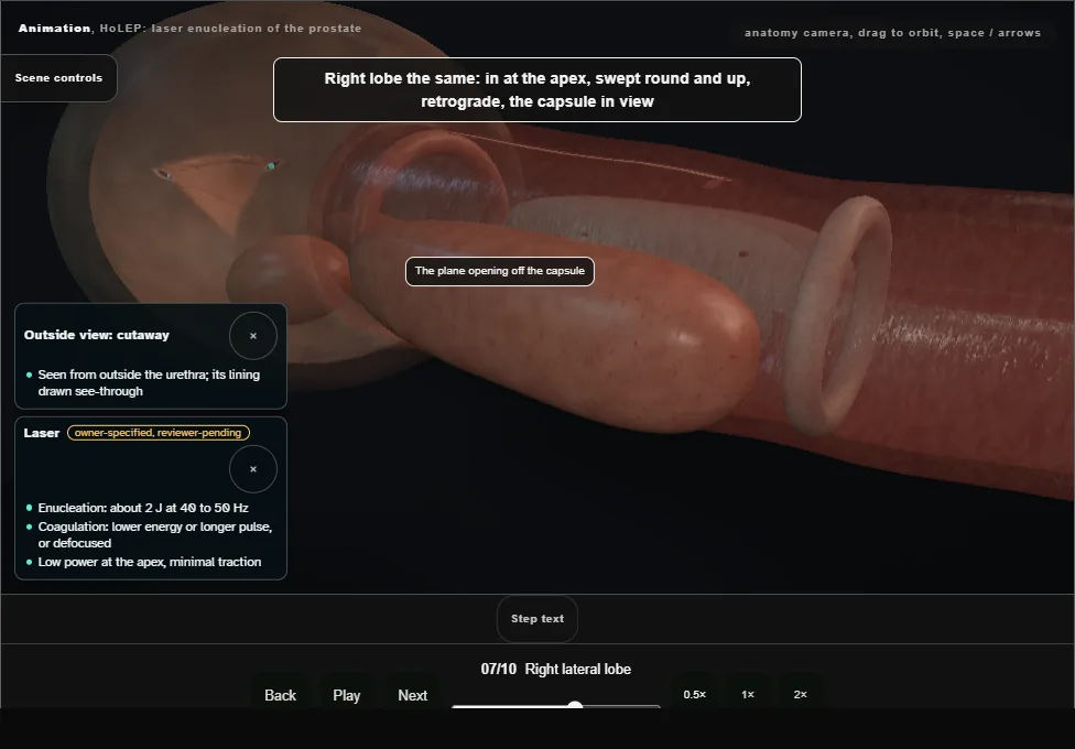
- The right lobe the same, the capsule kept in view as the safety line; then divided at the bladder neck and pushed into the bladder.
- 08
Haemostasis
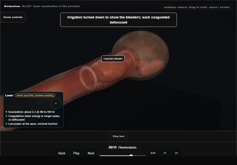
- Irrigation turned down to reveal the bleeders, each coagulated defocused; both orifices confirmed intact; a wide smooth cavity lined by capsule.
- 09
Morcellation
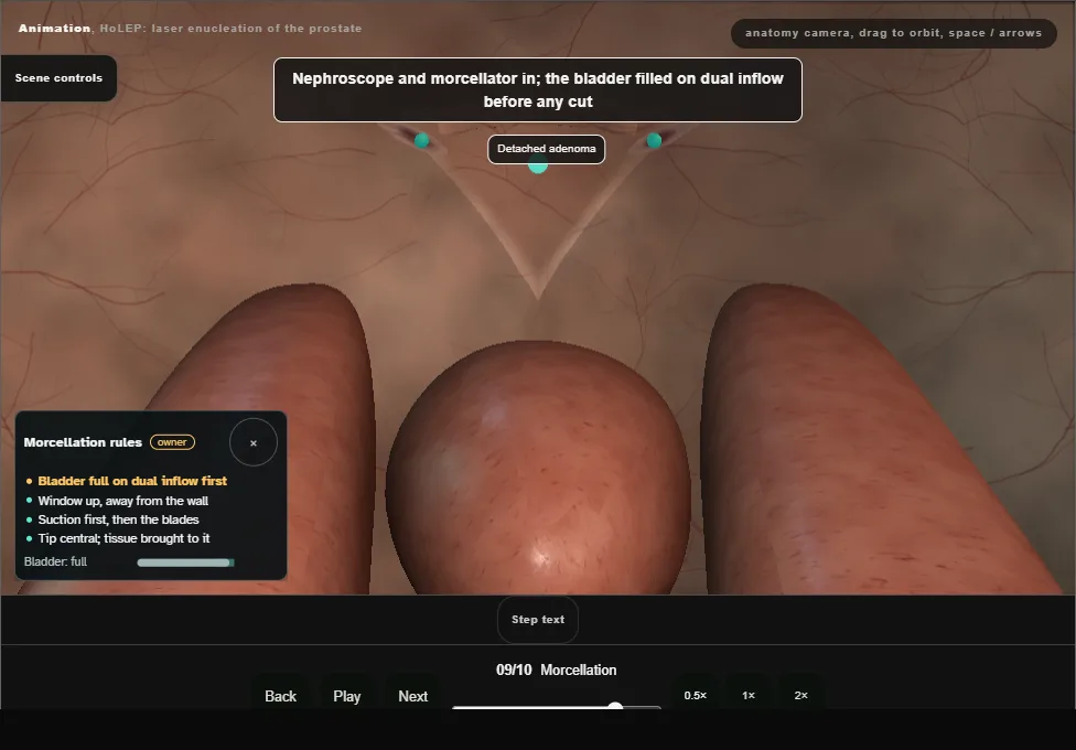
- A full bladder on dual inflow first.
- Window up, away from the wall; suction first, then the blades, only with adenoma in the window.
- The bladder wall is one blind bite away.
- 10
Finish
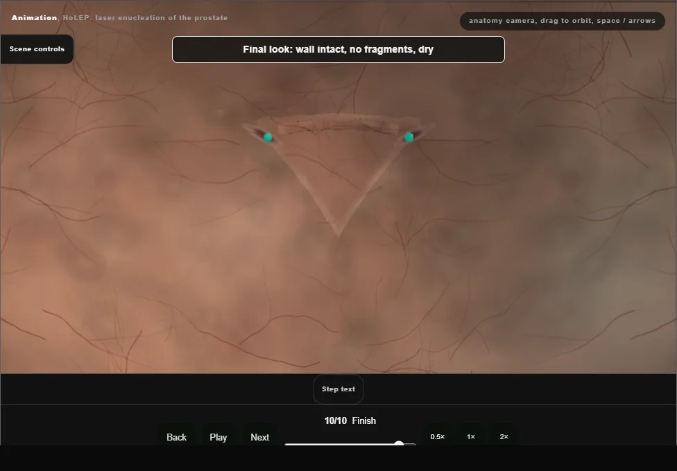
- Wall intact, no fragments, dry.
- A three-way 20 to 22 Fr catheter with continuous irrigation; traction only for bleeding at the bladder neck.
- The whole specimen goes to histology.
Derived from HolepScene.tsx at build time: every step title and caption above is the authored
ProcedureDef text, reproduced verbatim, and this page shares its readable-points renderer with the animation
itself. 10 of 10 steps have a captured frame.
Text fingerprint 9860dae2da3fc041. A caption edit without a re-capture fails the production
check, so the sheet and the animation cannot drift apart silently.