
Revision sheet
Mitrofanoff appendicovesicostomy
7 steps, each one the authored title and caption from the interactive animation, with the frame captured from that step. Read it before the case, or print it and work through the steps in order.
Teaching only, not for diagnosis or treatment decisions. The text on this sheet is the animation's own step copy, reproduced verbatim.
UrOops3D · UroRef · © Nity G
- 01
The principle
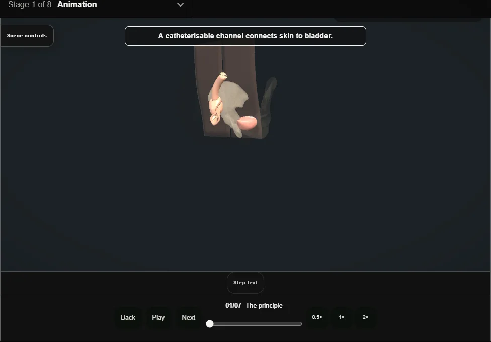
- A continent channel is a narrow tube laid in a long tunnel in the bladder wall: the full bladder compresses the tunnel closed, urine cannot escape, and the channel opens only for a catheter.
- The flap valve is the whole operation.
- 02
Isolate the appendix
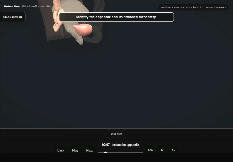
- The appendix is taken with a cuff of caecum and, critically, with its mesentery and its single artery, which will feed the channel for life.
- Skeletonised, the channel dies.
- 03
Create the detrusor tunnel
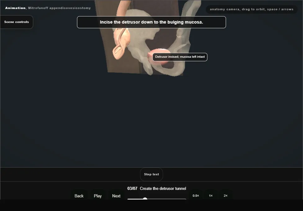
- From outside the bladder: the detrusor is incised down to the bulging mucosa and raised as flaps, leaving a trough three to four times as long as the channel is wide.
- Long enough for the flap valve, wide enough not to strangle the channel.
- 04
Implant + the valve test
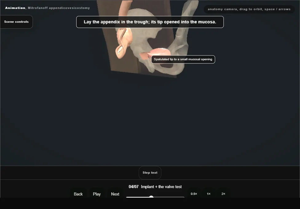
- The appendix is laid in the trough, its spatulated tip sewn to a small opening in the mucosa, and the detrusor closed over it.
- As the bladder fills, the tunnel compresses the channel shut: that is continence, tested on the table before the abdomen is closed.
- 05
Route to the skin
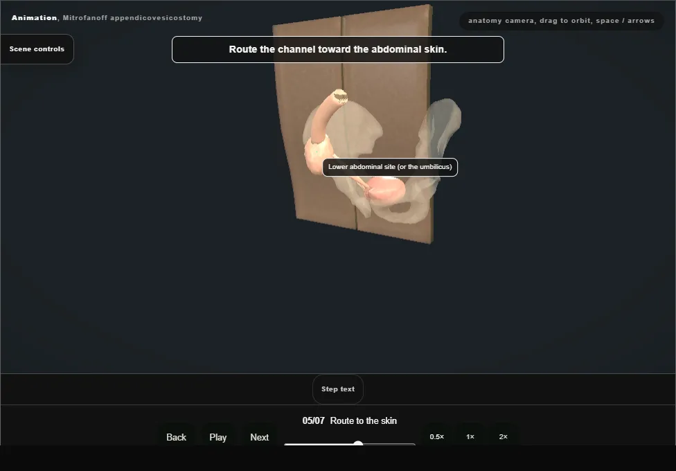
- The channel runs a short, straight course to the umbilicus or a right iliac fossa stoma site: no kinks, no tension, no acute angles for a catheter to negotiate.
- 06
Stoma maturation
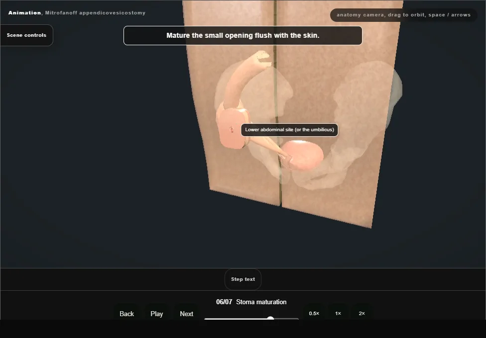
- A VQZ-type or umbilical stoma is matured flush to the skin: no spout to catch clothing, no buried opening to stenose.
- The first catheterisations are supervised before discharge.
- 07
Catheterisation teaching
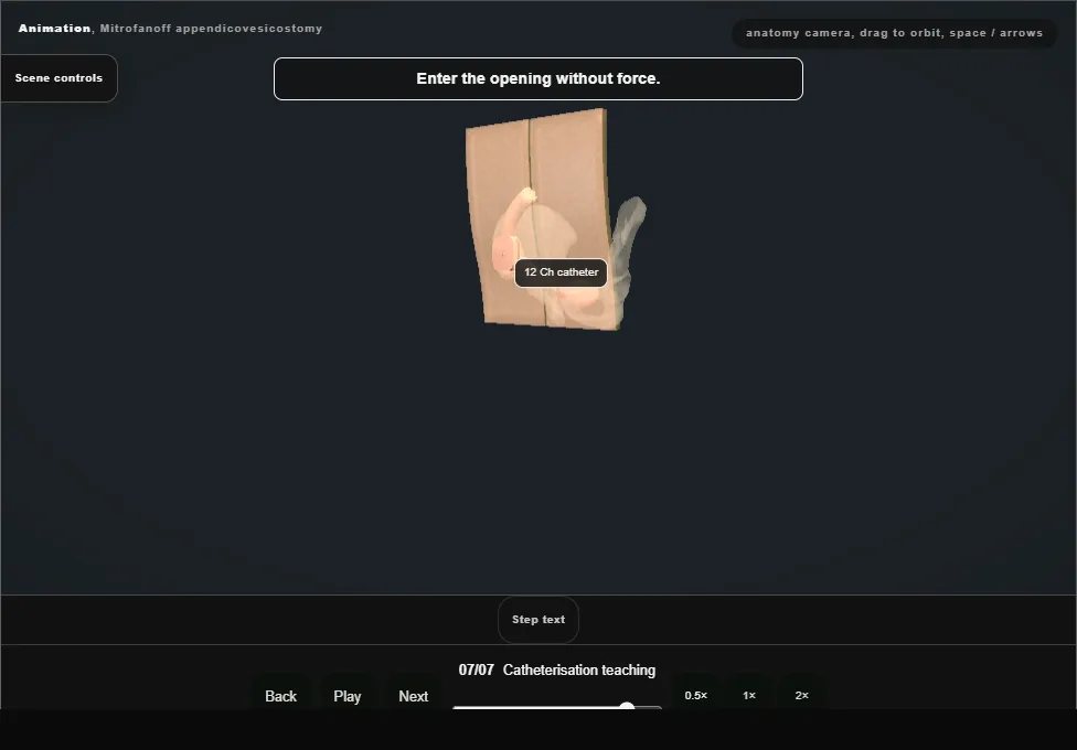
- The channel only works if it is used: lifelong clean intermittent catheterisation, taught before discharge, with a plan for who to call when catheterisation becomes difficult.
Derived from MitrofanoffScene.tsx at build time: every step title and caption above is the authored
ProcedureDef text, reproduced verbatim, and this page shares its readable-points renderer with the animation
itself. 7 of 7 steps have a captured frame.
Text fingerprint aeaa485192d28b62. A caption edit without a re-capture fails the production
check, so the sheet and the animation cannot drift apart silently.