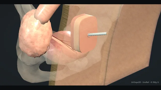
One isolated appendix, still supplied by its mesoappendix, is transferred as one continuous channel: one end implanted through a continent bladder-wall tunnel and the other delivered to a catheterisable skin stoma.
The animation now removes the native appendix before revealing the transferred channel, so it never depicts a duplicated or Y-shaped appendix.
Advanced tierFRCS reconstructive urology, paediatric urologyInteractive 3D
Along the one continuous channel, skin stoma to bladder. Schematic, not to scale; relationships only.
Follow one continuous channel.
The appendix is detached from the caecum with its mesoappendix intact;
the caecal stump closes, one appendiceal end is implanted through the bladder-wall tunnel and the other reaches the umbilicus or chosen abdominal-wall stoma without a kink or mesenteric twist.
The cutaway hides the abdominal wall by default so the viewer can verify that there is one tube, not a retained native appendix plus a second skin channel.
Continence is assessed in the context of a safe, compliant reservoir; a small high-pressure bladder is a danger to the upper tracts, not a stronger valve.
›Appendix + mesentery - the channel and its single arterial supply; harvested together, implanted together, and compromised together.
›Submucosal tunnel - the bladder-wall passage whose length is the continence mechanism; compressed shut as the bladder fills.
›Safe bladder reservoir - continence assumes a compliant, adequately capacious, low-pressure reservoir; high storage pressure threatens the upper tracts and must be treated, sometimes with augmentation.
›The stoma at the skin - umbilical or right iliac fossa, matured flush; the commonest long-term point of failure by stenosis.
›The catheter path - the straight line from stoma through channel into the bladder that intermittent self-catheterisation must travel several times a day for life.
The principleA Mitrofanoff channel is a narrow, compliant tube, usually the appendix, implanted into the bladder through a long submucosal tunnel. The tunnel is the continence mechanism: as the bladder fills, pressure rises within its wall and collapses the soft channel inside it, holding urine in until a catheter mechanically reopens the lumen. Every later step serves that one principle: a tube with an intact blood supply, a tunnel long relative to its calibre, and a stoma a catheter can reach in a straight line. where it bites: the tunnel is the continence mechanism: too short and it leaks, too tight and it strangles the channel.
Isolate the appendixThe appendix is harvested with a small cuff of caecum at its base and, above all, with its mesentery intact, so the appendicular artery travels with the channel for its whole course. The mesentery is handled by its edges and never stripped off the appendix to buy length, and the pedicle is checked untwisted at every move, before and after transfer. A channel carried on one artery has no reserve: skeletonising or twisting the mesentery at harvest is how a channel dies silently over the first days. where it bites: skeletonising or twisting the mesentery at harvest is how a channel dies silently over the first days.
Create the submucosal tunnelThe bladder is opened and the mucosa is lifted off the wall with fine scissors and light traction, raising a tunnel in the submucosal plane. The tunnel is made long relative to the channel calibre, because that length is the flap valve, yet roomy enough not to strangle the channel or its blood supply. The plane is submucosal and not intramuscular: a tunnel carved through muscle both bleeds and fails to compress evenly, so the valve never closes properly. where it bites: the plane is submucosal, not intramuscular: a tunnel carved through muscle both bleeds and fails to compress evenly.
Implant + the valve testThe channel is drawn through the tunnel with its mesentery lying untwisted, and its distal end is opened and sutured into the bladder lumen. The bladder is then filled: urine is seen to be held, the tunnelled segment is seen to collapse, and the stoma is checked to stay dry, because the valve is proven on the table and never assumed. A channel that leaks on the table will leak at home, so a leak means the tunnel is lengthened or the implantation refashioned before anything is closed. where it bites: a channel that leaks on the table will leak at home: lengthen or refashion before closing.
Route to the skinThe channel is laid along the shortest straight line to the skin, usually the umbilicus and sometimes a right iliac fossa site chosen with the patient sitting and standing. The course is checked for kinks where it leaves the bladder, crosses the abdominal wall and meets the skin, and the mesentery is checked again for tension or twist at the final position. The channel is fixed so a catheter meets no acute angle on the way in: a kinked or angled channel is the one that becomes impossible to catheterise at home. where it bites: a kinked or angled channel is the one that becomes impossible to catheterise at home.
Stoma maturationThe stoma is matured at the chosen site by VQZ flaps or an umbilical reconstruction, sutured flush with the skin so it hides in the contour and clears clothing. A catheter is passed through the completed channel before closure, confirming the whole line from skin to bladder. The first post-operative catheterisations are performed and supervised before discharge, because the skin level is the commonest site of long-term failure and its habits are set here. where it bites: a spouted or buried stoma catches clothing and stenoses: the skin level is the commonest site of long-term failure.
Catheterisation teachingBefore discharge, the patient and any carers are taught clean intermittent catheterisation through the channel until they can manage it unsupervised. They leave with spare catheters, a written drainage schedule, washout instructions for mucus, and a named contact for the day catheterisation becomes difficult. The teaching is part of the operation and not an afterthought: a channel that is not catheterised regularly closes, and the first resistance must be reported the day it appears, not at the next clinic. where it bites: a channel that is not catheterised regularly closes: teaching and follow-up are part of the operation, not an afterthought.
The spatial relationships the animation is built to make automatic. Before you read each one, ask yourself: at this point in the operation, what is about to go wrong, and what is my first move? The save for each sits in the Complications tab.
RiskThe mesentery and its artery - a single vessel on a pedicle that must not be skeletonised, stretched or twisted; the channel's entire blood supply.
RiskThe submucosal tunnel - length buys continence, excess length strangulates: the calibration of the tunnel is the calibration of the whole operation.
RiskThe skin-level stoma - the commonest long-term failure point; flush maturation and early gentle calibration protect it.
Trigger each of these live in the Animation tab, then read the save: the first move, the escalation trigger, and the line that goes in the op note.
Channel stenosis at the skin
the commonest long-term problem: the stoma narrows at the skin level, the catheter meets resistance, and the channel is lost if the narrowing is not treated early.
The save
Act on the first report of resistance: a channel that is harder to catheterise is narrowing now, not later.first move
Examine the stoma and pass a catheter gently in clinic to define the level, skin or deeper.
Skin-level stenosis is calibrated with gentle dilatation or a small meatoplasty in the early window, before the lumen is lost.
Keep the channel in use daily meanwhile: a rested channel closes by contracture.
Catheterisation impossible, or the lumen lost despite calibration → urgent specialist referral for endoscopic or surgical rescue.escalation trigger
Note: date resistance began, calibre that passes, interventions performed, and the daily schedule agreed.document
Incontinence through the channel
urine leaks continuously through the channel because the tunnel is too short to be compressed closed by the filling bladder; the valve, not the catheter, is the problem.
The save
Confirm the source: leak through the stoma with a full bladder and a closed native outlet is the channel.first move
Exclude the simple causes first: a bladder that is overfull from a missed drainage schedule, and a catheter left out that should be in.
Assess the mechanism with cystography and endoscopy: tunnel length versus a patulous implantation.
Definitive treatment is surgical: tunnel lengthening, reimplantation, or channel revision, because no catheter or device lengthens a tunnel.
Skin breakdown or social failure from uncontrolled leak → expedited revision rather than pad management alone.escalation trigger
Note: leak pattern, imaging of the tunnel, and the revision plan discussed with the patient.document
Channel ischaemia
the mesentery has been skeletonised, stretched or twisted, and the channel loses its supply: a dusky stoma, then a stricture, then a closed channel.
The save
Recognise it early: a dusky or sloughing stoma in the first days after formation.first move
Nothing rescues dead channel tissue: preserve what remains by keeping the bladder decompressed and the area rested.
Do not force catheterisation through a demised segment: a false passage here ends the channel.
Plan the salvage pathway deliberately, a delayed relook and, if needed, an alternative channel, once inflammation settles.
Necrosis extending toward the bladder, or systemic upset → return to theatre for assessment and diversion.escalation trigger
Note: the appearance at formation, the mesenteric handling, and the findings at each review.document
When the case turns: the problem, then the next move.
ActionAppendix absent, short or already used - move to an alternative channel: a re-tubularised ileal Monti or Yang-Monti channel, or a ureteric channel when a ureter is spare, planned before the abdomen is opened.
ActionCatheter will not pass at first attempt after surgery - stop, reassess position and technique with the team who taught the patient, and pass under imaging or endoscopy; force creates a false passage and ends the channel.
ActionChannel leaks in the early weeks - check the drainage schedule and bladder capacity first; persistent leak is the tunnel, and is assessed for revision rather than pad-managed indefinitely.
ActionMucus blocks the catheter or drains poorly - increase catheterisation frequency and teach bladder washouts; thick mucus is expected in bowel channels and prevents drainage rather than continence.
The examiner stems this scene prepares you for. Try to answer out loud first, then reveal, and learn the discriminator, the detail that turns a pass into a strong pass.
QExplain the principle of the Mitrofanoff channel. Why does the flap valve work?reveal answer
The Mitrofanoff principle is that a narrow tube implanted in the bladder wall, with a long intramural course, behaves as a valve.
The mechanism is pressure transmission.
When the bladder fills, pressure within its wall rises, and that pressure is transmitted to the segment of channel lying inside the wall, compressing its walls together and collapsing the lumen.
Urine inside the bladder presses outward against the tunnelled segment from the mucosal side at the same time, opposing any flow through the channel, which is what makes it a flap valve rather than a simple pinch valve: the wall of the channel is held against the bladder wall by the same pressure that would drive urine out.
A catheter defeats the valve mechanically, because the rigid catheter reopens the lumen and holds it open for the duration of drainage.
Three design consequences follow.
The channel must be narrow and compliant, which is why appendix and re-tubularised ileum work.
The tunnel must be long relative to the channel calibre, because continence is proportional to the length compressed.
And the bladder must generate and sustain wall pressure, which is why the procedure is combined with attention to bladder capacity and compliance, and why results differ between the neuropathic and normal bladder.
Strong pass
Say both
Explaining the flap valve as pressure transmitted through the bladder wall to collapse a compliant narrow tube
Naming tunnel length relative to channel calibre as the design variable
Examiner note
Explaining the flap valve as pressure transmitted through the bladder wall to collapse a compliant narrow tube, and naming tunnel length relative to channel calibre as the design variable.
QThe appendix has already been used or is absent. What are the alternatives?reveal answer
The underlying principle is what matters, not the organ: any narrow, compliant, well-vascularised tube with a reliable blood supply can be made into a channel.
The established alternative is the Monti procedure, in which a short segment of ileum is opened longitudinally along its antimesenteric border and re-tubularised around a catheter in the opposite direction, so that the mesentery arrives at the middle of a long narrow tube.
That tube is then implanted into the bladder through a submucosal tunnel exactly as the appendix would be.
The Yang-Monti modification joins two re-tubularised segments for a longer channel when the distance to the skin demands it.
A ureter can serve when a non-refluxing, expendable ureter exists, most often in the augment or neuropathic population, and it brings its own blood supply with it.
The choice is made before the abdomen is opened, from the operative history and imaging: previous appendicectomy, previous bladder surgery, the presence and state of both ureters, and the planned concomitant procedures, such as bladder augmentation, which share the same incision.
One further alternative deserves mention: in the patient in whom no channel is achievable, a continent ileovesicostomy or, at the other end of the spectrum, an incontinent vesicostomy preserves drainage while the reconstructive options are reviewed.
Strong pass
Say all three
Describing the Monti retubularisation geometrically
How the mesentery ends up mid-tube
Choosing the channel from the patient's previous surgery before the abdomen is opened
Examiner note
Describing the Monti retubularisation geometrically, how the mesentery ends up mid-tube, and choosing the channel from the patient's previous surgery before the abdomen is opened.
QHow do you select and prepare a patient for a Mitrofanoff channel?reveal answer
Selection is about the patient and their support as much as the anatomy.
The clinical indications are the inability to void or to catheterise urethrally: neuropathic bladder from spinal conditions, spina bifida and cord injury being the classic groups, urethral destruction or stricture disease, and patients for whom urethral catheterisation is impossible or unacceptable.
The patient or their carers must be able and willing to perform clean intermittent catheterisation on a fixed schedule, several times a day, indefinitely, because a channel that is not used closes.
That commitment is assessed honestly before surgery: hand function and dexterity, cognition, social support, and for children, the family's capacity to sustain the schedule through years of growth.
The bladder must be evaluated before the operation: capacity and compliance on urodynamics, because a small, high-pressure bladder will both leak through any channel and damage the upper tracts, and a hostile bladder is treated at the same sitting, with augmentation or medical therapy, as part of one plan.
The upper tracts are imaged, previous abdominal surgery is mapped for channel options, and the stoma site is chosen with the patient, sitting and standing, where their hands can reach.
The final preparation is teaching: patients who meet a patient who already lives with a channel, and who practise the concept before surgery, understand what they are consenting to.
Strong pass
Say both
Assessing the family's capacity to sustain a lifelong catheterisation schedule
Pairing the channel with bladder compliance assessment rather than treating it as a stand-alone operation
Examiner note
Assessing the family's capacity to sustain a lifelong catheterisation schedule and pairing the channel with bladder compliance assessment rather than treating it as a stand-alone operation.
QWhat are the long-term problems with a Mitrofanoff channel, and how are they followed up?reveal answer
The channel is a lifelong commitment with a lifelong complication profile, and follow-up is structured around catching problems before the channel is lost.
Stenosis at the skin level is the commonest long-term problem, presenting as increasing resistance to catheterisation, and it is treated successfully when caught early with calibration or minor revision, and lost when ignored.
Incontinence through the channel is the second, usually a short or failed tunnel, and it needs formal assessment for revision.
Difficulty catheterising from a false passage or an angled channel is the third, and an urgent one, because a channel that cannot be catheterised on a given day is an emergency until proven otherwise.
Beyond the channel itself, the bladder that the channel serves needs surveillance: compliance can deteriorate over years, especially in the neuropathic population, threatening the upper tracts, and bowel channels produce mucus that stones form on, so intermittent imaging of the tracts and surveillance for stones belong in the programme.
The follow-up architecture is therefore annual review with symptoms, catheterisation ease, continence status, renal imaging on a schedule, and open access in between, because the day catheterisation fails is not a clinic-day problem.
The consistent message to the patient is that the channel is maintained by use and by early reporting, and that most failing channels can be salvaged if the failure is met early.
Strong pass
Say both
Framing failed catheterisation as the emergency of the channel
Pairing channel surveillance with bladder compliance and upper tract monitoring in the same programme
Examiner note
Framing failed catheterisation as the emergency of the channel, and pairing channel surveillance with bladder compliance and upper tract monitoring in the same programme.
Say it aloud, then reveal. Reading is not remembering.
01The relationshipSay the critical spatial relationship out loud, then reveal.reveal
Memory frame
Follow one continuous channel.
Why?
The appendix is detached from the caecum with its mesoappendix intact; the caecal stump closes, one appendiceal end is implanted through the bladder-wall tunnel and the other reaches the umbilicus or chosen abdominal-wall stoma without a kink or mesenteric twist. The cutaway hides the abdominal wall by default so the viewer can verify that there is one tube, not a retained native appendix plus a second skin channel. Continence is assessed in the context of a safe, compliant reservoir; a small high-pressure bladder is a danger to the upper tracts, not a stronger valve.
02The first moveChannel stenosis at the skin. Your first move?reveal
First move
Act on the first report of resistance: a channel that is harder to catheterise is narrowing now, not later.
03The escalation triggerChannel stenosis at the skin. What makes you call for help?reveal
Escalate when
Catheterisation impossible, or the lumen lost despite calibration → urgent specialist referral for endoscopic or surgical rescue.
04The discriminatorWhat turns a pass into a strong pass?reveal
Strong pass
Say both
Explaining the flap valve as pressure transmitted through the bladder wall to collapse a compliant narrow tube
Naming tunnel length relative to channel calibre as the design variable
Examiner note
Explaining the flap valve as pressure transmitted through the bladder wall to collapse a compliant narrow tube, and naming tunnel length relative to channel calibre as the design variable.
05The pitfallWhere does this operation bite first?reveal
Pitfall
The mesentery and its artery
Why?
a single vessel on a pedicle that must not be skeletonised, stretched or twisted; the channel's entire blood supply.
Revision practice with self-marked answers, not an assessment of clinical competence. Kept in
this browser only; it does not sync between devices.