
Revision sheet
Nephroureterectomy + bladder cuff
7 steps, each one the authored title and caption from the interactive animation, with the frame captured from that step. Read it before the case, or print it and work through the steps in order.
Teaching only, not for diagnosis or treatment decisions. The text on this sheet is the animation's own step copy, reproduced verbatim.
UrOops3D · UroRef · © Nity G
- 01
Before the knife
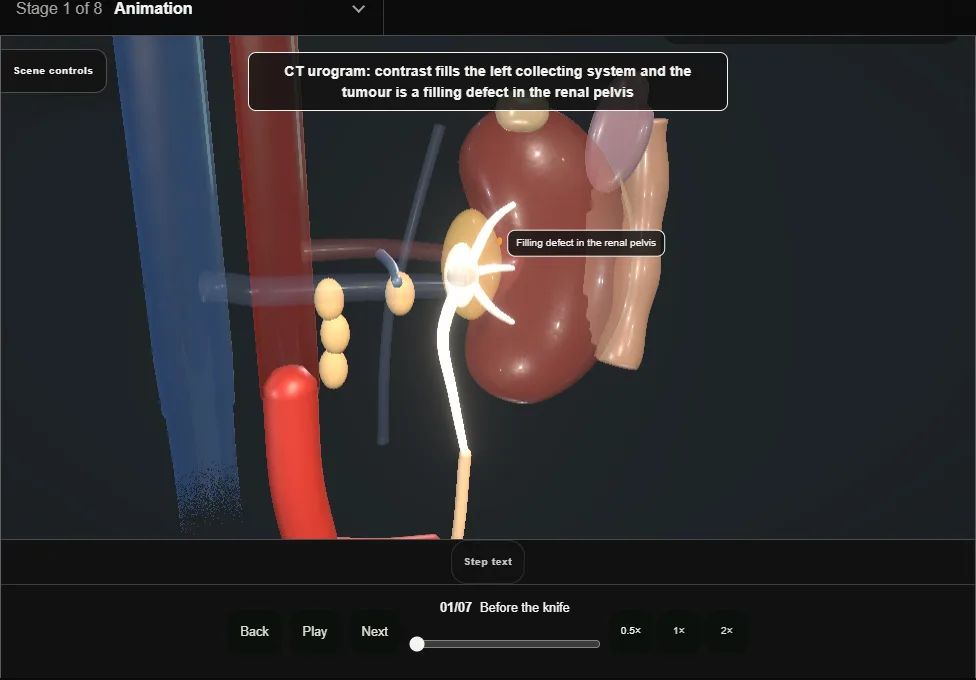
- Diagnosis by CT urogram and ureteroscopy with biopsy; high-risk features decide radical surgery over kidney-sparing treatment.
- Renal function is measured now, because losing a kidney decides who can have cisplatin later; neoadjuvant chemotherapy is considered in selected high-risk cases.
- 02
Access + colon reflection
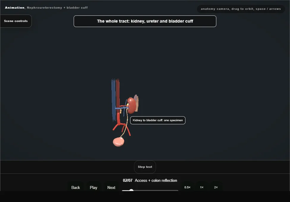
- Open, laparoscopic or robotic access to the left retroperitoneum; the colon is reflected medially along the line of Toldt and Gerota's fascia is kept intact over the tumour-bearing tract.
- Position and ports are planned so the distal ureter and bladder can be reached, or the patient is repositioned for the cuff.
- 03
Hilar control, artery first
- The renal artery is controlled and divided before the vein, exactly as in nephrectomy.
- Early arterial control devascularises the tumour-bearing kidney before any manipulation.
- The adrenal is spared unless it is involved.
- 04
No-touch mobilisation
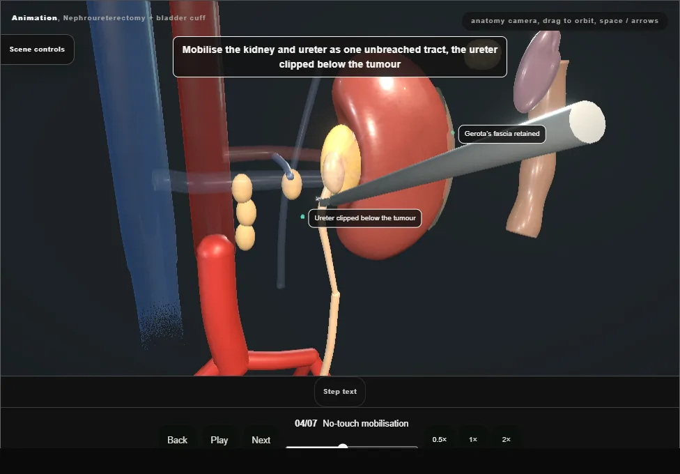
- Kidney and ureter are mobilised en bloc outside Gerota's fascia without breaching the collecting system.
- The ureter is clipped below the tumour early and is never squeezed or transected mid-course: tumour cells implant wherever they spill.
- 05
Bladder cuff
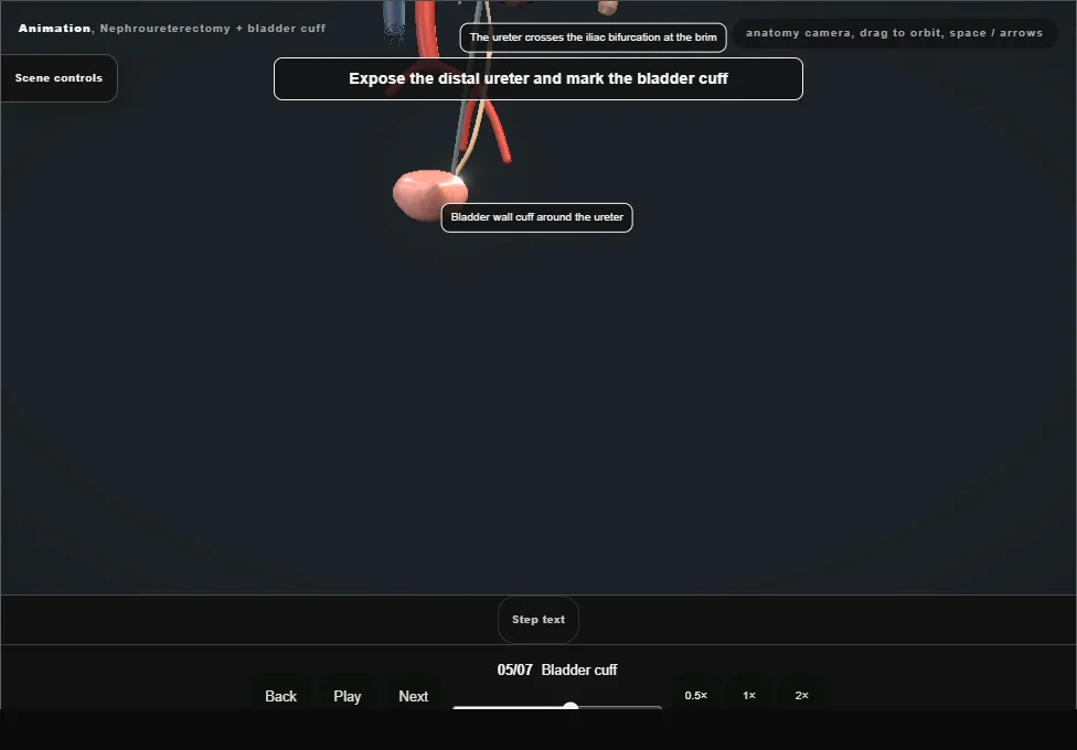
- A true cuff of bladder wall is excised around the ureteric orifice with the intramural ureter: open through a lower incision, or extravesically or intravesically in minimally invasive surgery.
- The ureter stays clipped so nothing spills into the bladder.
- The bladder is closed watertight, well clear of the other ureteric orifice.
- The cuff is the operation: a ligated stump is a recurrence waiting to happen.
- 06
Template nodes + specimen out
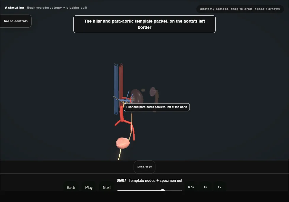
- Nodes are taken to a template set by side and tumour site: hilar and para-aortic on the left, hilar, paracaval and interaortocaval on the right, and ipsilateral pelvic nodes for the lower ureter.
- The whole specimen, kidney to cuff, leaves intact in a bag; morcellation is not an option in urothelial cancer.
- 07
Closure + catheter
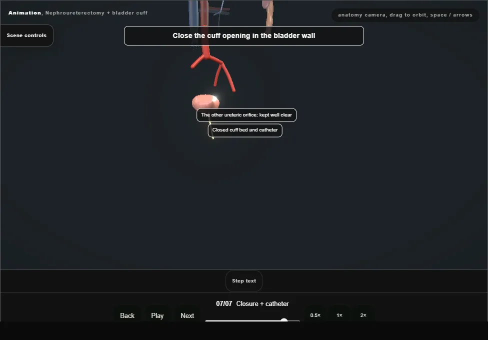
- The bladder is closed and drained with a catheter, with a cystogram before removal if the closure is in doubt.
- A single dose of intravesical chemotherapy after surgery lowers the risk of bladder recurrence.
- The surveillance plan is set before the patient leaves theatre: cystoscopy, imaging of the other upper tract and renal function; adjuvant platinum chemotherapy is discussed for invasive or node-positive disease.
- The whole urothelium remains at risk.
Derived from NephroureterectomyScene.tsx at build time: every step title and caption above is the authored
ProcedureDef text, reproduced verbatim, and this page shares its readable-points renderer with the animation
itself. 7 of 7 steps have a captured frame.
Text fingerprint 3312ccdfa9d57f1f. A caption edit without a re-capture fails the production
check, so the sheet and the animation cannot drift apart silently.