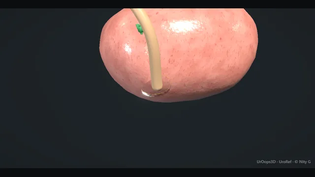
Upper tract urothelial cancer is removed as one continuous specimen: kidney in its fascia, ureter followed the whole length of the wound, and a true cuff of bladder wall around the ureteric orifice.
The no-touch principle governs every step, because tumour cells that spill implant wherever they land, and a ureteric stump left behind is a recurrence with an address on it.
Advanced tierFRCS oncology, upper tractInteractive 3D
Along the urothelial tube, kidney to bladder cuff. Schematic, not to scale; relationships only.
The specimen is a continuous tube of urothelium: renal pelvis to ureter to intramural tunnel to orifice, and the operation follows it like a thread.
The kidney sits high and lateral with its hilum facing the great vessels;
the ureter descends on psoas, crossing the pelvic brim over the iliac vessels to reach the bladder low and deep, where its oblique intramural course ends at the orifice.
The cuff is the deepest end of that thread: a disc of bladder wall whose margin is the difference between clearance and recurrence, approached through a lower incision exactly because the orifice cannot be trusted from above.
›Gerota's fascia - the plane the dissection stays outside of; breach it and the tumour-bearing kidney is exposed.
›Whole ureter - runs renal pelvis to orifice in continuity; never transected mid-course because spillage implants.
›Intramural ureter + orifice - the oblique tunnel through the bladder wall; the segment that recurs if it is left.
›Bladder cuff - the disc of bladder wall excised around the orifice; its margin is the margin that matters.
›Hilar vessels - artery first, vein second; early arterial control before any traction on the kidney.
Access + colon reflectionAccess is open or laparoscopic according to the plan made on imaging, positioned so the whole renal mobilisation and the ureteric course to the bladder can be followed without re-docking. The colon is reflected along the line of Toldt on the involved side until the retroperitoneum stands open from upper pole to pelvic brim. The dissection stays outside Gerota's fascia over the kidney and outside the ureteric sheath over the tract, taking the plane with blunt sweeps and keeping diathermy away from anything that bears tumour. The check is a continuous sheet of fascia still covering the whole tumour-bearing tract at the end of the exposure, because the first breach of that sheet is where cells start travelling. where it bites: the first breach of the plane around the ureter is where tumour cells start travelling; stay outside the fascia.
Hilar control, artery firstThe hilum is opened and the artery identified behind and slightly below the vein, skeletonised over a clean length and divided between clips or across a vascular stapler. Only then is the vein taken. Early arterial control matters twice in tumour surgery: it prevents the engorgement that thins the renal capsule under manipulation, and it empties the field of arterial inflow before anything is grasped. The check is a soft, dusky, non-pulsatile kidney before mobilisation begins; a turgid kidney means an accessory artery is still live, and it is found before the dissection moves on. where it bites: the artery lies behind and slightly below the vein at the hilum; a clip placed on the vein first engorges the kidney and thins its wall.
No-touch mobilisationThe kidney is mobilised inside Gerota's fascia and the ureter followed downward on its adventitial plane, the two carried as one continuous specimen from upper pole toward the bladder. Nothing touches the tumour-bearing segment: the ureter is never crushed in a grasper, never clipped mid-course and never divided above the bladder, and the collecting system is never opened for convenience. Traction is applied through the surrounding fat and fascia so the ureter is carried rather than held. Every breach of that discipline is tumour spillage, and spilled urothelial cells implant readily in the tract, the wound and the port sites. where it bites: every breach of the ureter is tumour spillage; the cells implant in the tract, the wound and the port sites.
Bladder cuffThe distal ureter is followed to the bladder through a lower incision, and the bladder is opened so the ureteric orifice can be seen from inside. A true disc of bladder wall is excised around the orifice with the intramural tunnel, and the specimen is delivered in continuity, the cuff arriving attached as the ureter is drawn up from below. The check is a visible rim of normal bladder around the excised disc, with the orifice and its tunnel unmistakably part of the specimen. A ligated stump left in the tunnel instead of an excised cuff is the classic technical failure of this operation, and it is the address of the next recurrence. The bladder is closed in layers and catheterised to drain it while it heals. where it bites: a ligated ureteric stump instead of a cuff is the classic technical failure of this operation and the address of the next recurrence.
Template nodes + specimen outLymph nodes are cleared to the template the side and stage demand: hilar and paracaval on the right, paraaortic on the left, with boundaries set from imaging before the case rather than negotiated from what the exposure happens to show. The dissection is nodal and stays off the great vessel walls unless staging requires more. The whole specimen, kidney, full-length ureter and cuff, is bagged intact under vision and delivered with the wound edges protected, the bag closed before it crosses the fascia. Morcellation or piecemeal delivery in urothelial cancer is spillage by another name, and it also sacrifices the pathology: an intact specimen is the only specimen that can be staged properly. where it bites: morcellation or piecemeal delivery in urothelial cancer is spillage by another name, and it sacrifices the pathology.
Closure + catheterThe bladder closure is filled and tested through the catheter before the wounds are closed, a drain is left to the cuff bed where the repair is deep or the field oozy, and the specimen is oriented for the pathologist with the cuff margin identified before the drapes come down. Before the patient leaves theatre the surveillance contract is written and owned by a named clinician: cystoscopic follow-up of the bladder, imaging of the contralateral tract, and the intervals at which both are due. The remaining urothelium stays at risk for years, and a plan that lives only in the op note is not a plan. where it bites: the remaining urothelium stays at risk for years; a surveillance plan that lives only in the op note is not a plan.
The spatial relationships the animation is built to make automatic. Before you read each one, ask yourself: at this point in the operation, what is about to go wrong, and what is my first move? The save for each sits in the Complications tab.
RiskThe ureteric wall itself - thin and tumour-bearing; crush, clip or breach it and cells implant in the tract and port sites.
RiskIliac vessels at the pelvic brim - the ureter crosses them directly in the deepest part of the mobilisation.
RiskThe intramural ureter - the oblique bladder tunnel; the segment a ligated-stump technique abandons and the stump then recurs.
Trigger each of these live in the Animation tab, then read the save: the first move, the escalation trigger, and the line that goes in the op note.
Tumour spillage / tract implantation
the collecting system breached or the specimen squeezed during mobilisation; cells implant at port sites and in the wound, presenting later as nodules at those exact sites.
The save
Prevent first: stay outside the fascia, handle the ureter by its adventitia, and never transect or crush the tumour-bearing segment.first move
If a breach happens, contain it: control the leak, wash the field copiously, and retrieve all spilled fragments under vision.
Convert the extraction to intact bagged removal with wound protection if it was not already planned that way.
Tell the pathologist and the MDT exactly what happened; the event changes the surveillance intensity.
A port-site or wound nodule during follow-up → biopsy and MDT discussion; metastatic territory, not a local tidy-up.escalation trigger
Op note: site of breach, washout, intact or piecemeal extraction, bagged or not.document
Positive bladder cuff margin
the pathology returns urothelium at the bladder edge of the cuff; the risk rises with a cuff taken too tight around the orifice or a ligated-stump technique.
The save
Know it early: orient the specimen yourself and ask the pathologist to ink and report the cuff margin specifically.first move
Re-cystoscope and resect the cuff bed with diathermy to destroy any residual urothelium at the resection base.
Consider a single induction course of intravesical therapy on MDT advice, since the field risk in the bladder is high in this disease.
Tighten cystoscopic surveillance for the bladder, which is where recurrence clusters after any cuff problem.
Deep residual disease at the cuff bed or unresectable involvement → partial cystectomy of the cuff bed in selected patients after MDT discussion.escalation trigger
Note: margin status, re-resection, intravesical plan and the revised surveillance schedule.document
Ureteric stump recurrence
a retained stump after incomplete distal excision; presents with haematuria and a visible lesion at the stump on cystoscopy, sometimes years later.
The save
Recognise the setup: a ligated or poorly visualised distal segment at index surgery, then haematuria with a lesion at the stump orifice.first move
Biopsy at cystoscopy and image the stump and surrounding tissue for depth and nodal status.
Completion excision of the stump with a proper bladder cuff, usually open, is the treatment; the resection needs the same margin discipline the first operation missed.
Survey the whole urothelium at the same time: the bladder and the contralateral tract are in the same field risk.
Invasive disease at the stump or nodal disease → MDT for possible partial cystectomy or systemic therapy rather than local excision alone.escalation trigger
Note: the original operation, the completion procedure and the long-term surveillance commitment.document
When the case turns: the problem, then the next move.
ActionThe ureter is torn during proximal mobilisation - do not chase the tear; control the segment, wash the field, and proceed to an intact en bloc retrieval with the breach contained.
ActionThe orifice cannot be identified from inside the bladder - fill and empty the bladder to watch the jet, use the ipsilateral trigone geometry, and take a slightly wider cuff rather than a blind cut.
ActionThe cuff will not come with the specimen - deliver the specimen from above while the assistant steadies the bladder: the cuff should arrive in continuity or the technique has failed.
ActionNodal disease encases the hilum at access - stop, image and stage, and take the case to the MDT; a frozen hilum in upper tract cancer changes the operation and possibly the sequence of systemic therapy.
The examiner stems this scene prepares you for. Try to answer out loud first, then reveal, and learn the discriminator, the detail that turns a pass into a strong pass.
QDoes the technique for the distal ureter and cuff actually matter?reveal answer
The principle is not negotiable: all of the at-risk urothelium comes out, kidney, whole ureter, and a disc of bladder wall around the orifice with the intramural segment.
Anything less leaves urothelium behind, and recurrence in a retained stump is a failure of the index operation rather than a new disease.
The debate is about technique, not principle.
The open cuff through a lower incision is the reference standard: direct vision, a true wall disc, secure closure, and a catheter.
Endoscopic alternatives exist to spare the incision: the pluck, freeing the intramural ureter endoscopically so it plucks away as the specimen is delivered, and intussusception or stripping variants.
Their criticisms are consistent: the orifice is opened into a field that is not controlled, cells can spill retroperitoneally or into the wound, and the bladder defect is left unclosed with a catheter trusting to healing.
My test for any technique is twofold: no residual intramural urothelium, and no open communication between bladder and operative bed while the specimen comes out.
A method that passes both in the positioned patient is defensible; one that does not loses to the open cuff, because the cheapest re-operation is the one avoided on the first day.
Strong pass
Say all three
Stating the two absolute requirements
Residual urothelium and a controlled bladder defect
Judging every technique against them rather than reciting the method names
Examiner note
Stating the two absolute requirements, residual urothelium and a controlled bladder defect, and judging every technique against them rather than reciting the method names.
QWhen is kidney-sparing endoscopic management of upper tract cancer reasonable?reveal answer
Selection is everything.
Endoscopic management is reasonable for tumours that look and behave low-risk: small, unifocal, low-grade on biopsy, without invasion on imaging, and crucially in patients in whom nephroureterectomy would mean dialysis or major renal compromise: a solitary kidney, bilateral disease, or established impairment.
It is a commitment, not a shortcut: serial ureteroscopic inspection, laser ablation, and indefinite imaging surveillance.
The risks I state honestly are under-staging, because biopsy grade understates final pathology in a proportion of patients, incomplete ablation, and progression under surveillance in the small proportion who turn out to harbour worse disease.
Segmental ureteric resection with reimplantation sits between the poles: defensible for distal ureteric tumours, where a wide distal segment plus a reimplanted ureter clears the lesion and keeps the kidney, and less defensible proximally, where the blood supply makes a tension-free join unreliable and recurrence harder to reach.
The failure mode of the whole strategy is drifting surveillance: the patients who do badly are those whose follow-up lapses.
So I document the surveillance contract before treatment begins, and offer radical surgery the moment the disease stops looking low-risk.
Strong pass
Say all three
Naming under-staging as the core risk
Framing kidney-sparing as a surveillance contract with an exit trigger
Rather than a softer operation
Examiner note
Naming under-staging as the core risk and framing kidney-sparing as a surveillance contract with an exit trigger, rather than a softer operation.
QDesign the surveillance schedule after nephroureterectomy.reveal answer
Surveillance follows two surfaces: the remaining urothelium and the retroperitoneum.
The bladder carries the dominant recurrent risk, so flexible cystoscopy is the anchor, front-loaded in the early years and lengthening only if it stays clear.
The contralateral upper tract is imaged on a schedule matched to grade and any carcinoma in situ, with CT urography as the workhorse.
The resection bed and nodes are covered by cross-sectional imaging in the early period to exclude nodal recurrence.
Renal function is followed after losing a kidney, particularly where medical comorbidity compounds a solitary remaining kidney.
Where a kidney-sparing approach was chosen instead, the treated tract needs direct endoscopic review as well as imaging, because ureteroscopy outperforms radiology for flat and small lesions.
Cytology is an adjunct where available, most useful as a high-grade signal, and never a clearance.
I write the schedule into the discharge summary and a surveillance system with a named responsible consultant, because the lesson of every upper tract series is that recurrence is common, late, and silent until it is not.
The patient who understands why cystoscopy continues for years is the patient who attends for it, so the plan is explained, not just posted.
Strong pass
Say all three
Separating the two surfaces
Urothelium and retroperitoneum
Assigning cystoscopy to the first and cross-sectional imaging to the second rather than treating surveillance as one list
Examiner note
Separating the two surfaces, urothelium and retroperitoneum, and assigning cystoscopy to the first and cross-sectional imaging to the second rather than treating surveillance as one list.
QHow do you prevent tumour spillage and tract implantation?reveal answer
Prevention is mechanical discipline, not luck.
The tumour-bearing collecting system is never entered: dissection stays outside Gerota's fascia and on the outside of the ureter, which is handled by its adventitia and never crushed, clipped or divided mid-course.
The kidney is devascularised early so manipulation cannot engorge and leak it.
The specimen is bagged intact under vision before extraction, the bag closed, and the wound protected during delivery.
Port sites are not dilated against the specimen, and morcellation is off the menu in urothelial cancer, because an intact specimen is also a reportable one.
If the collecting system is breached, the field is washed copiously, spilled fragments retrieved under vision, and the event documented for the pathologist and the MDT, since it changes surveillance intensity.
Port-site recurrence after spillage is real, avoidable, and poorly salvaged, which is why the effort belongs upstream in technique.
Finally I check the specimen myself with the pathologist in mind: kidney, full-length ureter and the cuff with its orifice identifiable, because a specimen that cannot be oriented cannot be reported, and an unreportable margin cannot be called clear.
The op note records intact extraction, bagged retrieval and wound protection.
Strong pass
Ending on specimen orientation and pathologist reporting: spillage prevention includes delivering a specimen whose margins can actually be assessed.
Say it aloud, then reveal. Reading is not remembering.
01The relationshipSay the critical spatial relationship out loud, then reveal.reveal
Memory frame
The specimen is a continuous tube of urothelium: renal pelvis to ureter to intramural tunnel to orifice, and the operation follows it like a thread.
Why?
The kidney sits high and lateral with its hilum facing the great vessels; the ureter descends on psoas, crossing the pelvic brim over the iliac vessels to reach the bladder low and deep, where its oblique intramural course ends at the orifice. The cuff is the deepest end of that thread: a disc of bladder wall whose margin is the difference between clearance and recurrence, approached through a lower incision exactly because the orifice cannot be trusted from above.
02The first moveTumour spillage / tract implantation. Your first move?reveal
First move
Prevent first: stay outside the fascia, handle the ureter by its adventitia, and never transect or crush the tumour-bearing segment.
03The escalation triggerTumour spillage / tract implantation. What makes you call for help?reveal
Escalate when
A port-site or wound nodule during follow-up → biopsy and MDT discussion; metastatic territory, not a local tidy-up.
04The discriminatorWhat turns a pass into a strong pass?reveal
Strong pass
Say all three
Stating the two absolute requirements
Residual urothelium and a controlled bladder defect
Judging every technique against them rather than reciting the method names
Examiner note
Stating the two absolute requirements, residual urothelium and a controlled bladder defect, and judging every technique against them rather than reciting the method names.
05The pitfallWhere does this operation bite first?reveal
Pitfall
The ureteric wall itself
Why?
thin and tumour-bearing; crush, clip or breach it and cells implant in the tract and port sites.
Revision practice with self-marked answers, not an assessment of clinical competence. Kept in
this browser only; it does not sync between devices.