
Revision sheet
Paediatric open inguinal hernia repair
8 steps, each one the authored title and caption from the interactive animation, with the frame captured from that step. Read it before the case, or print it and work through the steps in order.
Teaching only, not for diagnosis or treatment decisions. The text on this sheet is the animation's own step copy, reproduced verbatim.
UrOops3D · UroRef · © Nity G
- 01
Skin crease incision
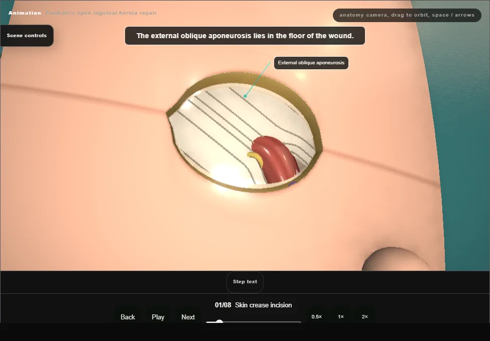
- Supine, the lower abdomen and genitalia prepared.
- The incision lies in the skin crease between the pubic tubercle and the anterior superior iliac spine, over the superficial ring.
- Skin and fat are opened, Scarpa’s fascia is divided with diathermy, and the external oblique aponeurosis is seen.
- 02
Open the canal
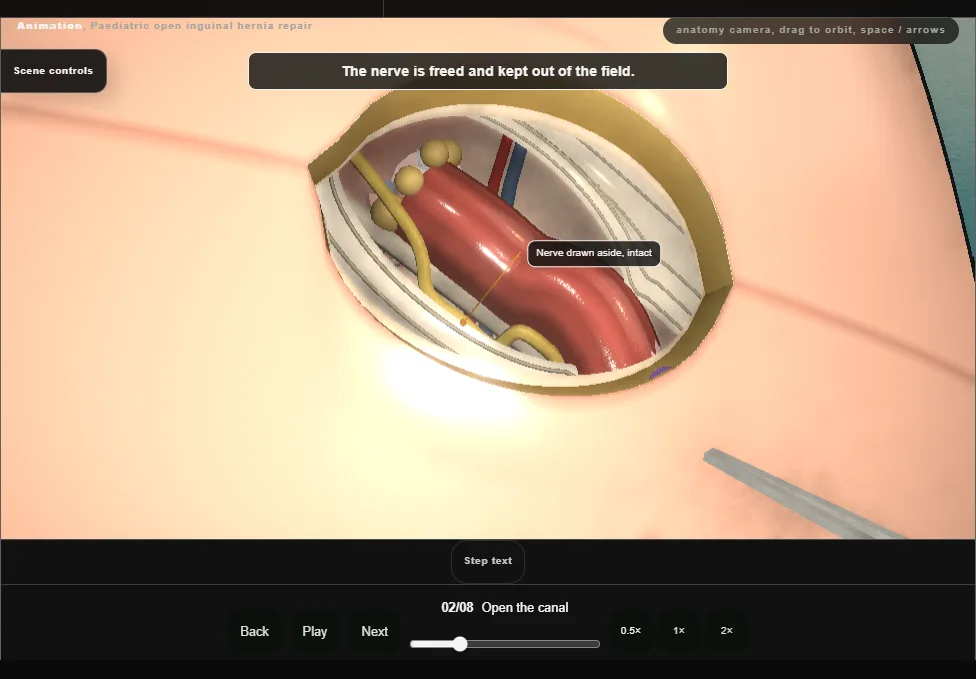
- The superficial ring is opened toward the deep ring, in the line of the fibres of the aponeurosis.
- The ilioinguinal nerve lies on the cord directly beneath: it is identified, freed and kept out of the field.
- 03
Lift the cord, find the sac
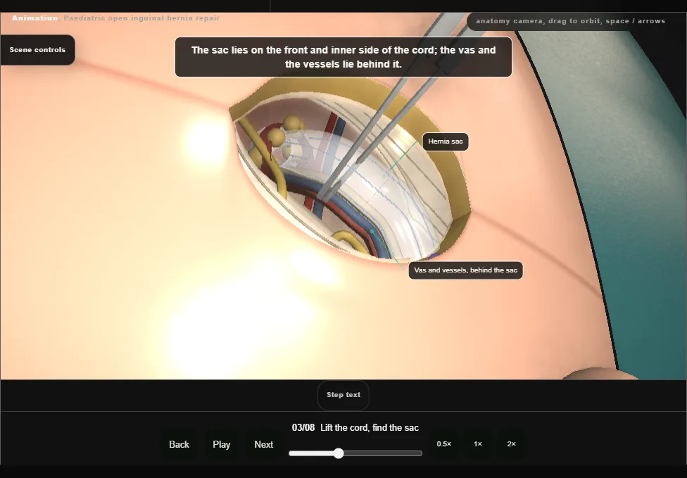
- The cremaster is opened and the cord is lifted into the wound, which confirms the sac.
- The sac lies on the front and inner side of the cord; the vas and the testicular vessels are identified behind it before any dissection.
- 04
Free the sac from the cord
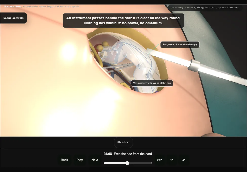
- With the edge of the sac held, the vas and the testicular vessels are dissected off its back wall under direct vision.
- They are swept, never grasped.
- The dissection is complete when the sac is clear all the way round and is seen to hold no bowel or omentum.
- 05
Follow the sac to the deep ring
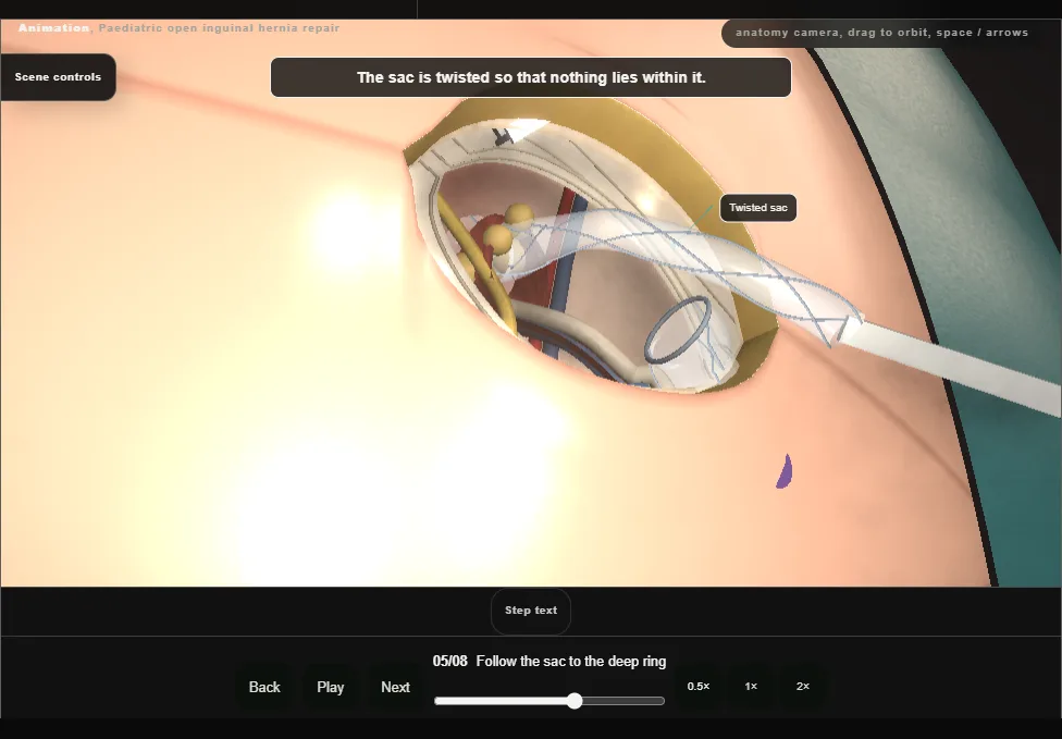
- The cleared sac is divided between two mosquitoes and its proximal part is followed up to the deep ring.
- Preperitoneal fat at the neck marks the level, with the inferior epigastric vessels just medial.
- The sac is twisted so that nothing lies within it.
- 06
High ligation
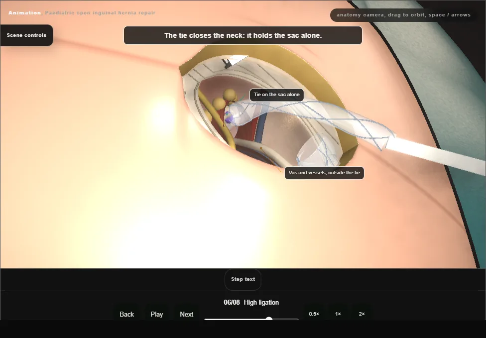
- The neck is transfixed and tied flush with the deep ring, on the sac alone: the vas and the vessels lie outside the tie.
- The sac beyond the tie is cut away and the stump draws back through the ring.
- The distal sac is left open.
- 07
Replace the cord and the nerve
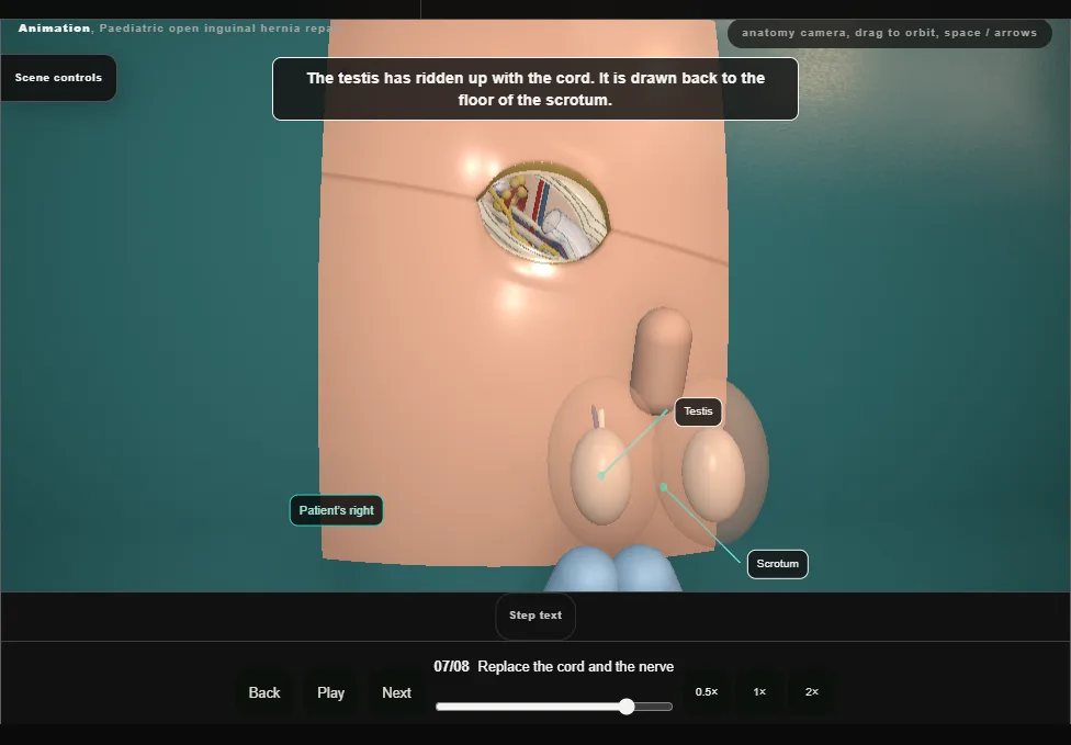
- The cord is laid back in the canal in its own orientation, and the canal is irrigated and checked for bleeding.
- The testis is drawn back to the floor of the scrotum.
- The ilioinguinal nerve is inspected, found intact and replaced beneath the aponeurosis.
- 08
Close in layers
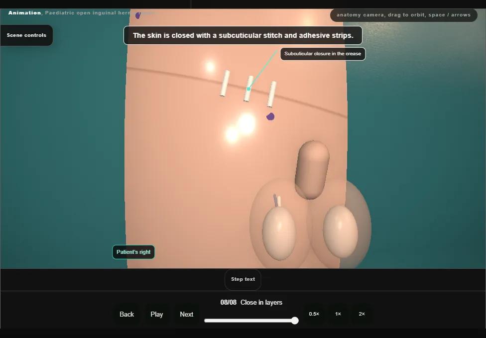
- The external oblique aponeurosis is closed with a running stitch that takes neither the nerve nor the cord and leaves the superficial ring its own size.
- Scarpa’s fascia is closed, then the skin with a subcuticular stitch and adhesive strips.
Derived from paediatric/InguinalHerniaScene.tsx at build time: every step title and caption above is the authored
ProcedureDef text, reproduced verbatim, and this page shares its readable-points renderer with the animation
itself. 8 of 8 steps have a captured frame.
Text fingerprint f49c0905f5857b54. A caption edit without a re-capture fails the production
check, so the sheet and the animation cannot drift apart silently.