
Revision sheet
Renal tumour ablation
9 steps, each one the authored title and caption from the interactive animation, with the frame captured from that step. Read it before the case, or print it and work through the steps in order.
Teaching only, not for diagnosis or treatment decisions. The text on this sheet is the animation's own step copy, reproduced verbatim.
UrOops3D · UroRef · © Nity G
- 01
Who
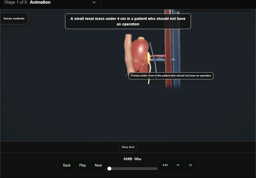
- A small renal mass under four centimetres in the patient who should not have an operation: age, comorbidity, a single kidney, a syndrome.
- Partial nephrectomy is the standard; ablation trades a little control for a lot less harm.
- 02
Biopsy first
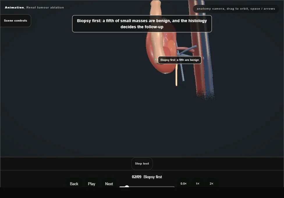
- At a separate sitting or the same one, coaxial, two cores through the track the probe will use.
- A fifth of small masses are benign, and the histology decides the follow-up.
- 03
Cryotherapy, radiofrequency or microwave
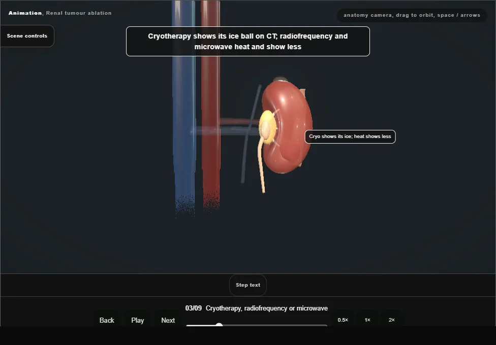
- Cryotherapy freezes and shows its ice ball on CT; radiofrequency and microwave heat and show less.
- Heat near the collecting system strictures the ureter; cold does not, so cryotherapy is chosen for the central mass.
- 04
Planning the approach on CT
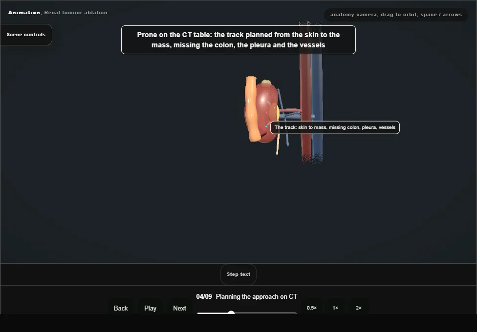
- Prone or oblique on the CT table under sedation or anaesthetic: the track from the skin to the mass planned to miss the colon, the pleura and the vessels.
- The colon behind a posterior kidney is the structure that changes the plan.
- 05
Hydrodissection
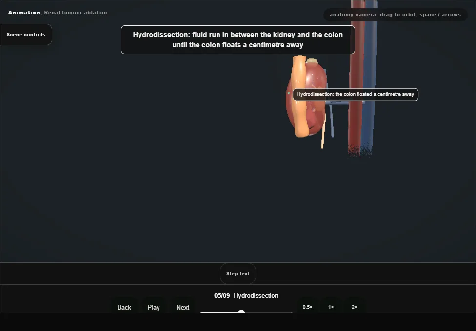
- A needle between the kidney and the colon, and dextrose or saline run in until the colon floats a centimetre away.
- Dextrose for radiofrequency, because saline conducts; carbon dioxide where fluid will not stay.
- 06
Placing the probes
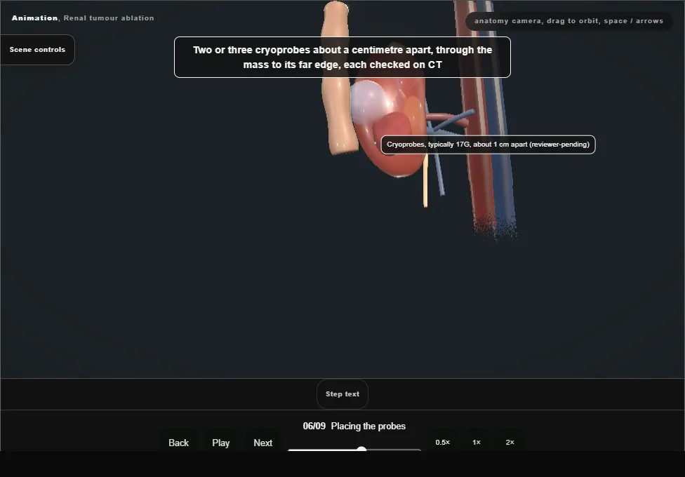
- Two or three cryoprobes a centimetre apart through the mass to its far edge, each checked on CT before the freeze; placed to cover the mass and its margin, not just its centre.
- 07
The ablation zone and its margin
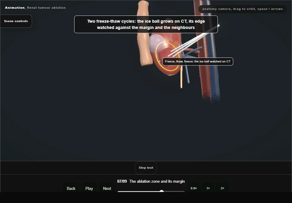
- Two freeze-thaw cycles, the ice ball growing on CT and its edge watched against the margin and the neighbours.
- Lethal ice is a few millimetres inside the visible edge: the ball must pass the margin by that much.
- 08
The collecting system and the ureter
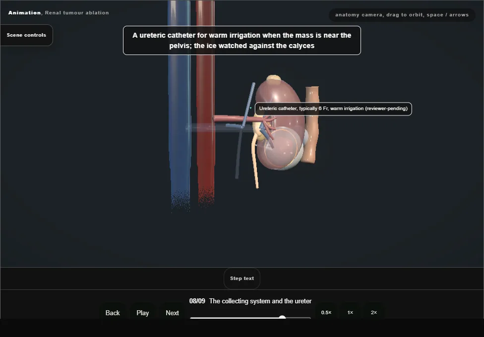
- A stent or a warm irrigation catheter for the mass near the pelvis, the ice watched against the calyces.
- Probes thawed and withdrawn with the track ablated behind them, and a final CT for bleeding and the pleura.
- 09
After
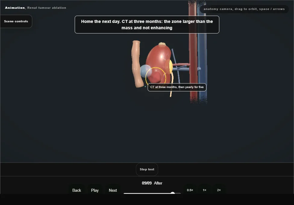
- Home the next day.
- CT at three months: the zone larger than the mass and not enhancing; then yearly for five years.
- Enhancement at the edge is residual tumour and a second ablation.
Derived from kidney/RenalAblationScene.tsx at build time: every step title and caption above is the authored
ProcedureDef text, reproduced verbatim, and this page shares its readable-points renderer with the animation
itself. 9 of 9 steps have a captured frame.
Text fingerprint 8e31cf69d273865e. A caption edit without a re-capture fails the production
check, so the sheet and the animation cannot drift apart silently.