
UrOops3D, danger-zone atlas
Operative animation, Renal tumour ablation

Percutaneous ablation of the small renal mass

The small enhancing mass under four centimetres in the patient who is too old, too unwell or too short of kidney to have it cut out: a needle through the skin under CT, and the tumour frozen or cooked in place with a margin, the colon and the ureter moved or protected first.

This page is the selection, the biopsy that comes before, the choice of energy, the plan on the CT, the probes, the zone and its margin, the collecting system, and the years of imaging that decide whether it worked.

Advanced tier FRCS uro-oncology, the small renal mass and nephron sparing Interactive 3D

Operative visualisation

Schematic scene preview: Renal tumour ablation
Preparing the 3D anatomy…

UrOops3D · UroRef · © Nity G

Click me next Section 2 of 8 Anatomy The structures this operation works between, and the one it must not cross.

The structures in the scene

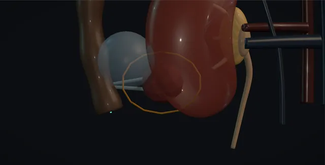
Renal tumour ablation: how the structures relateProne, the kidney from behind and what lies around the ablation zone. The small renal mass: lateral; moved with a fluid cushion or it perforates The colon and the bowel (danger). The small renal mass: within a centimetre: cooled, warmed or displaced The ureter and the renal pelvis (danger). The small renal mass: flowing blood spoils the margin at the central edge The renal sinus vessels (danger). The small renal mass: a path above the twelfth rib crosses it The pleura (danger). The small renal mass: medial; heat or cold numbs the groin and thigh Psoas and the genitofemoral nerve (danger). The small renal mass: placed a centimetre inside the edge The probe or antenna.The small renal mass!The colon and thebowel!The ureter and therenal pelvis!The renal sinusvessels!The pleura!Psoas and thegenitofemoral nerveThe probe or antennalateral; moved witha fluid cushion orit perforateswithin acentimetre: cooled,warmed or displacedflowing bloodspoils the marginat the central edgea path above thetwelfth rib crossesitmedial; heat orcold numbs thegroin and thighplaced a centimetreinside the edgeTargetDangerContext
Prone, the kidney from behind and what lies around the ablation zone. Schematic, not to scale; relationships only.
  • The patient is prone and the kidney is seen from behind through the posterior abdominal wall, the mass on its posterolateral surface with the colon lateral to it and the psoas medial.
  • The probes come in from the back below the twelfth rib, parallel and a centimetre apart, and the ice ball grows as a sphere around them, its edge a centimetre beyond the tumour.
  • Medially the renal sinus with the pelvis and the segmental vessels is the warm edge that resists the zone, and the ureter leaves the pelvis and runs down on the psoas.
  • ›The small renal mass - four centimetres or less; exophytic and posterior is easy, central and anterior is hard; the biopsy comes first.
  • ›The colon and the bowel - within a centimetre of the zone it is moved with a fluid cushion before any energy is applied.
  • ›The ureter and the renal pelvis - the structures that stricture; cooled or warmed through a ureteric catheter, or displaced, and cryotherapy chosen when close.
  • ›The renal sinus vessels - segmental vessels that warm the ice ball and sink the heat; a central mass is under-treated at the sinus edge.
  • ›The pleura - reflects at about the twelfth rib posteriorly; an upper pole path above it crosses the lung.
  • ›The psoas and the genitofemoral nerve - behind the lower pole; heat or cold on them gives groin and thigh numbness or weakness.
  • ›The probe or antenna - placed a centimetre inside the edge and a centimetre apart, tip in the deep half, checked in three planes before it is switched on.
Click me next Section 3 of 8 Steps The operation in order, with the landmark that guards each move.

Operative steps

  1. Who: the small mass in the wrong patient for surgery A solid enhancing renal mass of four centimetres or less, T1a, in a patient for whom partial nephrectomy carries too much risk: age, cardiorespiratory disease, anticoagulation that cannot be stopped, a solitary kidney or chronic kidney disease where every nephron matters, or hereditary multifocal disease such as von Hippel-Lindau where operations are counted over a lifetime. The alternatives are laid out at the MDT: active surveillance, which is right for many small masses in the elderly because most grow slowly and few metastasise; partial nephrectomy, which has the lowest local recurrence; and ablation, which sits between the two with a higher local recurrence than surgery but far less to recover from. The position, size and depth of the mass on CT decide whether a needle can reach it safely.
  2. Biopsy first A renal mass biopsy is taken before ablation, at a separate sitting or at the same one immediately before the probes go in, because a fifth to a quarter of small masses are benign, oncocytoma or fat-poor angiomyolipoma, and treating those is harm without benefit; and because once the tumour is ablated the histology is lost for good and the follow-up cannot be interpreted. Two or three 18 gauge cores through a coaxial needle under CT or ultrasound, from the periphery of the mass, avoiding necrosis, with the tract kept within the planned ablation zone. Seeding along the tract is very rare. A non-diagnostic biopsy is repeated; a benign result ends the plan.
    where it bites: a mass ablated without a biopsy is a mass with no diagnosis, no grade and no way to read the scans that follow.
  3. Cryotherapy, radiofrequency or microwave Cryoablation uses argon expanding in the probe tip to freeze the tissue: cell death is certain below about minus 40 degrees, the ice ball is visible on CT as a low-density sphere, and its visible edge sits at about 0 degrees, so the lethal zone is several millimetres inside the edge. Radiofrequency heats tissue by ionic agitation to above 60 degrees around the electrode, is not visible during treatment, and is weakened by flowing blood beside it, the heat sink. Microwave heats faster and hotter, is less affected by heat sink, and makes a larger zone. Cryotherapy is chosen for central and larger masses where the ice ball can be watched and for the mass beside the ureter; heat is chosen for small exophytic masses.
  4. Planning the approach on CT The patient lies prone or lateral on the CT table and a planning scan is taken with the breath held in the phase that will be used for the probes. The path is chosen to be short, to avoid the bowel, the liver, the spleen and the pancreas, to stay below the pleural reflection, which is at about the twelfth rib posteriorly, and to cross little normal parenchyma. The mass is measured in three planes and the number of probes decided: one cryoprobe treats about two centimetres, so a three centimetre mass takes two or three probes a centimetre apart and a centimetre inside the edge. The distance from the mass to the colon, the ureter and the renal sinus is measured, because each decides what protection is needed.
    where it bites: an upper pole mass approached from above the twelfth rib crosses the pleura; the path is planned below it or the pleura accepted with a chest drain on the trolley.
  5. Hydrodissection: moving the colon away Bowel within about a centimetre of the planned ablation zone is moved away before the probes go in. A fine needle is passed under CT into the fat between the kidney and the colon, and fluid is injected until a cushion of a centimetre or more separates them: five per cent dextrose for radiofrequency, because saline conducts the current, and saline or dextrose for cryotherapy, sometimes with a little contrast so it can be seen. Carbon dioxide or a balloon does the same work when fluid runs away. The cushion is rescanned and topped up during the treatment because it disperses. The same trick lifts the kidney off the psoas and the genitofemoral nerve, or separates the mass from the liver or spleen.
    where it bites: the colon that has not been moved sits against the ice ball or the heat and perforates, typically a week later, with a fistula or an abscess.
  6. Placing the probes Local anaesthesia with sedation, or a general anaesthetic where the patient cannot hold still or the breath. A skin nick, and each probe is advanced along the planned path under CT in steps, with a scan after each step, the tip aimed to lie in the deep half of the mass so that the zone extends beyond it. Probes are placed in parallel, a centimetre apart, a centimetre inside the tumour edge, and the last one is checked in all three planes before anything is switched on, because a probe that lies in the sinus or beside the ureter is repositioned now, not after. For a mass in contact with the collecting system, a ureteric catheter placed cystoscopically beforehand is connected for cooling or warming irrigation.
  7. The ablation zone and its margin For cryotherapy, a ten minute freeze, a passive or active thaw of five to eight minutes, and a second ten minute freeze: the double freeze-thaw is what kills, because the first freeze injures and the second, into tissue already damaged, is lethal. The ice ball is scanned every few minutes and grown until its visible edge lies five to ten millimetres beyond the tumour in every plane, because the lethal isotherm is inside the edge. For radiofrequency and microwave, the generator runs to the manufacturer's protocol for the size, usually eight to fifteen minutes per position, and the zone is inferred from the planning rather than seen. The endpoint in every case is a zone that encloses the whole tumour with a margin, and no more than the margin.
    where it bites: an ice ball that stops short at one edge, because a vessel or the sinus warms it, is the recurrence found at the first scan.
  8. The collecting system and the ureter The ureter and the renal pelvis are the structures that turn a good ablation into a stricture. For a lower pole or medial mass within a centimetre of the ureter, the ureteric catheter placed beforehand runs cold saline during heat ablation or warm saline during cryotherapy, protecting the urothelium, or the ureter is displaced with hydrodissection. A central mass abutting the collecting system is treated with cryotherapy, because frozen urothelium heals where heated urothelium sloughs; the ice ball is allowed to reach the sinus but not to cross it. Bleeding from the parenchyma is expected and small; the tract is ablated or thawed slowly on withdrawal, and a scan at the end looks for a haematoma, urine extravasation, pneumothorax and the position of the bowel.
  9. After: CT at three months and the years Observation for four to six hours or overnight, the haemoglobin checked if the tract bled, and home with the warning that fever, loin pain or frank haematuria in the next week is investigated. Contrast CT or MRI at three months is the first true assessment: a successfully treated zone does not enhance and slowly shrinks, and any nodular enhancement within or at the edge of the zone is residual tumour, retreated by a second ablation or, if the patient has become fit, by surgery. Then imaging at six and twelve months and yearly to five years and beyond, because recurrence after ablation appears later than after surgery. Local recurrence is higher than after partial nephrectomy but most is retreatable, and cancer-specific survival for T1a disease is similar.
Click me next Section 4 of 8 Danger zones Where this operation bites, and why it bites there.
Critical relationship

Danger zones

The spatial relationships the animation is built to make automatic. Before you read each one, ask yourself: at this point in the operation, what is about to go wrong, and what is my first move? The save for each sits in the Complications tab.

  • RiskThe collecting system and the ureter - heat sloughs urothelium and cold heals it; a ureter within a centimetre is cooled, warmed or displaced, or the patient has a stricture and a nephrostomy.
  • RiskThe colon - unmoved, it lies against the zone and perforates about a week later as a fistula or abscess.
  • RiskThe renal sinus - flowing blood warms the ice ball and sinks the heat; the margin fails at the central edge and the recurrence is there.
  • RiskThe pleura and the upper pole - a path above the twelfth rib gives a pneumothorax or a hydrothorax; the path goes below it or a chest drain is ready.
Practise it Take me into the scene Opens the animation at a danger moment and asks you to predict the mechanism and your first move before it reveals.
Click me next Section 5 of 8 Complications The save for each one: the first move, the escalation trigger, the op-note line.

Complications and immediate saves

Trigger each of these live in the Animation tab, then read the save: the first move, the escalation trigger, and the line that goes in the op note.

Bleeding and perirenal haematoma

  • a perirenal haematoma on the completion scan, frank haematuria, or a falling haemoglobin over the first day, from the tract or a segmental vessel in the zone; a pseudoaneurysm appears later.
The save
  1. Look at the completion CT before the patient leaves the table; a small haematoma is watched, a growing one is a decision. first move
  2. Bed rest, haemoglobin at six hours and the next morning, anticoagulants held, and a clotting screen if the patient bleeds out of proportion.
  3. Cryotherapy bleeds more than heat because it does not coagulate; the tract can be treated on withdrawal.
  4. A transfusion requirement, haemodynamic change, or a pseudoaneurysm on CT angiography: selective embolisation with interventional radiology; nephrectomy is the last resort. escalation trigger
  5. Note: the energy, the number of probes, the haematoma size on the completion scan and the haemoglobin trend. document

Collecting system injury, urine leak and ureteric stricture

  • a central mass or a lower pole mass beside the ureter: urine in the perinephric space on the scan, a urinoma, or a hydronephrosis weeks later from a stricture of the heated ureter.
The save
  1. Recognised at the time on the completion scan: a stent placed retrograde the same day or the next, and antibiotics. first move
  2. A urinoma: drain it percutaneously and stent the kidney; most leaks close over weeks with the collecting system decompressed.
  3. Ultrasound at six weeks after any central ablation, because a stricture is silent until the kidney is lost.
  4. Hydronephrosis, fever or a falling function: nephrostomy and an antegrade study; a long stricture needs reconstruction or, in the old patient, a nephrostomy for life. escalation trigger
  5. Note: the distance from the mass to the collecting system and the ureter, the protection used, and the imaging interval. document

Bowel or adjacent organ thermal injury

  • the colon, duodenum, liver, spleen or pancreas within the zone without a cushion: pain and fever at about a week, then a fistula or an abscess, or a pancreatic collection.
The save
  1. Never switch on with bowel within a centimetre; hydrodissect, rescan and top up the cushion during the treatment. first move
  2. Pain, fever or peritonism in the first two weeks: CT with oral and intravenous contrast, and the general surgeons told the same day.
  3. A contained collection is drained percutaneously; a colonic fistula to the kidney needs diversion and later repair.
  4. Free perforation or sepsis: laparotomy with the general surgeons, and the possibility of nephrectomy with the bowel resection. escalation trigger
  5. Note: the cushion, its volume and fluid, and the bowel distance on the final planning scan. document

Pneumothorax, pleural effusion and nerve injury

  • the upper pole path crossing the pleura, or the lower pole zone reaching the psoas: a pneumothorax on the completion scan, a hydrothorax later, or numbness and weakness in the groin and thigh.
The save
  1. Completion CT includes the lung bases; a pneumothorax of any size in a sedated patient is a chest drain decision made in the room. first move
  2. A small pneumothorax in a well patient is aspirated or watched with a repeat film at four hours.
  3. A genitofemoral or intercostal neuropathy is usually temporary; it is explained, and neuropathic analgesia is started.
  4. A tension pneumothorax, a hydrothorax with breathlessness, or a nerve deficit with motor loss: chest drain, respiratory review, neurology as needed. escalation trigger
  5. Note: the rib level of the path, the pleura seen on the scan, and any deficit at discharge. document

Residual tumour and local recurrence

  • nodular enhancement in or at the edge of the zone on the three month scan, or a new enhancing focus a year or more later, most often at the sinus edge of a central mass or from an ice ball that stopped short.
The save
  1. Contrast CT or MRI at three months read against the pre-treatment scan; the zone should not enhance and should not have grown. first move
  2. Enhancement at the edge in the first scan is residual disease, not a change in the zone; retreat by ablation, which is usually possible.
  3. Yearly imaging to five years and beyond, because recurrence after ablation presents later than after surgery.
  4. Recurrence after a second ablation, or a mass that has grown beyond ablation, in a patient who has become fit: partial or radical nephrectomy, which is harder after ablation because of the scar. escalation trigger
  5. Note: the zone size on the completion scan, the margin achieved in each plane, and the surveillance schedule with dates. document
Click me next Section 6 of 8 Troubleshooting When the case turns: the problem, then the next move.

Intra-operative troubleshooting

When the case turns: the problem, then the next move.

  • ActionThe colon lies against the mass and will not move with fluid - more fluid from a second needle position, carbon dioxide, or a balloon; if it still touches, the mass is treated laparoscopically with the bowel held away or not ablated.
  • ActionThe mass cannot be seen without contrast for the probes - contrast at planning with the probes placed on the landmarks of the nephrogram, or ultrasound fusion; a mass that cannot be targeted is not ablated blind.
  • ActionThe probe lies in the sinus on the check scan - withdraw and redirect before switching on; a probe frozen or heated in the sinus is a pelvic injury or a segmental vessel.
  • ActionThe ice ball will not extend past the medial edge - the sinus is warming it: a further probe medially, a longer freeze, or accept a smaller margin there and image early; heat would do worse.
  • ActionA pneumothorax on the completion scan - small and asymptomatic, aspirate through the tract and repeat the film; larger, a chest drain before the patient leaves the CT room.
  • ActionFrank haematuria on the ward - catheter and irrigation, haemoglobin, hold anticoagulants; if it persists, CT angiography and embolisation of the segmental vessel.
Click me next Section 7 of 8 FRCS viva Say it out loud, then mark yourself against the model answer.

FRCS viva questions

The examiner stems this scene prepares you for. Try to answer out loud first, then reveal, and learn the discriminator, the detail that turns a pass into a strong pass.

QA 78 year old with ischaemic heart disease has a three centimetre enhancing renal mass. What are the options?reveal answer
  • I would present three options at the MDT and to the patient.
  • Active surveillance, with imaging at three to six months and then six to twelve monthly, which is appropriate because most small renal masses grow at a few millimetres a year and the risk of metastasis from a mass under four centimetres is low, particularly in a patient whose competing mortality is high.
  • Ablation, percutaneous cryotherapy or a heat-based technique under CT, which removes the tumour with a single sedated procedure and preserves nephrons, at the cost of a local recurrence rate higher than surgery and a need for biopsy first and imaging for years.
  • Partial nephrectomy, which gives the lowest recurrence and a histological specimen, at the cost of a general anaesthetic and a major operation in a man with ischaemic heart disease.
  • I would recommend a renal mass biopsy whichever of the last two is chosen, because a fifth or more of these masses are benign.
  • For this man, if the mass is accessible percutaneously, I would offer ablation or surveillance depending on his wishes and his fitness after cardiology assessment, and I would explain that cancer-specific survival for T1a disease is similar across all three.
Strong pass

Say both

  • Surveillance offered as a real option with the natural history that justifies it
  • Biopsy stated as the step before ablation rather than after
Examiner note
Surveillance offered as a real option with the natural history that justifies it, and biopsy stated as the step before ablation rather than after.
QCompare cryoablation with radiofrequency and microwave ablation for renal tumours.reveal answer
  • Cryoablation freezes tissue with argon gas in the probe tip.
  • Cell death is reliable below about minus 40 degrees, and because the ice ball is visible on CT or ultrasound as a low-density sphere, the treatment can be monitored and grown to a margin, remembering that the visible edge is about 0 degrees and the lethal isotherm lies several millimetres inside it;
  • a double freeze-thaw cycle is used.
  • It does not coagulate, so it bleeds more, but it is kinder to urothelium and is the choice for central masses and those near the ureter, and multiple probes allow larger masses.
  • Radiofrequency heats tissue above 60 degrees by ionic agitation around an electrode;
  • it is cheap and fast but the zone is not visible during treatment, the size is limited by charring, and flowing blood near the sinus removes heat, the heat sink, which causes under-treatment centrally.
  • It requires non-conducting fluid such as five per cent dextrose for hydrodissection.
  • Microwave heats faster to higher temperatures with less heat sink and larger zones from one antenna, but its zone is less predictable near the collecting system.
  • In practice cryotherapy is favoured in the UK for its monitoring and for central tumours; heat suits small exophytic masses.
Strong pass

Say both

  • The lethal isotherm inside the visible ice ball edge
  • The heat sink explained as the reason heat under-treats central masses
Examiner note
The lethal isotherm inside the visible ice ball edge, and the heat sink explained as the reason heat under-treats central masses.
QHow do you protect the bowel and the collecting system during renal ablation?reveal answer
  • I would identify both risks on the planning CT and deal with each before any energy is applied.
  • For the bowel, colon or duodenum within about a centimetre of the intended zone is displaced by hydrodissection: a needle placed under CT into the fat between kidney and bowel and fluid injected until a cushion of at least a centimetre separates them, five per cent dextrose for radiofrequency because saline conducts current, saline or dextrose for cryotherapy, with a little contrast to see it.
  • Carbon dioxide or a balloon are alternatives if fluid disperses, and the cushion is rescanned and topped up during treatment.
  • If the bowel cannot be moved, the mass is treated laparoscopically with the bowel retracted or not ablated.
  • For the collecting system, a mass abutting the pelvis or within a centimetre of the ureter is treated with cryotherapy rather than heat, because frozen urothelium heals and heated urothelium sloughs and strictures;
  • a ureteric catheter placed cystoscopically beforehand allows warm saline irrigation during freezing or cold saline during heating;
  • and the ureter can be displaced with hydrodissection.
  • The completion scan looks for extravasation, and an ultrasound at six weeks looks for a silent stricture.
Strong pass

Say both

  • Dextrose rather than saline for radiofrequency with the reason
  • Cryotherapy chosen over heat beside the collecting system with the reason
Examiner note
Dextrose rather than saline for radiofrequency with the reason, and cryotherapy chosen over heat beside the collecting system with the reason.
QHow is a patient followed after renal ablation, and what does recurrence look like?reveal answer
  • The first imaging that means anything is a contrast CT or MRI at about three months, because the early zone is oedematous and haemorrhagic and cannot be read.
  • A successfully treated zone does not enhance, the treated tumour may remain as a non-enhancing scar after cryotherapy or be absorbed, and the zone shrinks slowly over years.
  • Residual tumour appears as nodular or crescentic enhancement within or at the margin of the zone, most often at the deep, central edge where the sinus vessels resisted the treatment;
  • it is usually retreated by a second ablation, which brings the overall local control close to that of surgery.
  • Imaging is then at six and twelve months and yearly thereafter to at least five years, and longer for the young or the hereditary syndromes, because recurrence after ablation presents later than after partial nephrectomy.
  • Renal function is tracked with eGFR.
  • New enhancement appearing after a clear scan is a local recurrence and is treated as a new small mass: repeat biopsy if the histology matters, repeat ablation, or surgery if the patient has become fit, remembering that surgery after ablation is harder.
  • Metastatic surveillance follows the stage and grade of the biopsy.
Strong pass

Say both

  • The three month scan named as the first interpretable one with the reason
  • Enhancement at the central edge explained by the heat sink or warming from the sinus
Examiner note
The three month scan named as the first interpretable one with the reason, and enhancement at the central edge explained by the heat sink or warming from the sinus.
Click me next Section 8 of 8 Evidence The guidance each of these answers is built on.

Evidence and guidance

Two minutes

Close the lap: recall it before you leave.

Say it aloud, then reveal. Reading is not remembering.

  1. 01 The relationship Say the critical spatial relationship out loud, then reveal. reveal
    Memory frame

    The patient is prone and the kidney is seen from behind through the posterior abdominal wall, the mass on its posterolateral surface with the colon lateral to it and the psoas medial.

    Why?
    The probes come in from the back below the twelfth rib, parallel and a centimetre apart, and the ice ball grows as a sphere around them, its edge a centimetre beyond the tumour. Medially the renal sinus with the pelvis and the segmental vessels is the warm edge that resists the zone, and the ureter leaves the pelvis and runs down on the psoas.
  2. 02 The first move Bleeding and perirenal haematoma. Your first move? reveal
    First move

    Look at the completion CT before the patient leaves the table; a small haematoma is watched, a growing one is a decision.

  3. 03 The escalation trigger Bleeding and perirenal haematoma. What makes you call for help? reveal
    Escalate when

    A transfusion requirement, haemodynamic change, or a pseudoaneurysm on CT angiography

    More detail
    A transfusion requirement, haemodynamic change, or a pseudoaneurysm on CT angiography: selective embolisation with interventional radiology; nephrectomy is the last resort.
  4. 04 The discriminator What turns a pass into a strong pass? reveal
    Strong pass

    Say both

    • Surveillance offered as a real option with the natural history that justifies it
    • Biopsy stated as the step before ablation rather than after
    Examiner note
    Surveillance offered as a real option with the natural history that justifies it, and biopsy stated as the step before ablation rather than after.
  5. 05 The pitfall Where does this operation bite first? reveal
    Pitfall

    The collecting system and the ureter

    Why?
    heat sloughs urothelium and cold heals it; a ureter within a centimetre is cooled, warmed or displaced, or the patient has a stricture and a nephrostomy.

Revision practice with self-marked answers, not an assessment of clinical competence. Kept in this browser only; it does not sync between devices.