
Revision sheet
Transureteroureterostomy
8 steps, each one the authored title and caption from the interactive animation, with the frame captured from that step. Read it before the case, or print it and work through the steps in order.
Teaching only, not for diagnosis or treatment decisions. The text on this sheet is the animation's own step copy, reproduced verbatim.
UrOops3D · UroRef · © Nity G
- 01
Both ureters at the bifurcation
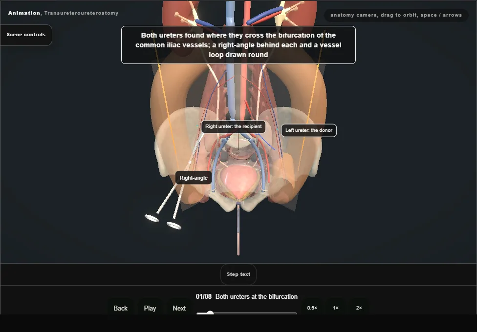
- Midline laparotomy.
- Both colons are mobilised along the white line of Toldt and lifted medially.
- The gonadal vessels are swept laterally.
- The ureters are found where they cross the bifurcation of the common iliac vessels and slung: the left is the donor, the right the recipient.
- 02
Donor mobilised and divided
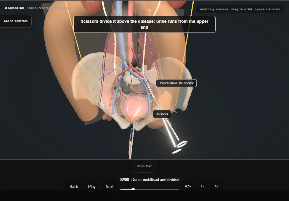
- The left ureter is dissected above the scarred part with its adventitia on, tied below and divided; a long tag stitch goes on the upper end and urine is seen to run.
- It is then freed cephalad, leaving the tissue on its medial side, which carries its blood supply.
- 03
The window above the artery
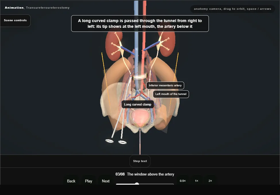
- The inferior mesenteric artery is found on the front of the aorta.
- The peritoneum is opened above it, in front of the aorta and the cava, to make a tunnel behind the root of the mesocolon, and a long curved clamp is passed through from right to left.
- 04
Donor across
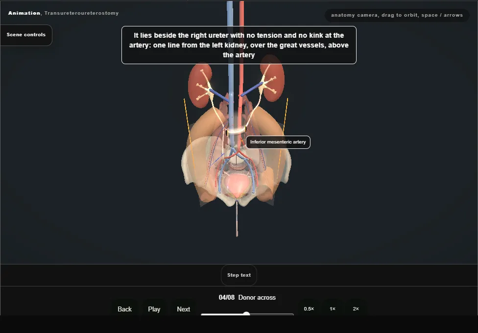
- The clamp takes the tag and the donor is drawn gently through the tunnel.
- It lies beside the right ureter without tension and without a kink at the artery: one line from the left kidney, over the great vessels, above the artery.
- 05
Recipient ureterotomy
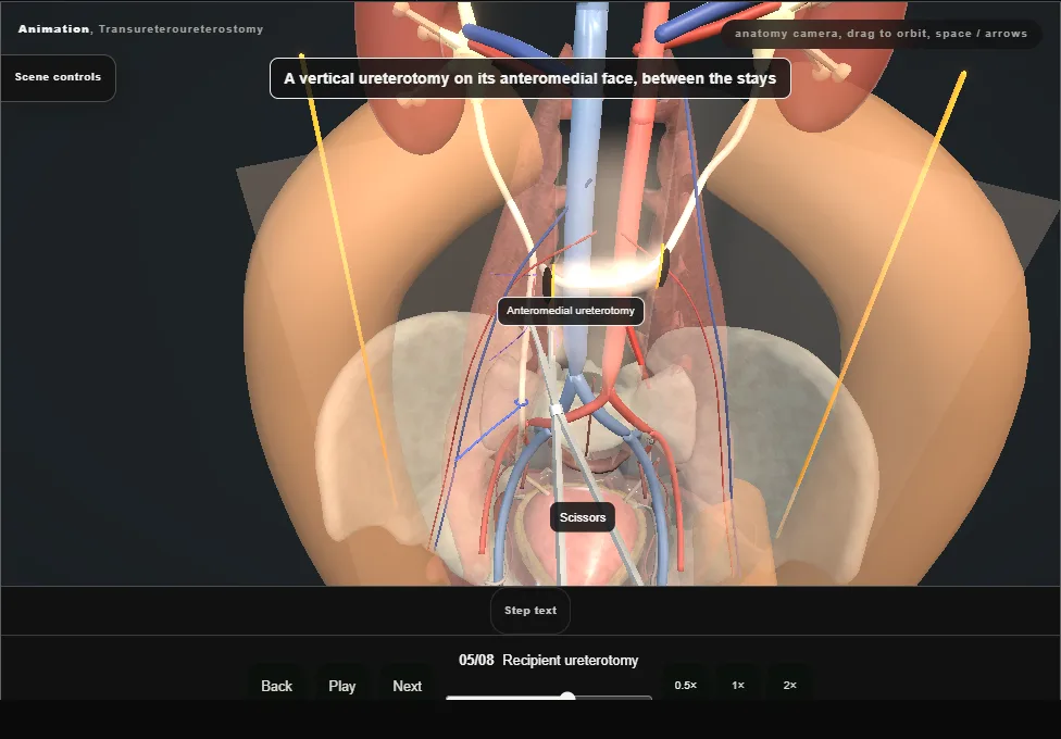
- The right ureter is mobilised as little as possible.
- A stay goes into its adventitia above the chosen site and one below, and a vertical ureterotomy is made between them on the anteromedial face.
- The slit gapes and the lumen shows.
- 06
Donor spatulated
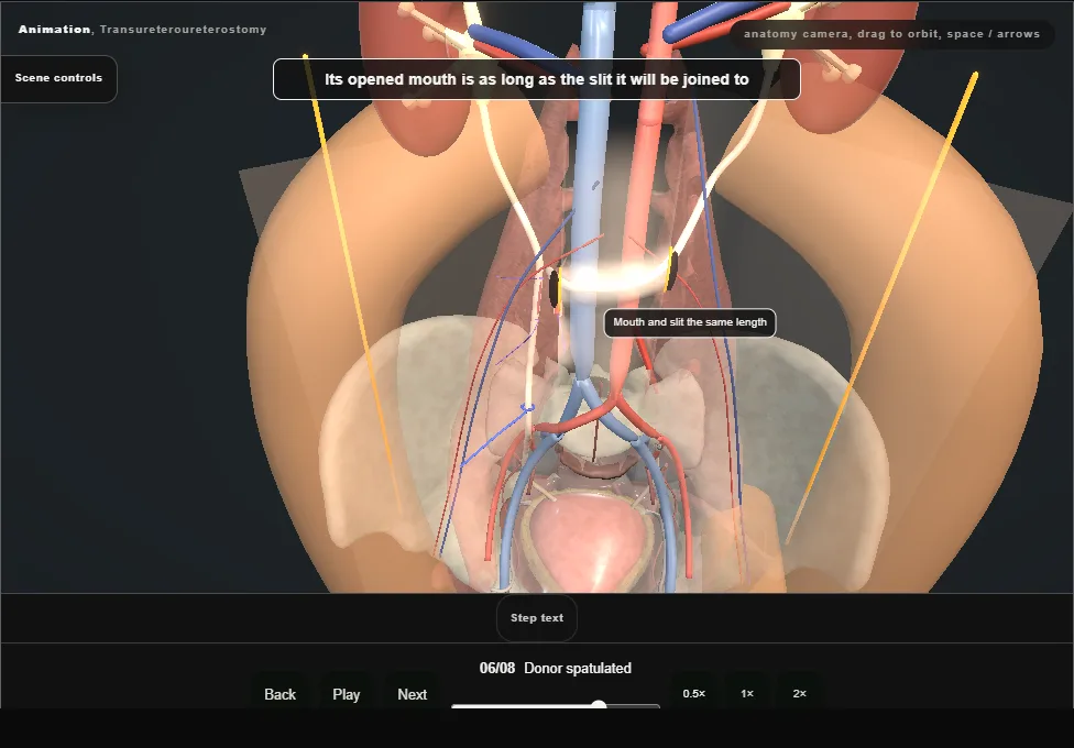
- The donor end is held up and spatulated on the side that will face the slit, until its opened mouth is as long as the ureterotomy it will be joined to.
- 07
End-to-side join
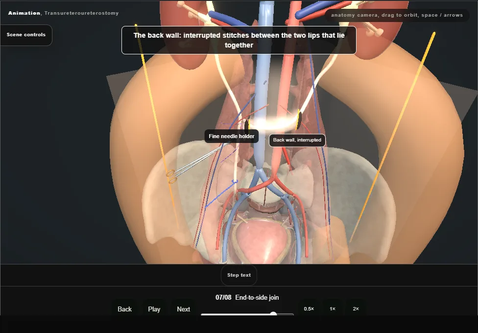
- Apex stitches top and bottom, then the back wall with interrupted absorbable stitches, mucosa to mucosa.
- The donor is then rolled onto the slit and the front wall is closed.
- Small bites on the recipient, and no tension.
- The recipient is never divided.
- 08
Close and drain
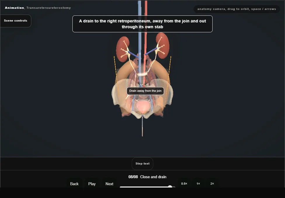
- The peritoneal window is closed loosely at each mouth so it cannot grip the ureter.
- A drain is laid in the right retroperitoneum away from the join and brought out through its own stab.
- Both kidneys now drain through the right ureter.
Derived from pelvis/TransureteroureterostomyScene.tsx at build time: every step title and caption above is the authored
ProcedureDef text, reproduced verbatim, and this page shares its readable-points renderer with the animation
itself. 8 of 8 steps have a captured frame.
Text fingerprint dac741615c043b78. A caption edit without a re-capture fails the production
check, so the sheet and the animation cannot drift apart silently.