
Revision sheet
Transurethral incision of a ureterocele
6 steps, each one the authored title and caption from the interactive animation, with the frame captured from that step. Read it before the case, or print it and work through the steps in order.
Teaching only, not for diagnosis or treatment decisions. The text on this sheet is the animation's own step copy, reproduced verbatim.
UrOops3D · UroRef · © Nity G
- 01
Into the bladder
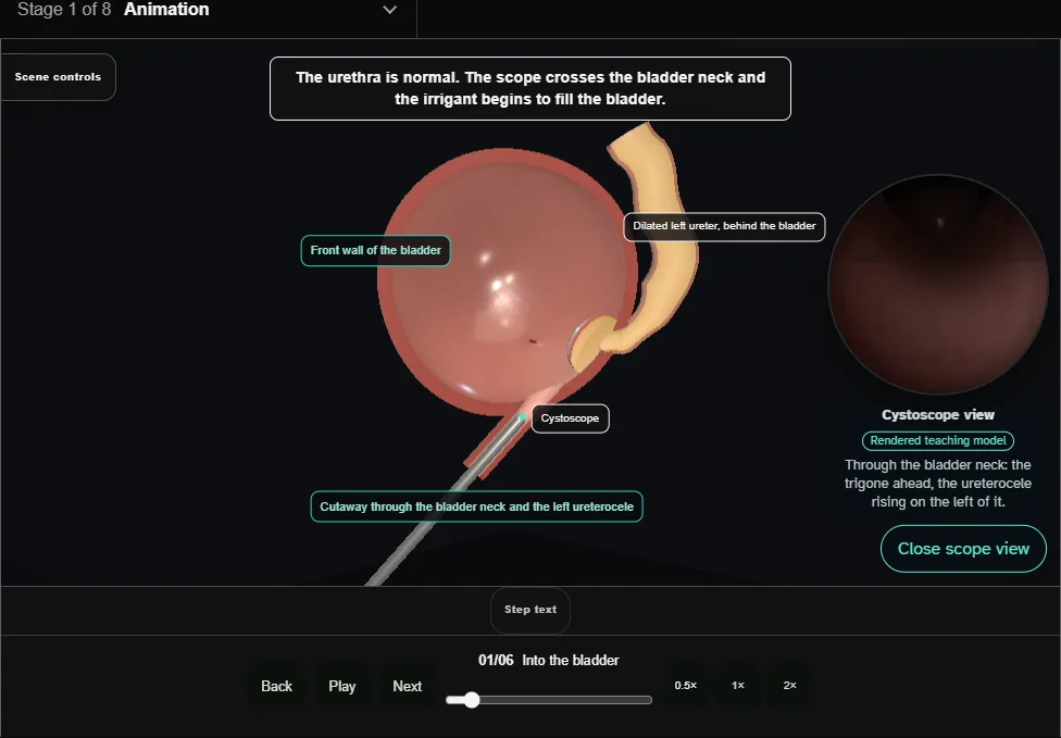
- Supine with the hips abducted; antibiotics are given.
- A rigid paediatric cystoscope with a 30 degree telescope is passed per urethra and advanced into the bladder under direct vision, the urethra inspected on the way.
- 02
Survey
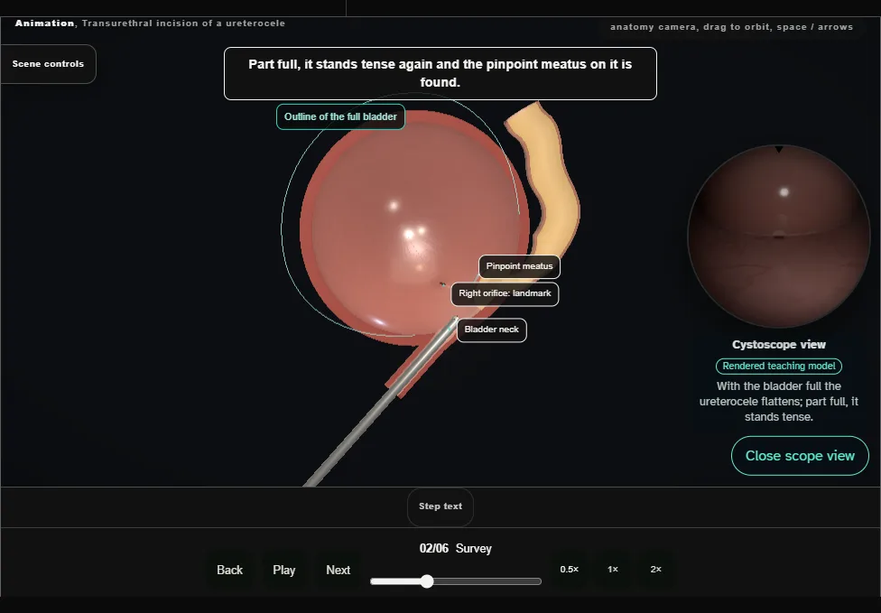
- The bladder is inspected with the 30 and the 70 degree telescopes as it fills: capacity, symmetrical expansion, trabeculation, diverticula.
- The ureterocele is seen on the left of the trigone and its pinpoint meatus is identified.
- The right orifice and the bladder neck are the landmarks.
- 03
Choose the site
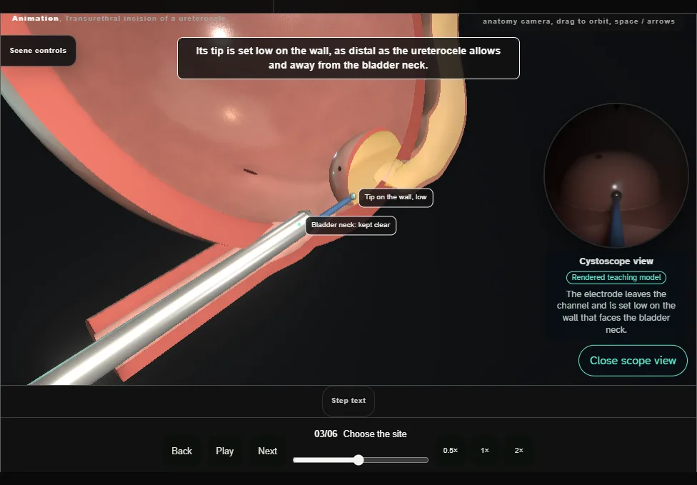
- A fine monopolar electrode (Bugbee) is passed down the working channel and set on the base of the ureterocele, on its distal wall: as low as it allows, and away from the bladder neck.
- 04
Incise
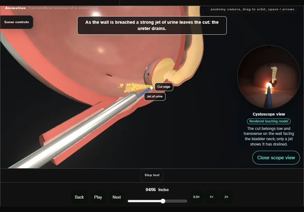
- With cutting current a transverse, full-thickness incision is made in the wall, as distal as possible but proximal to the bladder neck: placed there it helps to reduce later vesicoureteric reflux.
- A strong jet of urine from the cut confirms that the ureter drains.
- 05
Decompress
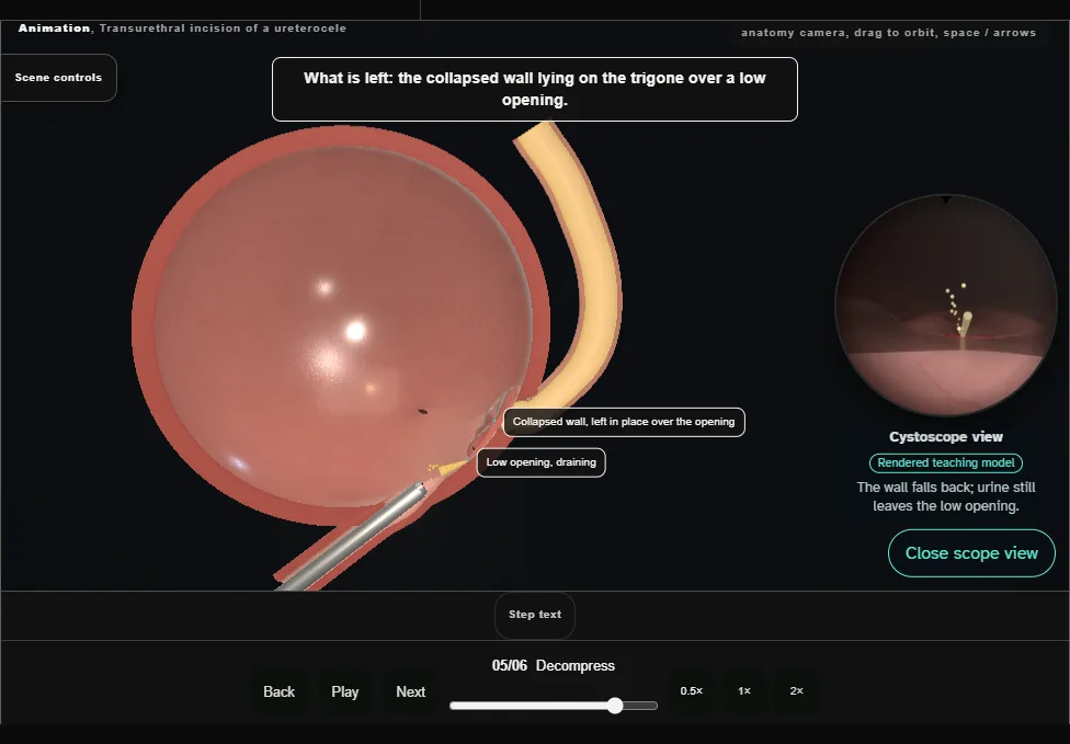
- The ureterocele empties and collapses, and the ureter behind the bladder narrows as it drains.
- A urine specimen is collected and sent for urinalysis and culture.
- The wall of the ureterocele is left in place.
- 06
Check and leave
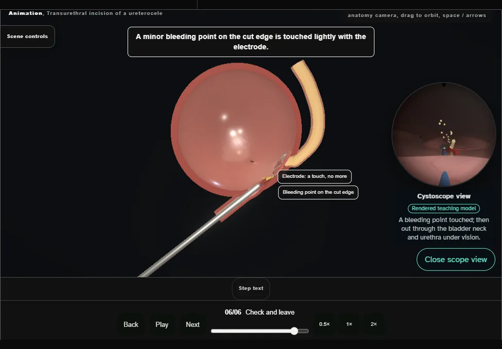
- Minor bleeding points are touched lightly with the electrode.
- The bladder is emptied and the cystoscope removed under direct vision, confirming that the bladder neck and the urethra are uninjured.
Derived from paediatric/UreteroceleScene.tsx at build time: every step title and caption above is the authored
ProcedureDef text, reproduced verbatim, and this page shares its readable-points renderer with the animation
itself. 6 of 6 steps have a captured frame.
Text fingerprint 9b980237e0375d73. A caption edit without a re-capture fails the production
check, so the sheet and the animation cannot drift apart silently.