UrOops3D, danger-zone atlas

Schematic child bladder: every dimension is an authoring estimate.

Operative animation, Ureterocele incision

Transurethral incision of a ureterocele

Indications

  • A ureterocele obstructing its kidney.
  • A ureterocele obstructing the bladder neck.
  • An ectopic ureterocele prolapsing as a vaginal mass.
  • An infected, obstructing ureterocele: the European guideline offers drainage.

Alternatives

  • Surveillance in a child without symptoms or significant obstruction.
  • Nephrostomy drainage in a severely ill child.
  • Laser puncture or cold-knife incision in place of the electrode.
  • Open intravesical repair, which duplex systems and ectopic ureters may need.

Risks

  • Bleeding and infection.
  • Persistent obstruction.
  • Vesicoureteric reflux into the decompressed ureter.
  • Injury to bladder, ureter, rectum or vagina; urinary incontinence.

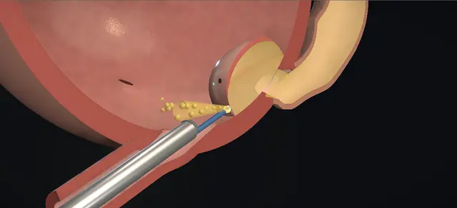
Endoscopic decompression of a ureterocele, the cystic and obstructed end of a ureter that balloons into the bladder. Shown is a left single-system ureterocele lying wholly within the bladder of a young child, with a dilated ureter behind it.

A paediatric cystoscope is passed, the ureterocele and its pinpoint meatus are identified, and a fine electrode makes one transverse cut through the full thickness of its wall, low on the side that faces the bladder neck. A jet of urine confirms drainage.

The lesson is where the cut goes and why: as distal as possible, to limit later reflux, and clear of the bladder neck.

Advanced tier FRCS paediatric urology, ureterocele, upper tract obstruction Interactive 3D

Operative visualisation

Schematic scene preview: Ureterocele incision
Preparing the 3D anatomy…

UrOops3D · UroRef · © Nity G

How to use this module. Play the six steps, or use Back and Next. The cutaway is the teaching view; the round picture is what the cystoscope sees. Watch the electrode set on the wall, the cut, the jet, the wall falling and the ureter narrowing. The triggers show reflux, a cut that did not drain, and bleeding.

Click me next Section 2 of 8 Anatomy The structures this operation works between, and the one it must not cross.

The structures in the scene

Ureterocele incision: how the structures relateA left intravesical ureterocele from the cystoscope, and what lies round it. Ureterocele: the tense dome on the trigone: its own narrow opening: the obstruction Pinpoint meatus on its slope. Ureterocele: the tense dome on the trigone: cut low and transverse, through the full wall Distal wall at the base: the cut. Ureterocele: the tense dome on the trigone: the cut stays proximal to it Bladder neck, just beyond the base (danger). Ureterocele: the tense dome on the trigone: across the trigone; seen, never touched Right ureteric orifice. Ureterocele: the tense dome on the trigone: drains into it; narrows once decompressed Dilated left ureter behind the bladder. Ureterocele: the tense dome on the trigone: reached through it, under vision Urethra of a young child: the scope must fit it (danger).Ureterocele: the tensedome on the trigonePinpoint meatus onits slopeDistal wall at thebase: the cut!Bladder neck, justbeyond the baseRight uretericorificeDilated left ureterbehind the bladder!Urethra of a youngchild: the scopemust fit itits own narrowopening: theobstructioncut low andtransverse, throughthe full wallthe cut staysproximal to itacross the trigone;seen, never toucheddrains into it;narrows oncedecompressedreached through it,under visionTargetDangerLandmarkContext
A left intravesical ureterocele from the cystoscope, and what lies round it. Schematic, not to scale; relationships only.
  • In the cutaway the bladder is closed and part full, and the half toward the patient’s left is removed along the plane that holds the urethra, the scope and the ureterocele.
  • The bladder neck is at the bottom, with the urethra leaving it downward and forward.
  • The trigone rises behind the neck, and the ureterocele stands on the left of it, its base a short way from the neck.
  • The left ureter runs down behind the bladder, crosses the wall and opens into the ureterocele.
  • The right orifice lies on the far wall.
  • In the cystoscope view the ureterocele rises from the floor ahead, and the cut is made across the low part of the wall that faces the lens.
  • ›Ureterocele - a cystic dilatation of the end of the ureter within the bladder wall; here on a single system and wholly inside the bladder.
  • ›Pinpoint meatus - the ureter’s own narrow opening on the ureterocele: the obstruction.
  • ›Base and distal wall - the part of the ureterocele nearest the bladder neck, where it meets the floor of the bladder; the cut is made here.
  • ›Bladder neck - just beyond the base; the cut stays proximal to it.
  • ›Trigone and interureteric bar - the floor the ureterocele stands on; the bar runs from it to the opposite orifice.
  • ›Opposite orifice - on the right of the trigone; a landmark that is not instrumented.
  • ›Dilated ureter - behind the bladder, wide and tortuous above the ureterocele; it drains through the cut.
  • ›Hiatus - where the ureter crosses the bladder wall and opens into the ureterocele.
  • ›Urethra - its size decides which scope and which cutting instrument will pass.
  • ›Intravesical and ectopic forms - an intravesical ureterocele lies wholly in the bladder and is mostly on a single system; an ectopic one reaches the bladder neck or urethra and usually drains the upper moiety of a duplex kidney.
Click me next Section 3 of 8 Steps The operation in order, with the landmark that guards each move.

Operative steps

  1. Into the bladder The child is supine with the hips abducted and secured, the pressure points padded. Intravenous antibiotics are given and, under general anaesthesia, the genitalia are prepared and draped. A rigid paediatric cystoscope with a 30 degree telescope is passed per urethra and advanced into the bladder under direct vision. The urethra is inspected on the way in.
    where it bites: a small urethra takes a small scope, advanced under vision and never forced.
  2. Survey The bladder is inspected with the 30 degree and the 70 degree telescopes as it fills: the medium is clear, the capacity is judged, the wall is seen to expand evenly, and trabeculation and diverticula are looked for. The ureterocele is seen on the left of the trigone, and the pinpoint ureteric meatus on it is identified. The opposite orifice and the bladder neck are noted before anything is cut.
    where it bites: a ureterocele that runs into the bladder neck or the urethra is an ectopic one, and the plan changes.
  3. Choose the site A fine monopolar electrode (Bugbee) is passed through the working channel of the cystoscope and positioned at the base of the ureterocele. The cut is planned on its distal wall: as distal as possible, and still proximal to the bladder neck.
    where it bites: the bladder neck lies just beyond the base; the electrode stays off it.
  4. Incise With cutting current, a full-thickness transverse incision is made distally in the wall of the ureterocele, away from the bladder neck. Placing it there helps to reduce the incidence of later vesicoureteric reflux. A strong jet of urine from the incised ureterocele confirms that the ureter is patent.
    where it bites: a cut that does not cross the wall leaves the obstruction in place; one carried toward the bladder neck injures it.
  5. Decompress The ureterocele decompresses as the urine leaves it, and the dilated ureter behind the bladder drains through the cut. A urine specimen is collected and sent for urinalysis and culture. Nothing is excised: the wall of the ureterocele is left where it lies.
    where it bites: no jet means no drainage, whatever the cut looks like.
  6. Check and leave Minor bleeding areas are gently coagulated with the electrode. The bladder is emptied and the cystoscope is removed under direct vision, confirming that the bladder neck and the urethra are uninjured.
    where it bites: heat spread over a thin wall in a small bladder is how the bladder and the ureter are injured; coagulation is a touch on the point.
Click me next Section 4 of 8 Danger zones Where this operation bites, and why it bites there.
Critical relationship

Danger zones

The spatial relationships the animation is built to make automatic. Before you read each one, ask yourself: at this point in the operation, what is about to go wrong, and what is my first move? The save for each sits in the Complications tab.

  • RiskThe bladder neck - just beyond the base of the ureterocele; the cut stops proximal to it.
  • RiskThe height of the cut - on the distal wall near the floor; an opening made high on the wall, or carried across it, invites reflux.
  • RiskThe thickness of the wall - the cut must cross all of it; a wall that is only scored still obstructs.
  • RiskThe opposite orifice - on the other side of the trigone; identified, and left alone.
  • RiskA small urethra - the instruments are chosen to fit it; nothing is forced through it.
  • RiskThin walls and heat - bladder, ureter, rectum and vagina are all listed as structures that can be injured; current is used in brief touches.
Practise it Take me into the scene Opens the animation at a danger moment and asks you to predict the mechanism and your first move before it reveals.
Click me next Section 5 of 8 Complications The save for each one: the first move, the escalation trigger, the op-note line.

Complications and immediate saves

Trigger each of these live in the Animation tab, then read the save: the first move, the escalation trigger, and the line that goes in the op note.

Vesicoureteric reflux

  • reflux into the decompressed ureter, found on later imaging; a listed complication, and the reason the cut is placed as distal as it can be.
  • A cut made high on the wall, or carried across it, leaves a wide opening with nothing lying over it.
The save
  1. On the table: keep the cut low, near the floor of the bladder, and stop once urine jets from it; do not lengthen it or move it up the wall. first move
  2. Afterwards: ultrasound to see the dilatation settle, and a cystogram where infection or persisting dilatation asks the question.
  3. Antibiotic prophylaxis is continued or stopped on those results, by the unit’s practice.
  4. Reflux that persists with infections goes back to the paediatric urology team for a planned decision about further surgery. escalation trigger
  5. Op note: the site and direction of the cut, how far it was carried, and that a jet was seen. document

Persistent obstruction

  • the ureterocele is still tense and nothing leaves the cut; a listed complication.
  • The usual cause is a cut that scored the wall without crossing it.
The save
  1. Go back to the same site and cut through the full thickness with cutting current until the jet is seen. first move
  2. If the ureterocele has gone flat and cannot be worked on, let some irrigant out: it stands up again as the bladder empties.
  3. After surgery, repeat the ultrasound: dilatation that does not improve means it is not draining.
  4. A child who stays septic or obstructed needs senior review and drainage by another route; the guideline names nephrostomy. escalation trigger
  5. Op note: whether a jet was seen, and how many cuts were made. document

Bleeding from the cut edge

  • bleeding is a listed complication.
  • The operative text expects minor bleeding points, touched with the electrode; in a small bladder even a little blood hides the view.
The save
  1. Irrigate until the point can be seen, then touch it lightly with the electrode. first move
  2. Coagulate the point and leave the wall round it alone: the wall is thin, and the opening is meant to stay small.
  3. A view that will not clear, or bleeding that continues, means stopping and asking for senior help. escalation trigger
  4. Op note: the bleeding point, how it was controlled, and that the view was clear at the end. document
Click me next Section 6 of 8 Troubleshooting When the case turns: the problem, then the next move.

Intra-operative troubleshooting

When the case turns: the problem, then the next move.

  • ActionThe ureterocele has gone flat, or cannot be found - the bladder is too full, and a full bladder presses a ureterocele flat. Let irrigant out and look again at a lower volume. Caione and colleagues, describing laser puncture, kept the bladder at about 30 per cent of capacity so that the ureterocele did not collapse backwards.
  • ActionThe scope will not pass the urethra - it is not forced. A range of instruments is kept ready according to the size of the urethra, and a smaller one is taken.
  • ActionThe cut has been made and there is no jet - it has not crossed the wall. Deepen it at the same site until urine comes; do not lengthen it or move up the wall.
  • ActionThe ureterocele runs into the bladder neck or the urethra - it is ectopic, usually on the upper moiety of a duplex kidney. The European guideline reports more secondary reflux and more reoperation after incision in that group, and open intravesical repair may be required.
  • ActionThe patient is an older child or an adult - a resectoscope loop, a bipolar (Collins) knife or a laser fibre can be used for the incision.
  • ActionA laser is available - the European guideline reports superior results for laser puncture over electrosurgical incision in orthotopic ureteroceles, for secondary reflux and for reoperation.
  • ActionThere is a ureterocele on each side - the identical procedure is done on the other side, and its jet is seen as well.
Click me next Section 7 of 8 FRCS viva Say it out loud, then mark yourself against the model answer.

FRCS viva questions

The examiner stems this scene prepares you for. Try to answer out loud first, then reveal, and learn the discriminator, the detail that turns a pass into a strong pass.

QAn infant has a febrile urinary infection. Ultrasound shows a dilated left ureter ending in a cystic swelling inside the bladder. What is it, and what do you do?reveal answer
  • This is a ureterocele: a cystic dilatation of the end of the ureter, here obstructing a single system.
  • I would treat the infection and resuscitate the child first, then complete the assessment with ultrasound of both kidneys and the bladder, a cystogram for reflux and for the bladder neck, and a functional study of the kidney once the child is well.
  • The European paediatric guideline recommends immediate drainage where there is urosepsis, pyonephrosis or obstruction at the bladder neck, and offers endoscopic decompression or nephrostomy for an infected obstructing ureterocele.
  • In a child fit for a brief anaesthetic I would offer transurethral incision.
  • I would pass a paediatric cystoscope under vision and inspect the urethra and the bladder.
  • I would identify the ureterocele, its pinpoint meatus, the opposite orifice and the bladder neck.
  • With a fine electrode on cutting current I would then make one transverse, full-thickness cut at its base, on the distal wall.
  • A jet of urine tells me that it drains.
  • I would send urine for culture, touch any bleeding point, empty the bladder and withdraw under vision.
  • Afterwards I would follow the dilatation on ultrasound and look for reflux, and I would warn the family that a second procedure is sometimes needed.
Strong pass

Say both

  • Draining an infected obstructed system before anything else
  • Naming the two routes the guideline offers: endoscopic decompression or nephrostomy
Examiner note
Draining an infected obstructed system before anything else, and naming the two routes the guideline offers: endoscopic decompression or nephrostomy.
QWhere exactly do you make the incision, and why there?reveal answer
  • I make it at the base of the ureterocele on its distal wall: transverse, through its whole thickness, as distal as possible while staying proximal to the bladder neck.
  • That is how the operative text I follow puts it, and its stated reason is to reduce the incidence of later vesicoureteric reflux.
  • The explanation usually given goes back to Rich and colleagues, who described a low transverse incision for single-system intravesical ureteroceles in 1990 and wrote that the flap-valve antireflux mechanism can be preserved in most children.
  • Later authors, Caione and colleagues among them, describe the collapsed wall of the ureterocele acting as that flap.
  • Rich reported seven ureteroceles in five children: the dilatation decreased in all of them and reflux appeared in one kidney, and the authors recommended a cut of 2 to 3 millimetres.
  • That is a small series and I would quote it as one.
  • In practice I do not unroof the ureterocele, and I stay clear of the bladder neck, because injury there is the price of chasing the lowest possible cut.
Strong pass

Say all three

  • Transverse and full thickness
  • Low on the distal wall
  • Saying whose observation the flap-valve explanation rests on and how small that series was
Examiner note
Transverse and full thickness, low on the distal wall, and saying whose observation the flap-valve explanation rests on and how small that series was.
QWhich ureteroceles do less well after endoscopic incision?reveal answer
  • The ectopic ones and those on a duplex system.
  • I would start from the European guideline’s classification.
  • The intravesical ureterocele lies wholly within the bladder, is mostly on a single system and is diagnosed more often in older children and adults.
  • The ectopic one reaches the bladder neck or the urethra, is the commoner form and usually drains the upper moiety of a duplex kidney.
  • The guideline cites two systematic reviews in which secondary reflux and reoperation after a primary incision were more frequent in ectopic ureteroceles and in duplex systems.
  • A 2024 meta-analysis by Salehi-Pourmehr and colleagues gives the size of it: reoperation after incision was more likely in ectopic than in intravesical ureteroceles, with a relative risk of 2.42 across 16 studies, and in duplex than in single systems, with a relative risk of 2.50 across 9 studies.
  • So I would tell the family of a child with an ectopic duplex ureterocele that incision is often a first step.
  • Armenakas and colleagues make the same point from the other side: duplicated systems and ectopic ureters may need open intravesical repair.
Strong pass

Say both

  • Separating the intravesical single-system ureterocele from the ectopic duplex one
  • Knowing that reflux and reoperation follow the second group
Examiner note
Separating the intravesical single-system ureterocele from the ectopic duplex one, and knowing that reflux and reoperation follow the second group.
QYou have made your cut and there is no jet. What now?reveal answer
  • No jet means I have not decompressed it, whatever the cut looks like.
  • The dictation I follow asks for a full-thickness incision and takes a strong jet as the proof that the ureter is patent, so the likeliest reason is a cut that has scored the wall without crossing it.
  • I would check the view first.
  • A very full bladder presses a ureterocele flat, so I would let some irrigant out until it stands up again.
  • Caione and colleagues, describing laser puncture, kept the bladder at about 30 per cent of capacity for that reason.
  • Then I would go back to the same site and deepen the cut with cutting current until urine comes.
  • I would not move higher on the wall or lengthen the cut to find the lumen, because that is how the opening is made too large.
  • If I still could not drain it, or the child were septic and unstable, I would stop and get senior help; the guideline’s other route to drainage is a nephrostomy.
  • I would record whether a jet was seen.
Strong pass

Say all three

  • Treating the jet as the end point
  • Checking how full the bladder is
  • Deepening the cut at the same site instead of cutting higher or longer
Examiner note
Treating the jet as the end point, checking how full the bladder is, and deepening the cut at the same site instead of cutting higher or longer.
QWhat could you use instead of the electrode, and is anything better?reveal answer
  • The operative text lists what to have ready according to the size of the urethra: a paediatric cystoscope, a Bugbee electrode, a resectoscope with a loop, and a laser.
  • In older children and adults it allows a resectoscope loop, a bipolar Collins knife or a laser fibre for the incision.
  • The European guideline names cold-knife incision, electrosurgical incision and laser puncture, and says that in orthotopic ureteroceles laser puncture has shown superior results to electrosurgical incision for secondary reflux and for reoperation.
  • One comparison of this kind is retrospective.
  • Caione and colleagues compared 64 children treated by multiple holmium laser punctures at the base with 26 earlier children treated by diathermy incision, most of them with ectopic duplex ureteroceles.
  • Reflux was found in 8 of the 27 laser patients who had a cystogram, against 16 of the 26 controls, and further surgery was needed in 12 of the 64 against 13 of the 26.
  • The groups were historical and unrandomised, so I would call the result encouraging and unproven.
  • Whatever the energy, I would keep to the same rules: a small opening low on the wall that faces the bladder neck, and a jet seen.
Strong pass

Say both

  • Knowing the named alternatives
  • That the comparison favouring laser puncture is retrospective with historical controls
Examiner note
Knowing the named alternatives, and that the comparison favouring laser puncture is retrospective with historical controls.
Click me next Section 8 of 8 Evidence The guidance each of these answers is built on.

Evidence and guidance

Operative Dictations in Urologic Surgery
Armenakas NA, Fracchia JA, Golan R. Transurethral incision of ureterocele. Chapter 120. Wiley, 2019
The indications; the two essential steps (see the ureterocele with a range of instruments ready according to the size of the urethra; incise it transversely, as distal as possible but proximal to the bladder neck, to help reduce later reflux); the template dictation followed step by step here; the variations; and the complication list: bleeding, infection, injury to bladder, ureter, rectum or vagina, persistent obstruction, vesicoureteric reflux, urinary incontinence. The chapter’s scope and electrode sizes are kept off the page. It gives no length for the cut, does not say how full the bladder is kept and leaves no catheter. It lists complications without their management: the first, escalate and document lines and the danger lines are general practice, with the guideline cited where it speaks. The scene is schematic: the cut is drawn much shorter than the base of the ureterocele, and the bladder is shown full and then part full during the survey.
Open guideline
EAU Guidelines
Paediatric Urology, 2026: chapter 19, Ureterocele and ectopic ureter, sections 19.1.2, 19.1.4 and 19.3, read 13 October 2026
The intravesical (orthotopic) ureterocele lies wholly in the bladder, is mostly on a single system and is diagnosed more often in older children or adults; the ectopic form is the commoner. Immediate drainage is recommended for urosepsis, pyonephrosis or obstruction at the bladder neck, and nephrostomy can be a first-line treatment in a severely sick child. Decompression is by cold-knife incision, electrosurgical incision or laser puncture; in orthotopic ureteroceles laser puncture shows superior results to electrosurgical incision for secondary reflux and reoperation; two systematic reviews suggest more secondary reflux and reoperation after primary incision in ectopic ureteroceles and in duplex systems. Offer drainage, endoscopic or by nephrostomy, for infected obstructing ureteroceles (strong). The chapter does not say where on the ureterocele to cut, how full to keep the bladder, or whether to leave a catheter.
Open guideline
The Journal of Urology
Rich MA, Keating MA, Snyder HM, Duckett JW. Low transurethral incision of single system intravesical ureteroceles in children. J Urol 1990;144:120-121
Abstract read 13 October 2026. Seven symptomatic single-system ureteroceles in five children treated by low transverse endoscopic incision: hydroureteronephrosis decreased in all, and reflux after incision was seen in one kidney. The authors recommend a short (2 to 3 mm) low transverse incision and say that the flap-valve antireflux mechanism can be preserved in most children. A small series, quoted in the viva as theirs. No length is shown on screen.
Open guideline
Frontiers in Pediatrics
Caione P, Gerocarni Nappo S, Collura G, et al. Minimally invasive laser treatment of ureterocele. Front Pediatr 2019;7:106
Full text read 13 October 2026. A retrospective single-centre comparison: 64 children treated by multiple holmium laser punctures at the base of the ureterocele (49 ectopic, 53 on duplex systems) against 26 earlier children treated by diathermy incision. Reflux was found in 8 of the 27 laser patients who had a cystogram (29.7%) against 16 of the 26 controls (61.5%); further surgery was needed in 12 (18%) against 13 (50%). The bladder was kept partly filled at low pressure, about 30% of capacity, so that the ureterocele did not collapse backwards: the source for the part-full bladder in the scene. The authors give the collapsed wall acting as a flap valve as the reason for a low opening. Historical controls, mostly ectopic duplex cases.
Open guideline
Journal of Pediatric Urology
Salehi-Pourmehr H, Lotfi B, Mohammad-Rahimi M, Tahmasbi F. Surgical patterns in the endoscopic management of pediatric ureterocele: a systematic review and meta-analysis. J Pediatr Urol 2024;20:731-740
Abstract read 13 October 2026. Eighty-three studies and 3,022 patients in the review. Reoperation after incision was more likely in ectopic than in intravesical ureteroceles (relative risk 2.42, 95% CI 1.89 to 3.11; 16 studies) and in duplex than in single systems (relative risk 2.50, 95% CI 1.60 to 3.91; 9 studies).
Open guideline
Journal of Pediatric Urology
Gnech M, Bebi C, Rotondi G, et al. Endoscopic incision of ureterocele. J Pediatr Urol 2024;20:1221-1222
Abstract read 13 October 2026; a video article. In an intravesical ureterocele the punctures are made just above the junction between the ureterocele and the bladder floor; an overaggressive incision or puncture could increase the chance of secondary reflux; prophylaxis is stopped once decompression is shown and reflux excluded. Behind the wording on a small opening near the floor and on prophylaxis.
Open guideline

Curriculum. Supports learning relevant to the ISCP Urology Curriculum syllabus topic Paediatric urology. Not a workplace-based assessment and not evidence of competence.

Two minutes

Close the lap: recall it before you leave.

Say it aloud, then reveal. Reading is not remembering.

  1. 01 The relationship Say the critical spatial relationship out loud, then reveal. reveal
    Memory frame

    In the cutaway the bladder is closed and part full, and the half toward the patient’s left is removed along the plane that holds the urethra, the scope and the ureterocele.

    Why?
    The bladder neck is at the bottom, with the urethra leaving it downward and forward. The trigone rises behind the neck, and the ureterocele stands on the left of it, its base a short way from the neck. The left ureter runs down behind the bladder, crosses the wall and opens into the ureterocele. The right orifice lies on the far wall. In the cystoscope view the ureterocele rises from the floor ahead, and the cut is made across the low part of the wall that faces the lens.
  2. 02 The first move Vesicoureteric reflux. Your first move? reveal
    First move

    On the table: keep the cut low, near the floor of the bladder, and stop once urine jets from it; do not lengthen it or move it up the wall.

  3. 03 The escalation trigger Vesicoureteric reflux. What makes you call for help? reveal
    Escalate when

    Reflux that persists with infections goes back to the paediatric urology team for a planned decision about further surgery.

  4. 04 The discriminator What turns a pass into a strong pass? reveal
    Strong pass

    Say both

    • Draining an infected obstructed system before anything else
    • Naming the two routes the guideline offers: endoscopic decompression or nephrostomy
    Examiner note
    Draining an infected obstructed system before anything else, and naming the two routes the guideline offers: endoscopic decompression or nephrostomy.
  5. 05 The pitfall Where does this operation bite first? reveal
    Pitfall

    The bladder neck

    Why?
    just beyond the base of the ureterocele; the cut stops proximal to it.

Revision practice with self-marked answers, not an assessment of clinical competence. Kept in this browser only; it does not sync between devices.