
UrOops3D, danger-zone atlas
Operative animation, Paediatric pyeloplasty

Paediatric pyeloplasty for pelvi-ureteric junction obstruction

The dilated renal pelvis found on the twenty-week scan, followed with ultrasound and a MAG3 renogram, and operated on only when the function falls, the pelvis grows or the child becomes symptomatic.

Then a dismembered pyeloplasty: the narrow junction excised, the ureter spatulated and joined to the most dependent part of a tailored pelvis in front of any crossing vessel, over a stent, with 6-0 or 7-0 suture and loupes.

This page is the selection, the timing, the approach and the anastomosis that is expected to work in nineteen out of twenty.

Advanced tier FRCS paediatric urology, upper tract obstruction Interactive 3D

Operative visualisation

Schematic scene preview: Paediatric pyeloplasty
Preparing the 3D anatomy…

UrOops3D · UroRef · © Nity G

Click me next Section 2 of 8 Anatomy The structures this operation works between, and the one it must not cross.

The structures in the scene

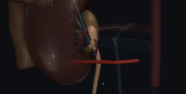
Paediatric pyeloplasty: how the structures relateThe retroperitoneum, lower pole above and the dilated pelvis below. Pelvi-ureteric junction: its most dependent part becomes the new junction Renal pelvis, dilated and thin-walled. Pelvi-ureteric junction: handled by adventitia only; stripped, it strictures Proximal ureter and its adventitia (danger). Pelvi-ureteric junction: cross in front; never divided, the join goes anterior Crossing lower pole vessels (danger). Pelvi-ureteric junction: opened over the lower pole to find the pelvis Gerota's fascia and the lower pole. Pelvi-ureteric junction: swept medially; bowel a diathermy stroke away Colon and peritoneum (danger). Pelvi-ureteric junction: runs through the join toward the bladder Stent.Pelvi-ureteric junctionRenal pelvis,dilated andthin-walled!Proximal ureter andits adventitia!Crossing lower polevesselsGerota's fascia andthe lower pole!Colon and peritoneumStentits most dependentpart becomes thenew junctionhandled byadventitia only;stripped, it…cross in front;never divided, thejoin goes anterioropened over thelower pole to findthe pelvisswept medially;bowel a diathermystroke awayruns through thejoin toward thebladderTargetDangerLandmarkContext
The retroperitoneum, lower pole above and the dilated pelvis below. Schematic, not to scale; relationships only.
  • The scene is the retroperitoneum with the lower pole of the kidney above and the dilated pelvis below and medial to it, funnelling down to a narrow junction and a normal-calibre ureter that runs down over the psoas.
  • A lower pole artery and vein cross in front of the junction from the aorta and the cava toward the lower pole.
  • When the junction is excised the ureter is brought forward in front of the vessels and its spatulated end is joined to the lowest part of the pelvis, with the stent running from the pelvis through the join and down toward the bladder.
  • ›The renal pelvis - dilated and thin-walled, held by a stay suture; cut obliquely so the most dependent part becomes the new junction, and reduced if it is redundant.
  • ›The pelvi-ureteric junction - the narrow aperistaltic segment, high insertion or kink that is excised and sent for histology.
  • ›The proximal ureter - spatulated laterally for one to one and a half centimetres and handled only by its adventitia, where its blood supply runs.
  • ›The crossing lower pole vessels - anterior to the junction in about a third of older children; an end artery, never divided, the anastomosis transposed in front of it.
  • ›Gerota's fascia and the lower pole - opened over the lower pole to find the pelvis; the kidney itself is left in its fat.
  • ›The colon and the peritoneum - swept medially in the open approach, reflected or windowed laparoscopically; bowel a diathermy stroke from the pelvis on the left.
  • ›The stent - JJ down the ureter into the bladder or a transanastomotic tube out through the skin; a written date for its removal.
Click me next Section 3 of 8 Steps The operation in order, with the landmark that guards each move.

Operative steps

  1. Who: antenatal hydronephrosis and the MAG3 Antenatal hydronephrosis is found on about one to two per cent of pregnancy scans and most of it resolves. Pelvi-ureteric junction obstruction is the commonest cause of the significant cases. After birth the ultrasound is repeated from about day three to seven, not in the first day or two when the baby is oliguric and the pelvis reads falsely small, and again at four to six weeks. The anteroposterior diameter of the pelvis is the working measure: under fifteen millimetres is usually watched, over about twenty to thirty is likely to need surgery. A MAG3 renogram with furosemide at four to six weeks gives the split function and the drainage curve. Reflux is excluded by a micturating cystogram where the ultrasound or a urine infection suggests it.
  2. When to operate The indications are a split function below about forty per cent, a fall in function of more than about five to ten per cent on serial renograms, a pelvis that is growing on serial ultrasound or is very large, and symptoms: loin pain in the older child, urinary infection, stones or haematuria. A dilated pelvis with good function and a stable diameter is watched with ultrasound at intervals for years, because many improve. An infected obstructed kidney in a baby is drained with a nephrostomy first and repaired later. When surgery is indicated in an infant it is usually done at a few months of age rather than in the neonatal period, unless the function is falling fast.
    where it bites: a renogram in a young infant reads obstruction easily because the immature kidney drains slowly; the decision rests on function and on the trend, not on one curve.
  3. Open, laparoscopic or robotic In the infant the open dismembered pyeloplasty remains the standard: a small muscle-splitting anterior subcostal incision or a dorsal lumbotomy, loupes, fine suture and a stay of a day or two, with success rates of about ninety-five per cent that the minimally invasive approaches are measured against. In the older child and adolescent the laparoscopic transperitoneal or retroperitoneal repair, and the robot where it is available, give the same result with smaller scars and a faster return to school; the robot makes the intracorporeal suturing easier and is used from about a year of age in units that have it. Endoscopic endopyelotomy has almost no place in children. The choice is the unit's and the surgeon's, made honestly against the size of the child.
  4. Exposure and the pelvis Open: the child lies with the side raised on a roll, and through the incision the muscles are split, the peritoneum swept medially and Gerota's fascia opened over the lower pole to find the dilated pelvis, which is followed down to the junction and the proximal ureter. Laparoscopic: the colon is reflected medially, or a window is made in the mesocolon on the left, and the pelvis found behind it. In either case the pelvis is held with a stay suture rather than forceps, the ureter is handled by its adventitia only, and the junction is inspected for its cause: an intrinsic aperistaltic segment, a high insertion, a kink, or a vessel crossing in front of it. The pelvis is not skeletonised beyond what the repair needs.
  5. The crossing vessel A lower pole artery and vein crossing anterior to the junction is found in about a third of older children and adolescents and less often in infants. It is an end artery to the lower pole and is never divided: the repair is transposed in front of it. The vessels are freed gently from the pelvis and the ureter with the ureter kept in its adventitia, and once the junction is dismembered the ureter is brought anterior to the vessels so that the new anastomosis lies in front of them with the vessels running behind, free of tension and kink. If the vessels are the only cause and the junction is otherwise healthy, some surgeons transpose without excising, but the standard is still to dismember and re-anastomose.
    where it bites: the crossing lower pole vessel is an end artery; divided, the lower pole infarcts and bleeds, and the child loses part of the kidney the operation was meant to save.
  6. Dismembering the junction Stay sutures are placed on the pelvis above the junction and on the ureter below it, marking the lateral wall of the ureter so it cannot rotate. The junction is excised with the narrow segment, cutting the pelvis obliquely so that the most dependent part becomes the new opening, and a redundant pelvis is reduced by excising the surplus wall and closing the upper part of the pelvis with a running 6-0 PDS. The excised segment goes for histology. The ureter is inspected to confirm that the cut end is healthy, wide and bleeding, and a catheter or a fine feeding tube is passed down it with saline to confirm free drainage to the bladder, because a second narrowing below the first is missed if this is not done.
  7. Spatulation and the anastomosis The ureter is spatulated on its lateral wall for one to one and a half centimetres, so that the anastomosis is wide and oblique and the apex of the spatulation will lie at the most dependent point of the pelvis. The join is made with 6-0 or 7-0 PDS, interrupted or two running sutures from the apex, under loupes or the laparoscope, with the knots outside and the mucosa apposed and the ureter handled only by its adventitia. The posterior wall is completed first, the stent placed, then the anterior wall and the rest of the pelvis closed. The anastomosis must be dependent, tension free, watertight and in front of any crossing vessel; a join that satisfies three of the four fails.
    where it bites: the ureteric blood supply runs in the adventitia; a ureter grasped with forceps, stripped or twisted at the anastomosis strictures.
  8. The stent or the nephrostomy Three ways to drain the repair, and the unit chooses one. An internal JJ stent, 3 to 4.7 French in an infant and 4.7 to 6 in an older child, passed antegrade over a wire down the ureter into the bladder before the anterior wall is closed, its lower coil confirmed by the return of blue-dyed saline from a bladder filled with methylene blue, and removed cystoscopically under a second anaesthetic at four to six weeks. An externalised transanastomotic stent or nephrostomy, brought out through the pelvis and the skin, clamped after a few days, removed on the ward at about a week without a second anaesthetic. Or no stent, in a healthy well-sutured repair. A drain in the retroperitoneum is optional and removed when dry.
  9. After Most children go home within one or two days after an open repair and the same day or the next after a laparoscopic one, on simple analgesia, with prophylactic antibiotics while a stent is in and the parents told what a stent does: haematuria, frequency and loin twinges on voiding. The stent comes out at four to six weeks. An ultrasound at about three months and a MAG3 at six to twelve months show a smaller pelvis and improved drainage; the function rarely rises much but stops falling, which is the aim. Success is about ninety-five per cent. Recurrent obstruction, in about five per cent, presents as a pelvis that grows again or a child in pain and is treated by redo pyeloplasty.
Click me next Section 4 of 8 Danger zones Where this operation bites, and why it bites there.
Critical relationship

Danger zones

The spatial relationships the animation is built to make automatic. Before you read each one, ask yourself: at this point in the operation, what is about to go wrong, and what is my first move? The save for each sits in the Complications tab.

  • RiskThe crossing lower pole vessels - an end artery to the lower pole; divided, the pole infarcts and bleeds, and the repair behind the vessel kinks.
  • RiskThe ureteric adventitia - the whole blood supply of the proximal ureter; grasped, stripped or twisted it strictures at the join.
  • RiskThe colon and the spleen on the left - across the pelvis in the transperitoneal approach; the mesocolic window is made away from its vessels.
  • RiskThe pleura - above the twelfth rib in a high flank incision in a small child; a pneumothorax found on the recovery film.
Practise it Take me into the scene Opens the animation at a danger moment and asks you to predict the mechanism and your first move before it reveals.
Click me next Section 5 of 8 Complications The save for each one: the first move, the escalation trigger, the op-note line.

Complications and immediate saves

Trigger each of these live in the Animation tab, then read the save: the first move, the escalation trigger, and the line that goes in the op note.

Urinary leak and urinoma

  • urine from the drain or a collection around the kidney in the first week: an anastomosis that was not watertight, a stent that has blocked or moved, or a pelvis closure that opened.
The save
  1. Check the stent or the tube is draining and the bladder is empty; a catheter in the bladder reduces reflux up a JJ stent and lets the join rest. first move
  2. Ultrasound for a collection and the position of the stent; drain fluid creatinine if there is a drain.
  3. A small leak with good drainage settles; the drain stays until dry and antibiotics cover the urinoma.
  4. A large or infected urinoma, or a leak with no stent in place: percutaneous drainage of the urinoma and a nephrostomy or a stent under anaesthetic. escalation trigger
  5. Op note: the suture, whether the join was watertight on the saline test, the drainage used and the stent removal date. document

Recurrent or persistent obstruction

  • a pelvis that grows again on the three-month ultrasound or a child in pain months to years later: scar at the join, a kink behind a vessel, an ischaemic ureter, or a second narrowing that was never seen.
The save
  1. Ultrasound at three months and MAG3 at six to twelve months for every child, with the pre-operative figures alongside. first move
  2. A pelvis that has not improved: repeat MAG3 for the drainage curve and the function, and exclude a retained stent or a stone.
  3. Symptoms with obstruction on the renogram: nephrostomy and an antegrade study to define the level and length.
  4. A confirmed recurrence: redo pyeloplasty, open or robotic, with ureterocalycostomy where the pelvis is scarred and intrarenal; endopyelotomy only in the older child with a short stricture. escalation trigger
  5. Note: the imaging dates, the function on each renogram and the trigger agreed for intervention. document

Crossing vessel injury and lower pole bleeding

  • the lower pole artery or vein torn while it is freed from the pelvis, or divided in error: bleeding in a small field, and an infarcted lower pole if the artery is lost.
The save
  1. Pressure with a small swab and better exposure; the vessel is small and the bleeding stops with patience, not with a blind clip. first move
  2. A venous tear is repaired with a fine suture or the vein ligated; the artery is repaired if at all possible with 7-0 Prolene.
  3. If the artery is lost, the lower pole is watched for infarction on the post-operative Doppler and the parents told.
  4. Bleeding that cannot be controlled laparoscopically: convert to open; a lower pole that infarcts and becomes infected or hypertensive may need a partial nephrectomy. escalation trigger
  5. Op note: the vessel, the repair or ligation, the transposition and the Doppler plan. document

Urinary infection and pyelonephritis

  • a febrile child with a stent in a system that was obstructed: stasis, the foreign body and a residual leak, in a kidney that cannot afford another scar.
The save
  1. Urine culture and blood cultures, and antibiotics started at once by the local paediatric policy, intravenous if the child is unwell. first move
  2. Ultrasound for obstruction above a blocked stent and for a collection; the stent may need changing or removing.
  3. Prophylactic antibiotics while the stent is in and until the first ultrasound, as the unit prescribes.
  4. An infected obstructed kidney above a failed repair: nephrostomy now, and the renogram after recovery to see what the infection has cost. escalation trigger
  5. Note: the organism, the antibiotics, the stent status and the imaging arranged. document

Stent migration, blockage and the retained device

  • a JJ stent that has slipped up the ureter or down into the bladder, a blocked externalised tube, or the stent that nobody removed because the date was never written.
The save
  1. Confirm the stent position on the first ultrasound or a plain film, and give the parents the removal date in writing. first move
  2. A migrated stent with a draining kidney is retrieved cystoscopically at the planned time; a stent above the join needs ureteroscopy or an antegrade approach.
  3. An externalised tube that stops draining is flushed gently with a small volume of saline, and removed if the repair is at a week.
  4. A forgotten stent found encrusted at review: a stone service, with the stent registered and the parents told what happened. escalation trigger
  5. Note: the stent type and size, the date placed, the date for removal and who is responsible for it, on a register. document
Click me next Section 6 of 8 Troubleshooting When the case turns: the problem, then the next move.

Intra-operative troubleshooting

When the case turns: the problem, then the next move.

  • ActionThe pelvis cannot be found behind the colon - follow the gonadal vein or the ureter up from below on the psoas; on the left a mesocolic window is quicker than reflecting the whole colon.
  • ActionA vessel crosses the junction - free it gently, dismember, and bring the ureter anterior to it; never divide it, and check the anastomosis is not kinked over it at the end.
  • ActionThe ureter will not reach the dependent pelvis - mobilise the ureter down its adventitia without stripping, reduce the pelvis less, or drop the kidney a little in its fat; a tensioned join strictures.
  • ActionSaline will not pass down the ureter - a second narrowing or a kink below; pass a fine feeding tube under fluoroscopy and revise the plan before the anastomosis, not after.
  • ActionThe blue does not return from the bladder - the stent is not in the bladder; check with fluoroscopy or reposition it over the wire before the anterior wall is closed.
  • ActionThe pelvis is intrarenal and there is nothing to sew to - a ureterocalycostomy to the lower pole calyx, or a flap pyeloplasty; senior help before the pelvis is cut.
Click me next Section 7 of 8 FRCS viva Say it out loud, then mark yourself against the model answer.

FRCS viva questions

The examiner stems this scene prepares you for. Try to answer out loud first, then reveal, and learn the discriminator, the detail that turns a pass into a strong pass.

QA baby has antenatal hydronephrosis. How would you investigate it after birth, and when would you operate?reveal answer
  • I would arrange an ultrasound between about day three and day seven, avoiding the first two days when the neonate is oliguric and the dilatation is underestimated, and repeat it at four to six weeks, recording the anteroposterior diameter of the pelvis, the calyces, the parenchyma, the ureter and the bladder.
  • A dilated ureter or an abnormal bladder would raise reflux or posterior urethral valves and lead to a micturating cystogram, with antibiotic prophylaxis in the interim.
  • For isolated pelvic dilatation I would arrange a MAG3 renogram with furosemide at four to six weeks for the split function and the drainage curve, interpreting drainage cautiously in the young infant whose kidney empties slowly.
  • I would operate for a split function below about forty per cent, a fall of more than about five to ten per cent on serial scans, a pelvis that is enlarging or exceeds about thirty millimetres, and for symptoms, infection or stones.
  • I would watch a stable pelvis with good function with ultrasound at intervals over years, because much of it resolves.
  • An infected obstructed kidney I would drain with a nephrostomy first.
  • The parents would understand that the operation protects the function that remains rather than restoring what has been lost.
Strong pass

Say both

  • The ultrasound timed after day three with the reason
  • The decision built on function and trend rather than on a single drainage curve
Examiner note
The ultrasound timed after day three with the reason, and the decision built on function and trend rather than on a single drainage curve.
QDescribe an Anderson-Hynes dismembered pyeloplasty in a child.reveal answer
  • Through a small anterior subcostal muscle-splitting incision in an infant, or laparoscopically or robotically in the older child, I would open Gerota's fascia over the lower pole, identify the dilated pelvis and follow it to the junction and the proximal ureter, handling the pelvis with a stay suture and the ureter by its adventitia.
  • I would look for a crossing lower pole vessel and, if present, free it and plan to transpose the repair anterior to it.
  • I would place stay sutures marking the lateral ureteric wall, excise the junction and the narrow segment obliquely so that the most dependent part of the pelvis forms the new opening, reduce a redundant pelvis and close its upper part, and pass a fine tube down the ureter to confirm free drainage to the bladder.
  • I would spatulate the ureter laterally for one to one and a half centimetres and anastomose it to the dependent pelvis with 6-0 or 7-0 PDS under loupes, posterior wall first, then place an antegrade JJ stent confirmed in the bladder by the return of methylene blue, or a transanastomotic tube, and complete the anterior wall.
  • The join would be dependent, tension free, watertight and anterior to any vessel.
  • I would send the segment for histology and plan the stent removal and the three-month ultrasound.
Strong pass

Say both

  • The distal ureter tested before the anastomosis
  • The four properties of the join named together with the transposition in front of the vessel
Examiner note
The distal ureter tested before the anastomosis, and the four properties of the join named together with the transposition in front of the vessel.
QWhat is the significance of a crossing vessel at the pelvi-ureteric junction?reveal answer
  • A lower pole artery and vein crossing anterior to the junction is found in about a third of older children and adolescents with pelvi-ureteric junction obstruction and less often in infants, where an intrinsic aperistaltic segment is the usual cause.
  • The vessel can be the cause of the obstruction, compressing or kinking the junction over it as the pelvis fills, or an associated finding on top of an intrinsic lesion, and the distinction is often made only at operation.
  • Its importance is twofold.
  • First, it is an end artery to the lower pole with no collateral supply, so it must never be divided: a divided lower pole artery infarcts the segment it supplies, with loss of the very parenchyma the operation is protecting.
  • Second, the repair must be placed in front of it: after dismembering the junction I would bring the ureter anterior to the vessels and make the anastomosis there, so that the new junction cannot be compressed as the pelvis fills, and I would check at the end that the join lies without kink over the vessels.
  • Because a crossing vessel is not seen well on ultrasound, I would consider it in the older child with intermittent loin pain, and in the laparoscopic approach it is looked for before the pelvis is opened.
Strong pass

Say both

  • The end artery status stated as the reason it is never divided
  • The anastomosis transposed anterior with the reason
Examiner note
The end artery status stated as the reason it is never divided, and the anastomosis transposed anterior with the reason.
QWhat are the complications of paediatric pyeloplasty, and how are the results followed up?reveal answer
  • Early: urinary leak and urinoma from an anastomosis that was not watertight or a stent that blocked, managed by ensuring drainage, draining the collection if it is large, and a nephrostomy or stent if there is none;
  • urinary infection with a stent in place, treated on culture with the kidney checked for obstruction;
  • bleeding from a crossing vessel or the pelvis;
  • and the approach-specific complications, bowel or spleen injury laparoscopically and pneumothorax after a high flank incision.
  • Device: stent migration, blockage and the retained stent, prevented by a written removal date on a register.
  • Late: recurrent or persistent obstruction in about five per cent from scar, kink, ischaemia or a second narrowing, presenting as a growing pelvis or pain, and treated by redo pyeloplasty or ureterocalycostomy.
  • I would present the success rate as about ninety-five per cent.
  • Follow-up is by imaging: ultrasound at about three months for the pelvic diameter, and a MAG3 at six to twelve months for drainage and function, compared against the pre-operative scans.
  • The pelvis is expected to shrink and drainage to improve; the function usually stabilises rather than rises.
  • A stable, improved kidney is discharged with the parents told the symptoms of recurrence, and a kidney that has not improved is investigated for the cause before it is reoperated.
Strong pass

Say both

  • The retained stent named as a complication with the register that prevents it
  • Function expected to stabilise rather than recover as the measure of success
Examiner note
The retained stent named as a complication with the register that prevents it, and function expected to stabilise rather than recover as the measure of success.
Click me next Section 8 of 8 Evidence The guidance each of these answers is built on.

Evidence and guidance

Two minutes

Close the lap: recall it before you leave.

Say it aloud, then reveal. Reading is not remembering.

  1. 01 The relationship Say the critical spatial relationship out loud, then reveal. reveal
    Memory frame

    The scene is the retroperitoneum with the lower pole of the kidney above and the dilated pelvis below and medial to it, funnelling down to a narrow junction and a normal-calibre ureter that runs down over the psoas.

    Why?
    A lower pole artery and vein cross in front of the junction from the aorta and the cava toward the lower pole. When the junction is excised the ureter is brought forward in front of the vessels and its spatulated end is joined to the lowest part of the pelvis, with the stent running from the pelvis through the join and down toward the bladder.
  2. 02 The first move Urinary leak and urinoma. Your first move? reveal
    First move

    Check the stent or the tube is draining and the bladder is empty; a catheter in the bladder reduces reflux up a JJ stent and lets the join rest.

  3. 03 The escalation trigger Urinary leak and urinoma. What makes you call for help? reveal
    Escalate when

    A large or infected urinoma, or a leak with no stent in place: percutaneous drainage of the urinoma and a nephrostomy or a stent under anaesthetic.

  4. 04 The discriminator What turns a pass into a strong pass? reveal
    Strong pass

    Say both

    • The ultrasound timed after day three with the reason
    • The decision built on function and trend rather than on a single drainage curve
    Examiner note
    The ultrasound timed after day three with the reason, and the decision built on function and trend rather than on a single drainage curve.
  5. 05 The pitfall Where does this operation bite first? reveal
    Pitfall

    The crossing lower pole vessels

    Why?
    an end artery to the lower pole; divided, the pole infarcts and bleeds, and the repair behind the vessel kinks.

Revision practice with self-marked answers, not an assessment of clinical competence. Kept in this browser only; it does not sync between devices.