
Revision sheet
Enterocele repair with uterosacral vault suspension
9 steps, each one the authored title and caption from the interactive animation, with the frame captured from that step. Read it before the case, or print it and work through the steps in order.
Teaching only, not for diagnosis or treatment decisions. The text on this sheet is the animation's own step copy, reproduced verbatim.
UrOops3D · UroRef · © Nity G
- 01
The prolapse
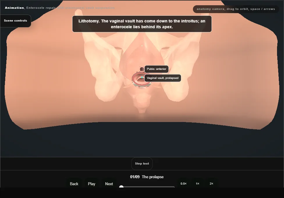
- Lithotomy.
- A urethral catheter drains the bladder and it is kept empty throughout.
- A weighted speculum and a self-retaining retractor are placed and the anterior wall is infiltrated.
- Two clamps grasp the prolapsed vault and bring it out through the introitus.
- 02
Open the wall
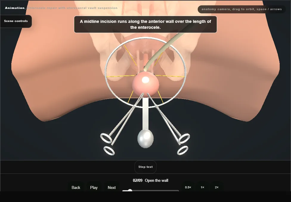
- A midline incision is made in the anterior vaginal wall along the length of the enterocele.
- The wall is dissected off the pubocervical fascia and off the enterocele sac, staying superficial.
- 03
Open the sac
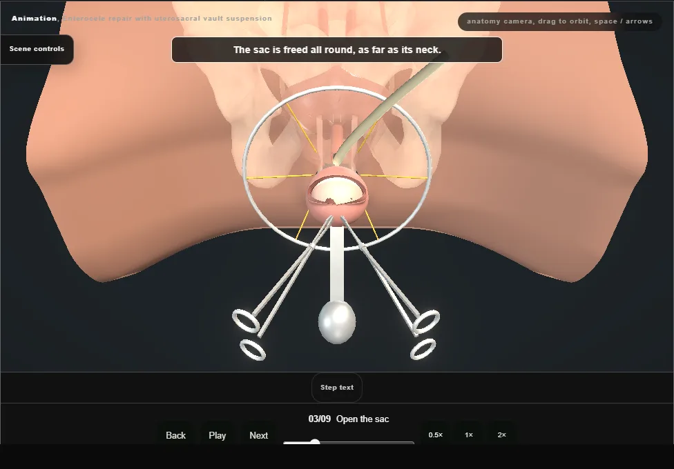
- The sac is isolated all round to its neck by sharp and blunt dissection, then opened at its far end and the peritoneal cavity entered.
- Small bowel lies inside it.
- 04
Hold the bowel up
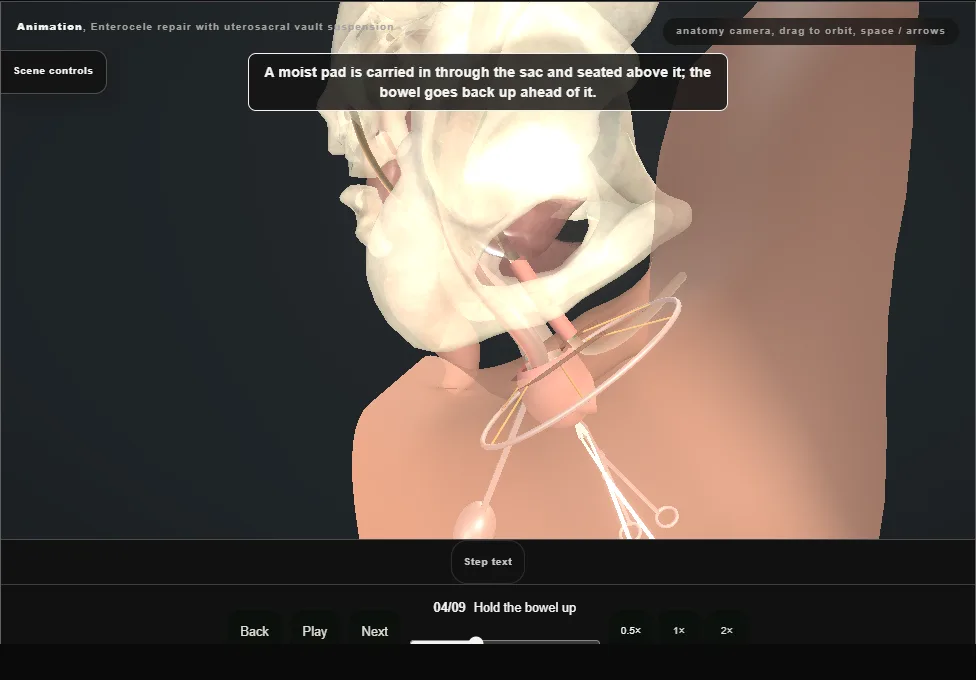
- A moist pad is placed through the opened sac and the bowel goes back up ahead of it.
- A retractor placed anteriorly holds the pad, so that no bowel lies where the needle will go.
- 05
Find the ligaments
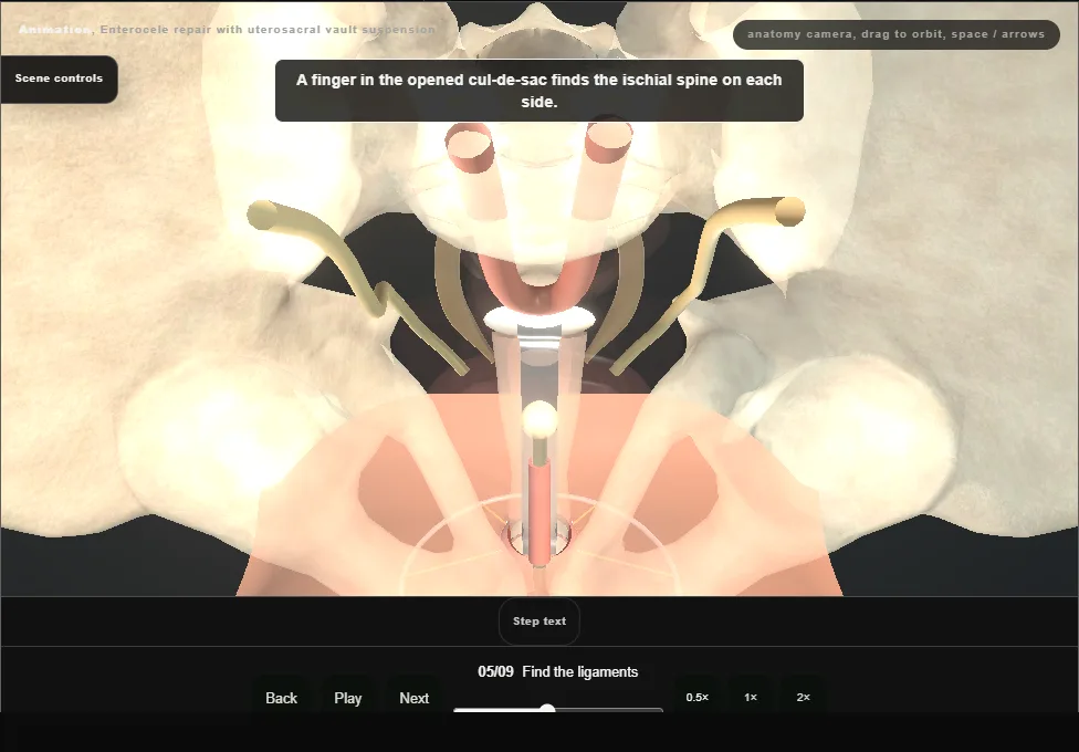
- The ischial spines are felt on each side.
- With traction on the vault each uterosacral ligament tightens and is felt posteromedial to the spine, at four and eight o'clock on the side wall.
- The ureter runs on the side wall lateral to the ligament, closest to it near the vault.
- 06
Sutures through the ligaments
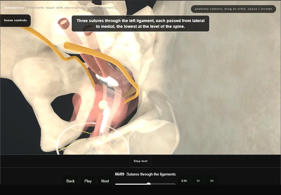
- Three double-armed sutures are passed through each ligament, from lateral to medial, the lowest at the level of the spine and the others above it.
- Each bite takes the ligament itself and stays away from the ureter.
- 07
Fix and tie
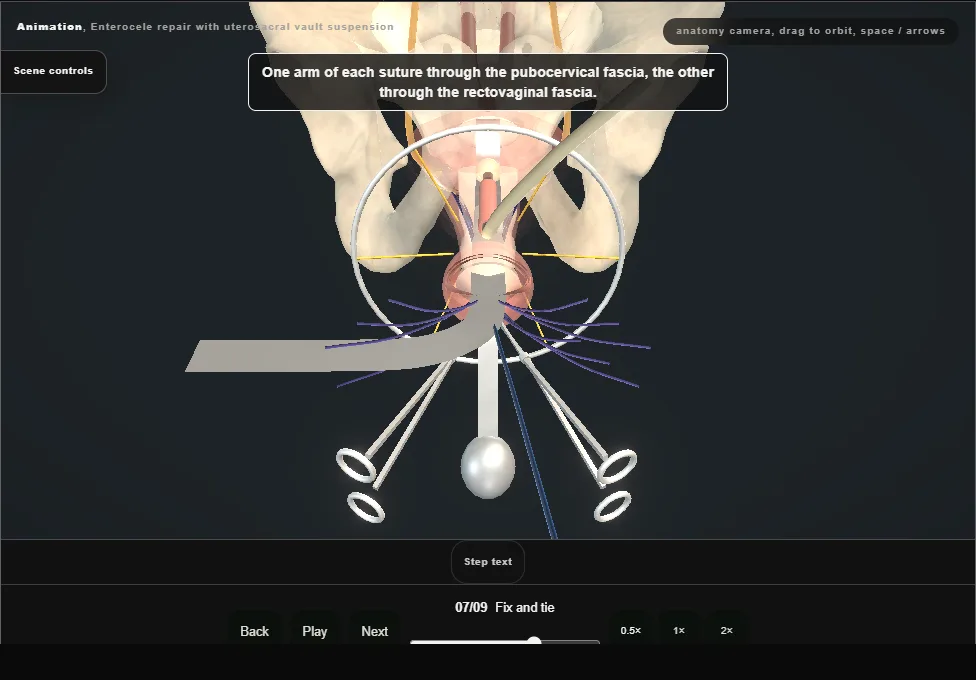
- One arm of each suture is passed through the pubocervical fascia and the other through the rectovaginal fascia.
- The pad and the retractor are removed and each suture is tied in turn: the vault goes back through the introitus and is drawn up to the ligaments.
- 08
Cystoscopy
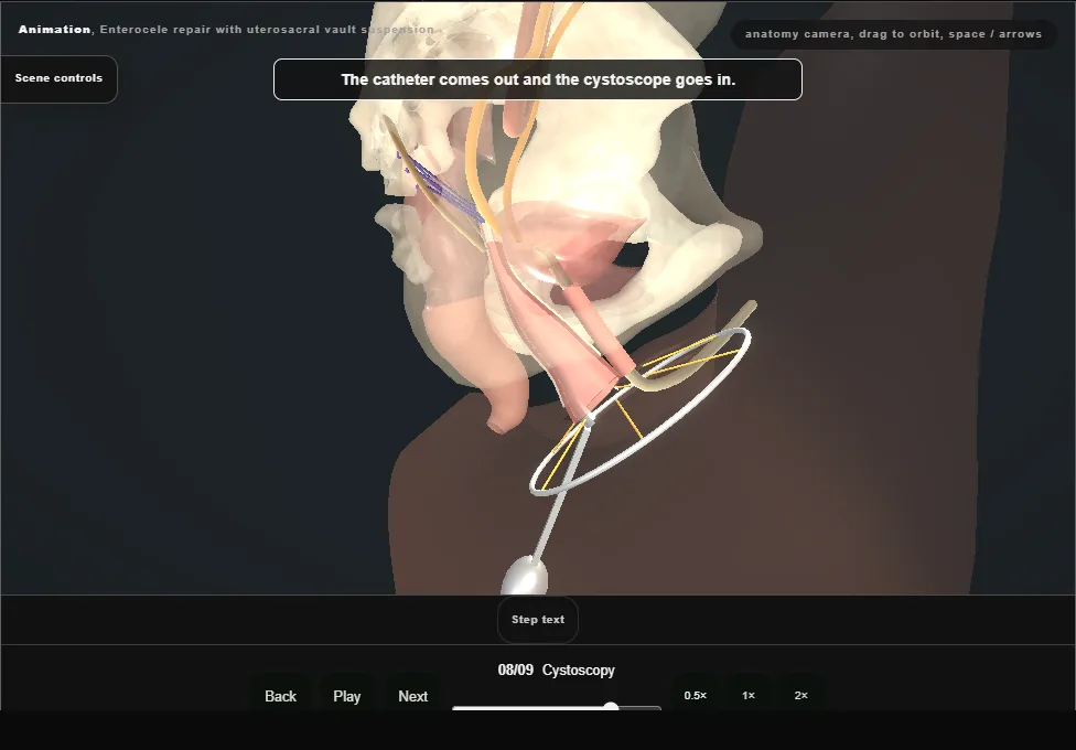
- The catheter is removed and the bladder filled.
- The bladder and urethra are intact, clear efflux is seen from both ureteric orifices and there is no suture in the bladder.
- The scope comes out and the catheter goes back.
- 09
Trim and close
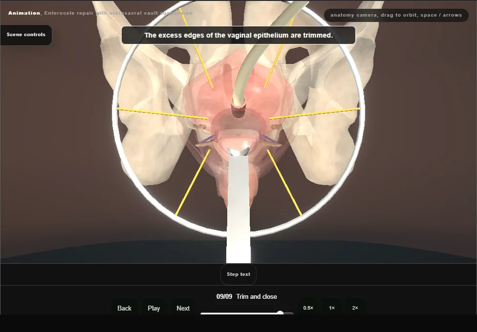
- The excess edges of the vaginal epithelium are trimmed and the wall is closed in the midline with a running absorbable suture.
- The retractor and the speculum come out and the catheter is left on drainage.
Derived from female/UterosacralSuspensionScene.tsx at build time: every step title and caption above is the authored
ProcedureDef text, reproduced verbatim, and this page shares its readable-points renderer with the animation
itself. 9 of 9 steps have a captured frame.
Text fingerprint 603d4f3055d5258f. A caption edit without a re-capture fails the production
check, so the sheet and the animation cannot drift apart silently.