
Revision sheet
York-Mason rectourethral fistula repair
9 steps, each one the authored title and caption from the interactive animation, with the frame captured from that step. Read it before the case, or print it and work through the steps in order.
Teaching only, not for diagnosis or treatment decisions. The text on this sheet is the animation's own step copy, reproduced verbatim.
UrOops3D · UroRef · © Nity G
- 01
Cystoscopy and a catheter through the fistula
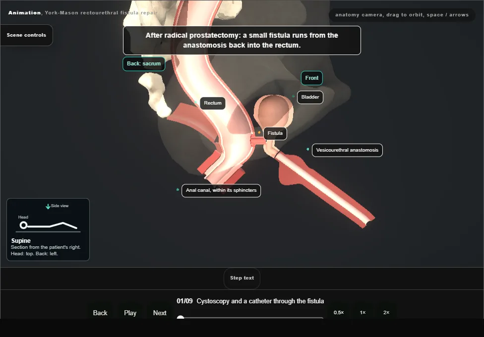
- A small rectourethral fistula after radical prostatectomy.
- With the patient supine, a cystoscope finds the opening at the bladder neck and a small open-ended catheter is passed through the fistula into the rectum.
- The scope is replaced by a urethral catheter on free drainage.
- 02
Prone jackknife and the incision
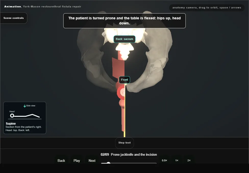
- The patient is turned prone on chest rolls and the table is flexed, hips up and head down.
- A vertical incision from the coccyx to the anal verge is taken through the subcutaneous tissue with diathermy and a self-retaining retractor is set.
- 03
Divide the sphincters, layer by layer
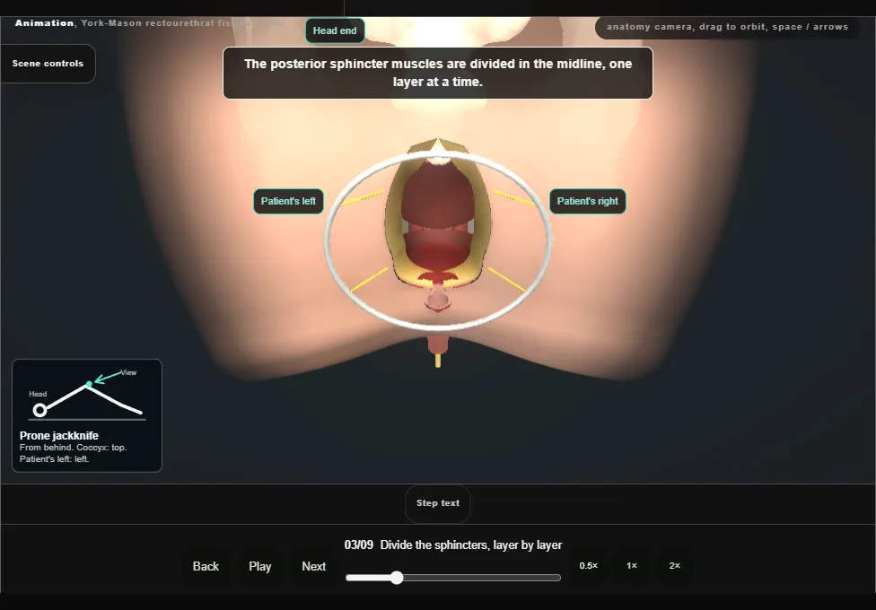
- The posterior anal sphincter is identified and its muscular layers are divided in the midline one at a time.
- Paired sutures are placed at each level as it is divided, so that every layer can later be matched to its other half.
- 04
Open the posterior rectal wall
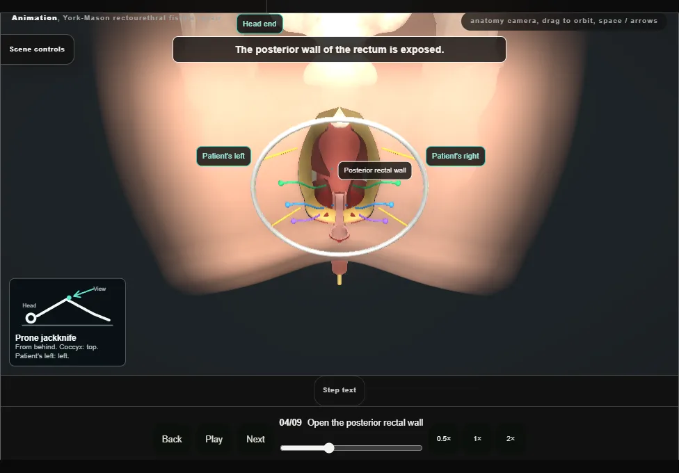
- The posterior rectal wall is exposed, incised lengthwise in the midline and retracted laterally.
- The open-ended catheter is seen coming out of the fistula on the anterior rectal wall.
- 05
Excise the fistula, separate the urethra
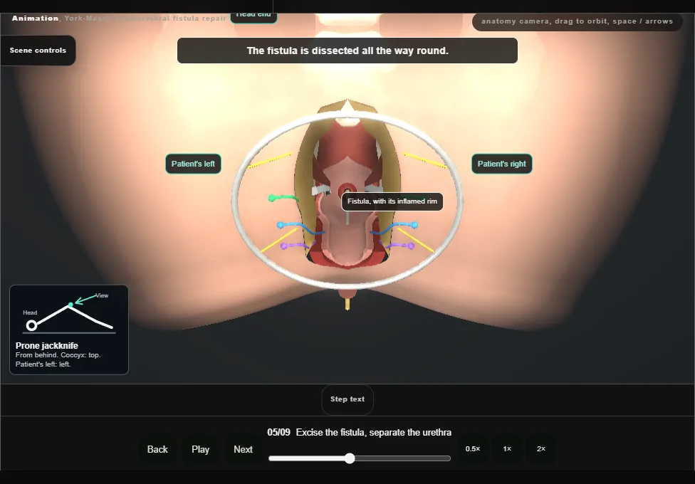
- The fistula is dissected all round and mobilised.
- The whole track and the inflamed tissue round it are excised and the small catheter is removed.
- The urethra is then separated further from the rectal wall by sharp dissection.
- 06
Close the urethra
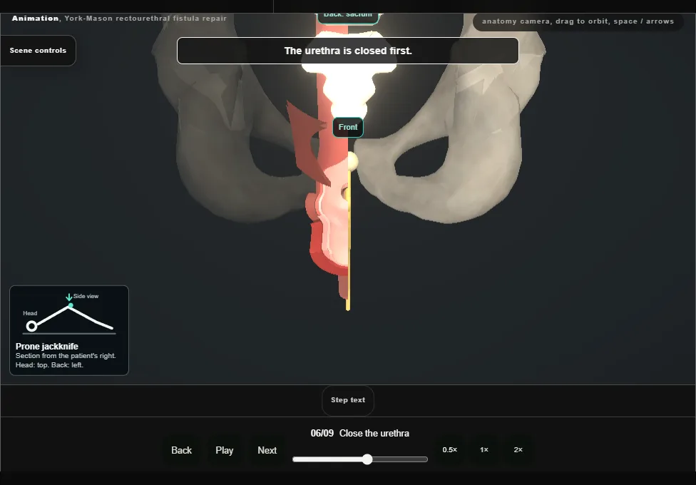
- The urethra is closed in one layer, with a running stitch, over the urethral catheter placed at the start.
- 07
Close the anterior rectal wall in two layers
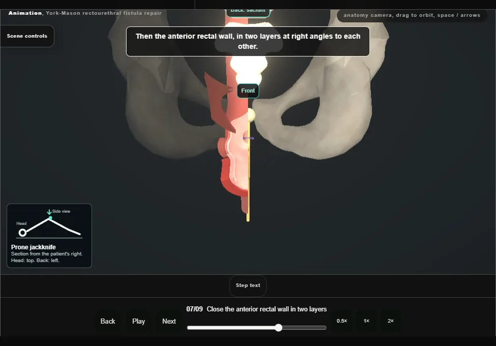
- The anterior rectal wall is closed in two layers at right angles to each other, with interrupted stitches.
- The deep layer takes the muscle and the submucosa.
- The lining is then brought over that row, overlapping it, so that no suture line lies along another.
- 08
Posterior wall, then the sphincters by their pairs
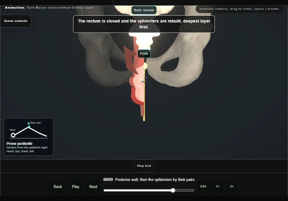
- The posterior rectal wall is closed in two running layers.
- The marking sutures are then tied one pair at a time, the deepest layer first, so the muscle is restored layer for layer in the reverse of the order it was divided.
- 09
Drain and closure
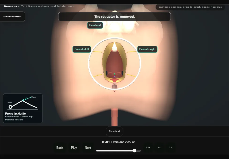
- A drain is placed under the presacral fascia and brought out through a separate stab incision.
- The subcutaneous layer and the skin are closed.
- The urethral catheter stays in.
Derived from pelvis/YorkMasonScene.tsx at build time: every step title and caption above is the authored
ProcedureDef text, reproduced verbatim, and this page shares its readable-points renderer with the animation
itself. 9 of 9 steps have a captured frame.
Text fingerprint 99e00d0951d65fc5. A caption edit without a re-capture fails the production
check, so the sheet and the animation cannot drift apart silently.