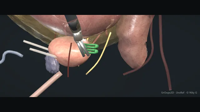
A tour of one fascia and two nerves: Denonvilliers’ plane between prostate and rectum, the neurovascular bundles on the posterolateral pedicles, the ureters inside the bladder neck and the sphincter at the urethra being divided. Every step protects one of them.
Advanced tierFRCS uro-oncology, prostate cancerInteractive 3D
Drop the bladder: space of RetziusThe retropubic fat is swept off the anterior abdominal wall until the endopelvic fascia is seen as a glistening line spanning from pubis to prostate. The puboprostatic ligaments, the dorsal venous complex and the balloon of the catheter are fixed as the landmarks every later step returns to. The bladder is peeled posteriorly off the pubis in an avascular plane, and fat that refuses to sweep usually marks accessory pudendal vessels, which are preserved rather than divided. Establishing these reference points early keeps the apex dissection oriented when the view tightens later in the case.
Bladder neck dissectionThe plane between bladder and prostate is entered anteriorly at 12 o'clock, where the fat line between smooth bladder wall and prostatic surface marks the junction. Scissors and bipolar develop the plane laterally until the catheter can be hooked through and used as traction. The neck is then transected exactly at the prostate, cutting close to the gland all the way round. The orifices are protected by that discipline and by looking for efflux as soon as the neck is open. A median lobe pushes the neck posteriorly, so the plane is re-found anterior to it rather than hunted for inside the bladder. where it bites: too wide catches the ureteric orifices; too narrow opens into the adenoma.
Seminal vesicles + vasaThe vasa are clipped and divided within the pelvis and each seminal vesicle is freed toward its tip, where the small artery entering it is controlled before the gland is lifted. Working close to the vesicles keeps the dissection away from the pelvic sidewall and everything crossing it. Once both vesicles are delivered, their tips mark the entry to Denonvilliers’ fascia, and the posterior layer is incised there to expose the pearly plane below. Starting the posterior dissection anywhere other than this defined point is how the plane is lost before it is found.
Denonvilliers’ planeThe posterior layer of Denonvilliers’ fascia is incised between the vesicle tips and the plane between the prostate-vesicle packet and the rectum is developed with blunt sweeping, keeping the fascia on the specimen. The rectum falls away as pale, avascular tissue opens, and fat that looks lobular or yellow means the plane has strayed, so the dissection stops and re-enters where the tissue is clean. Development continues to the apex so the rectum is safely off the specimen before the lateral work begins. Every posterior error in this operation is made one layer too deep, which is why the plane is entered under direct vision and widened, never forced. where it bites: the rectum is the next layer down; stay on the fascia.
Neurovascular bundles releasedEach lateral pedicle is clipped and divided athermally in short steps, cutting between clips rather than using energy, so heat never crosses to the bundle running on its posterolateral surface. The bundle is peeled off the prostatic fascia with gentle blunt dissection, kept under no traction and followed from the pedicle to the apex. Clips go on the prostatic side and the pedicle is released so the bundle drops back into the pelvis with its blood supply intact. Tissue that refuses to peel is read as disease at the capsule, and the plane on that side is converted to wide excision. where it bites: thermal or traction injury to a bundle that should have been spared; sacrifice is a cancer decision, not an accident.
DVC + urethra at the apexThe dorsal venous complex is ligated or suture-controlled anterior to the urethra and divided close to the prostate, with the field kept dry rather than diathermised through the complex. The urethra is then divided flush with the prostatic apex with a cold blade, as far distally as the cancer allows, so maximal functional length and rhabdosphincter fibres stay with the patient. The remaining posterior attachments are released and the specimen is delivered. The stump left behind carries both the continence mechanism and the future anastomosis, which is why the cut is made once, deliberately, in a dry field. where it bites: the continence and the anastomosis both live on this stump.
Vesicourethral anastomosisThe bladder neck is tailored to match the urethral stump and a running suture joins bladder mucosa to urethral mucosa circumferentially over a new catheter. Each pass takes a generous ring of bladder and a full thickness of urethra so the anastomosis is mucosa-to-mucosa and tension-free, and the suture ends are tied only once the running closure is complete. The bladder is filled through the catheter to test the closure watertight and a pelvic drain is left dependent in the pelvis. Leak and contracture are both born here: a leak from an imperfect closure and a contracture from an ischaemic or overlapping one, which is why the anastomosis is finished in a dry, unhurried field.
The spatial relationships the animation is built to make automatic. Before you read each one, ask yourself: at this point in the operation, what is about to go wrong, and what is my first move? The save for each sits in the Complications tab.
RiskThe posterior plane - one layer wrong at Denonvilliers’ and the case becomes a rectal repair.
RiskThe lateral pedicles - thermal energy here is a nerve injury; clips, not cautery, beside the bundles.
RiskThe apex - DVC control, urethral length and sphincter preservation compete in millimetres.
When the case turns: the problem, then the next move.
ActionCannot find the plane at the neck - re-identify the catheter balloon and come back at 12; a median lobe pushes the neck posteriorly, the plane is anterior to it.
ActionBleeding from the DVC - raise pneumoperitoneum, tamponade, suture-control the complex rather than diathermising blindly.
ActionBundles stuck to the gland - stop and reassess: adhesions from biopsies or tumour; a wide plane is safer than a torn capsule at tumour site.
ActionRectum does not fall away - you are in the wrong layer; return to a clean area and re-enter Denonvilliers’ plane under direct vision.
The examiner stems this scene prepares you for. Try to answer out loud first, then reveal, and learn the discriminator, the detail that turns a pass into a strong pass.
QHow do you decide between nerve-sparing and non-nerve-sparing?reveal answer
The decision is oncological first: nerve-sparing is offered when the imaging, biopsy pattern and clinical stage indicate tumour confined away from the capsule at that side, and it is bilateral, unilateral or absent depending on where the risk sits.
High-grade disease, palpable tumour, MRI showing extracapsular extension at the bundle, or multiple positive cores on one side push toward sacrificing that side.
Patient factors matter: age, baseline function, and the patient’s stated priorities after honest counselling about the trade of margin against function.
Operatively, the choice is executed athermally with clips on the pedicles, because the bundles are injured as much by heat as by scissors.
Where the anatomy or findings change on the table, taking the bundle on that side is the safe default; a positive margin buys a functional outcome that is harder to reverse than the other way round.
Strong pass
Laterality per side, and the intra-operative downgrade rule
Per side→Not a single binary decision
Rule→Name the intra-operative downgrade
Examiner note
Anchoring the decision in laterality per side rather than a single binary, and naming the intra-operative downgrade rule.
QYou are entering Denonvilliers’ plane and the tissue suddenly looks yellow and lobular. What is it and what do you do?reveal answer
Yellow lobular tissue at the posterior prostate means one of two things: periprostatic fat, which means the plane has strayed anteriorly outside the fascia, or mesorectal fat, which means the plane has gone through Denonvilliers’ into the rectal mesentery and the rectal wall is next.
I would stop, re-grasp in a clean area, and re-enter the pearly avascular plane under direct vision rather than dissecting further in an unidentified layer.
I would alert the team that the rectum is close, complete the posterior dissection with the rectum in view at all times, and inspect the rectal wall at the end with the pelvis filled, considering an air leak test.
If the wall is breached, that is a recognised, repairable event; the catastrophic version is the unrecognised one, so the response to doubt is inspection, not optimism.
Converting doubt into a formal inspection and leak test
Examiner note
Reading yellow fat directionally (mesorectal fat = posterior danger) and converting doubt into a formal inspection and leak test.
QWhat defines continence preservation at the apex?reveal answer
Three operative decisions.
First, the dorsal venous complex is controlled with minimal distal dissection, so the sphincter is not skeletonised before the urethra is even cut.
Second, the urethra is divided as far from the sphincter as oncologically allowed, preserving maximal functional urethral length and the rhabdosphincter fibres with their anterior support.
Third, the anastomosis is tension-free and mucosa-to-mucosa, because a disrupted or ischaemic anastomosis scars into the bladder-neck contracture that presents as obstruction and mimics incontinence failure.
Around the operation itself, catheter time is kept short where the anastomosis allows, and pelvic-floor rehabilitation starts early.
The honest framing for patients is that continence returns along a curve over months, and the operative technique sets the ceiling.
Strong pass
Say both
Listing urethral length and DVC dissection discipline as operative choices
Separating contracture (obstruction) from sphincter failure in the late failure analysis
Examiner note
Listing urethral length and DVC dissection discipline as operative choices, and separating contracture (obstruction) from sphincter failure in the late failure analysis.
QWhich patients would you not offer radical prostatectomy?reveal answer
Oncologically: those with demonstrable distant disease, or locally advanced disease better served by radiotherapy and hormones where surgery adds morbidity without a survival argument in most settings.
Functionally and medically: significant comorbidity with limited life expectancy, since the benefits of surgery accrue over a decade;
untreated clotting or anticoagulation states that cannot be bridged;
and patients whose priority profile, after honest counselling about urinary and sexual outcomes, points them to alternative modalities.
Age alone is not a criterion, fitness is.
There are also relative surgical contraindications: previous pelvic radiation or extensive pelvic surgery, where the planes may not exist and the complication profile changes fundamentally.
The decision belongs in an MDT with the patient in the room, not in the theatre list.
Strong pass
Separating absolute oncological exclusions from priority-of-outcome decisions that remain the patient’s to make.
Say it aloud, then reveal. Reading is not remembering.
01The relationshipSay the critical spatial relationship out loud, then reveal.reveal
Memory frame
Front to back: pubis, dorsal venous complex, urethra, prostate, Denonvilliers', rectum
Anterior→Every error bleeds
Posterior→Every error leaks
Bundles→The posterolateral corners of the stack
Why?
In the sagittal axis the structures stack front to back: pubis, dorsal venous complex, urethra, prostate, then Denonvilliers’ fascia, then rectum. The bundles run along the posterolateral corners of that stack, which is why every anterior error bleeds and every posterior error leaks.
02The first moveRectal injury. Your first move?reveal
First move
Recognise it on the table
The wall thins
Mucosa pouts
Bowel content appears
On-table recognition is the save.
More detail
Recognise it openly: the wall thins, mucosa pouts or bowel content appears; on-table recognition is the save.
03The escalation triggerRectal injury. What makes you call for help?reveal
Escalate when
Washout, wide drainage, then the stoma decision with general surgery
Bowel preparation
Quality of the repair
Degree of contamination
More detail
Washout, drain widely; decide on defunctioning stoma with general surgery based on preparation, repair quality and contamination.
04The discriminatorWhat turns a pass into a strong pass?reveal
Strong pass
Laterality per side, and the intra-operative downgrade rule
Per side→Not a single binary decision
Rule→Name the intra-operative downgrade
Examiner note
Anchoring the decision in laterality per side rather than a single binary, and naming the intra-operative downgrade rule.
05The pitfallWhere does this operation bite first?reveal
Pitfall
One layer wrong at Denonvilliers'
The case becomes a rectal repair.
Why?
The posterior plane: one layer wrong at Denonvilliers’ and the case becomes a rectal repair.
Revision practice with self-marked answers, not an assessment of clinical competence. Kept in
this browser only; it does not sync between devices.