
UrOops3D, danger-zone atlas
Operative animation, Adrenalectomy

Laparoscopic adrenalectomy, left and right

A gland the size of a thumb, sitting on the upper pole of the kidney against the cava on the right and behind the pancreas on the left, that makes the hormones which raise the blood pressure and can stop the heart on the table.

The operation finds the vein before the gland is touched and takes the gland whole.

This page is the selection, the blockade and the endocrinologist, the choice of approach, the exposure on each side, the vein first, the gland in a bag, and the steroids and the metanephrines after.

Advanced tier FRCS adrenal surgery, endocrine and uro-oncology Interactive 3D

Operative visualisation

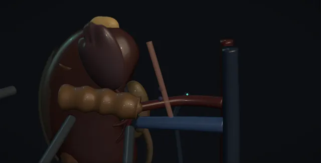
Schematic scene preview: Adrenalectomy
Preparing the 3D anatomy…

UrOops3D · UroRef · © Nity G

Click me next Section 2 of 8 Anatomy The structures this operation works between, and the one it must not cross.

The structures in the scene

Adrenalectomy: how the structures relateLaparoscopic, the adrenal above the upper pole on either side. Adrenal gland, capsule intact: short and wide into the cava; torn, a caval hole Right adrenal vein and the cava (danger). Adrenal gland, capsule intact: drops to the renal vein; clipped first Left adrenal vein to the left renal vein. Adrenal gland, capsule intact: across the front; hurt by retraction or heat Spleen and tail of the pancreas (danger). Adrenal gland, capsule intact: crus is the medial wall; a breach is a pneumothorax Crus, diaphragm and pleura (danger). Adrenal gland, capsule intact: sits medial and superior to the pole Upper pole of the kidney. Adrenal gland, capsule intact: lifted to show the cava and the gland behind it Liver, lifted on the right.Adrenal gland, capsuleintact!Right adrenal veinand the cavaLeft adrenal vein tothe left renal vein!Spleen and tail ofthe pancreas!Crus, diaphragm andpleuraUpper pole of thekidneyLiver, lifted on therightshort and wide intothe cava; torn, acaval holedrops to the renalvein; clipped firstacross the front;hurt by retractionor heatcrus is the medialwall; a breach is apneumothoraxsits medial andsuperior to thepolelifted to show thecava and the glandbehind itTargetDangerLandmarkContext
Laparoscopic, the adrenal above the upper pole on either side. Schematic, not to scale; relationships only.
  • On the left the camera looks up from below the costal margin with the spleen and the pancreatic tail rolled medially and Gerota opened over the upper pole;
  • the adrenal sits medial and superior to the pole with the renal vein running below it and the adrenal vein dropping from the gland to the vein's upper border.
  • On the right the liver is lifted and the cava runs vertically with the adrenal tucked behind its lateral border above the renal vein, the short adrenal vein passing straight into the cava.
  • In both, the crus of the diaphragm is the medial wall.
  • ›The adrenal gland - golden-orange and firm in the fat above the upper pole; lifted by its fat, never grasped, and taken with its capsule intact.
  • ›The left adrenal vein - runs from the inferomedial gland down to the superior border of the left renal vein, joined by the inferior phrenic; clipped first.
  • ›The right adrenal vein - a centimetre or less, wide, entering the posterolateral cava directly; the reason the right is the harder side.
  • ›The spleen and the tail of the pancreas - rolled medially on the left after the ligaments are divided; the pancreas lies across the front of the gland.
  • ›The inferior vena cava - the medial border of the right adrenal sits on it and behind it; followed upward from the renal vein.
  • ›The liver and the diaphragm - the right lobe retracted up after the triangular ligament is divided; the crus is the medial limit on both sides.
  • ›The upper pole of the kidney - the inferior border of the gland is freed from it; a small lower adrenal artery comes from the renal artery.
Click me next Section 3 of 8 Steps The operation in order, with the landmark that guards each move.

Operative steps

  1. Who: functioning, large or suspicious Three reasons to remove an adrenal. Function: a phaeochromocytoma with raised plasma or urinary metanephrines, an aldosterone-producing adenoma in primary hyperaldosteronism after adrenal venous sampling has lateralised it, or a cortisol-secreting adenoma with Cushing's syndrome. Size and appearance: an incidentaloma above four centimetres, or one of any size with a non-contrast density above 10 Hounsfield units and poor contrast washout, irregular margins or growth on repeat imaging, because the risk of adrenocortical carcinoma rises with size. And suspicion of malignancy: a primary adrenal carcinoma, which is removed open and en bloc, or a solitary adrenal metastasis in a patient whose primary is controlled. A non-functioning lesion under four centimetres with benign imaging is watched, not removed.
  2. Preparation: alpha blockade and the endocrinologist Every adrenal is discussed with an endocrinologist and screened for function before it is removed, because the preparation differs. A phaeochromocytoma is alpha blocked with phenoxybenzamine or doxazosin for at least ten to fourteen days, the dose increased until the blood pressure is controlled and the patient is postural, with a high salt intake to refill the circulation; a beta blocker is added only after alpha blockade if there is tachycardia, never first, because unopposed alpha stimulation drives a hypertensive crisis. Cushing's syndrome means hydrocortisone cover from induction and a taper after, because the other adrenal is suppressed. Conn's means potassium corrected and spironolactone before surgery. The anaesthetist meets the phaeochromocytoma patient in advance and has an arterial line, magnesium, phentolamine or a nitroprusside infusion and noradrenaline drawn up.
    where it bites: a beta blocker started before the alpha blocker, or a phaeochromocytoma missed on screening, is a hypertensive crisis when the gland is touched.
  3. Laparoscopic transperitoneal or retroperitoneal Most adrenals come out laparoscopically. The lateral transperitoneal approach, patient in the lateral position with the table broken, gives the familiar landmarks of the spleen, the colon, the kidney and the cava and room for a gland up to six centimetres or more; it is the standard in urological practice. The posterior retroperitoneoscopic approach, patient prone with the ports below the twelfth rib and the space held open at a higher pressure of about 20 millimetres of mercury, avoids the peritoneum and old adhesions and suits small glands and bilateral disease, at the cost of a small working space and unfamiliar landmarks. Open surgery is reserved for the suspected carcinoma, the gland above about eight centimetres, and the tumour that invades. The robot adds articulation for the retrocaval right gland.
  4. Exposure on the left: spleen, pancreas and the upper pole Right lateral position, three or four ports below the left costal margin. The splenic flexure is taken down and the splenocolic, splenorenal and splenophrenic ligaments divided up to the diaphragm, so that the spleen and the tail of the pancreas roll medially with gravity and the assistant does not have to retract them. The plane behind the pancreas leads onto Gerota's fascia over the upper pole of the kidney. Gerota is opened over the upper pole and the medial border of the kidney, and the golden-orange adrenal is found in the fat above the upper pole, medial to it, distinguished from the fat by its colour and its firmness. The left renal vein is identified along the upper border of the hilum, because the adrenal vein runs down to it.
    where it bites: the tail of the pancreas lies across the field in front of the adrenal; a retractor or a hot instrument on it is a pancreatic fistula.
  5. The adrenal vein first The left adrenal vein leaves the inferomedial aspect of the gland and runs down to enter the superior border of the left renal vein, often joined by the inferior phrenic vein on the way. It is found by following the renal vein medially along its upper border, dissected free with the hook or the bipolar for a centimetre, and taken between clips before the gland is handled, so that catecholamines released by manipulation cannot reach the circulation. The anaesthetist is told when the vein is about to be clipped, because the blood pressure falls when it is, and the noradrenaline is started as the clip goes on. In a phaeochromocytoma the gland is handled as little as possible before this point, and the vein is the first thing sought.
    where it bites: traction on the gland before the vein is clipped pours catecholamines into the circulation; the pressure hits 250 and the anaesthetist has no warning.
  6. Mobilising the gland With the vein clipped the gland is lifted by the fat around it, never grasped directly because it is friable and tears, and the arterial supply is taken as it is met: small branches from the inferior phrenic artery above, the aorta medially and the renal artery below, sealed with the bipolar or the ultrasonic shears clear of the gland. The medial border is freed from the crus and the aorta, the superior pole from the diaphragm, and the inferior border from the upper pole of the kidney, outside the capsule and with a cuff of fat so that a suspicious lesion is not breached. The dissection stays close enough to avoid the pancreas and the splenic vessels, which lie a centimetre or two in front.
  7. Extraction in a bag The gland is placed in a retrieval bag as soon as it is free, the bag closed, and delivered through the enlarged camera port or a port site extended for a larger gland, intact and unmorcellated, so that the pathologist has the whole gland and its capsule and so that nothing is spilt. The bed is inspected at low pressure for bleeding from the phrenic branches and the renal vein stump, and the spleen and the pancreatic tail are looked at before the ports come out. A drain is not usually needed unless the pancreas was handled. The specimen is orientated and weighed, and in a phaeochromocytoma the genetics are requested, because a third or more are hereditary.
  8. The right side: the cava and the short vein Left lateral position, the ports below the right costal margin, and the right lobe of the liver retracted upward by a fan or a Nathanson through an epigastric port after the triangular ligament is divided. The duodenum is kocherised if it lies over the field. The lateral border of the cava is found and followed upward, and the adrenal sits behind and lateral to it above the right renal vein, partly retrocaval. The right adrenal vein is short, a centimetre or less, wide, and enters the posterolateral cava directly; it is found by dissecting the medial border of the gland off the cava from below upward, and clipped twice on the caval side with the cava seen throughout. The gland is then mobilised as on the left.
    where it bites: the right adrenal vein torn from the cava is a hole in the cava that retracts; it is clipped under vision with the cava seen, or the operation is opened.
  9. After A phaeochromocytoma patient goes to a monitored bed: hypotension in the first day from the loss of catecholamines and the residual alpha blockade is treated with fluid and noradrenaline, and hypoglycaemia is looked for because the insulin suppression is gone. Plasma metanephrines are repeated at two to six weeks and then yearly for life. A Cushing's adenoma patient is on hydrocortisone, tapered over months while the suppressed contralateral adrenal recovers, with a morning cortisol guiding withdrawal. A Conn's patient stops spironolactone, has the potassium checked, and is warned that hypertension improves but may not resolve. After bilateral adrenalectomy the patient has hydrocortisone and fludrocortisone for life with a steroid card and sick day rules. Every patient sees the endocrinologist with the histology.
Click me next Section 4 of 8 Danger zones Where this operation bites, and why it bites there.
Critical relationship

Danger zones

The spatial relationships the animation is built to make automatic. Before you read each one, ask yourself: at this point in the operation, what is about to go wrong, and what is my first move? The save for each sits in the Complications tab.

  • RiskThe right adrenal vein and the cava - short, wide and posterior; torn, it leaves a hole in the cava that retracts and bleeds where the clip cannot reach.
  • RiskThe tail of the pancreas and the splenic vessels - across the front of the left gland; injured by retraction or heat, and presenting as a fistula or a bleed days later.
  • RiskThe gland before the vein is clipped - in a phaeochromocytoma, manipulation is a catecholamine surge; the vein is found first and the anaesthetist told.
  • RiskThe diaphragm and the pleura - the superior pole and the posterior approach reach the diaphragm; a breach is a pneumothorax under pressure.
Practise it Take me into the scene Opens the animation at a danger moment and asks you to predict the mechanism and your first move before it reveals.
Click me next Section 5 of 8 Complications The save for each one: the first move, the escalation trigger, the op-note line.

Complications and immediate saves

Trigger each of these live in the Animation tab, then read the save: the first move, the escalation trigger, and the line that goes in the op note.

Major vascular bleeding

  • the right adrenal vein avulsed from the cava, the left renal vein torn at the adrenal vein junction, or the inferior phrenic vessels or splenic vein bleeding in a field that fills in seconds.
The save
  1. Press on the point with a swab or the suction tip, raise the pressure briefly, and tell the anaesthetist and the scrub team; do not clip blind into a pool. first move
  2. A hole in the cava or the renal vein: hold pressure two minutes, then a clip on a seen edge or a 5-0 polypropylene suture laparoscopically if the skill is there.
  3. The inferior phrenic or the splenic vein: bipolar or clips under vision after the field is cleared with a second suction.
  4. Bleeding not controlled in minutes, or the cava opened: convert to open through a subcostal incision with the assistant holding pressure, and call for vascular help. escalation trigger
  5. Op note: the vessel, the control, the blood loss and transfusion, and whether conversion occurred. document

Hypertensive crisis and post-resection hypotension

  • the phaeochromocytoma handled before its vein is clipped: pressure above 200, tachyarrhythmia, pulmonary oedema or a stroke; then the collapse of pressure when the vein is clipped and the catecholamines are gone.
The save
  1. Stop touching the gland, let the pneumoperitoneum down, and let the anaesthetist bring the pressure down with phentolamine, nitroprusside or magnesium before anything else. first move
  2. Resume only when the pressure is controlled; find and clip the vein first with the anaesthetist warned as the clip goes on.
  3. After the vein: fluid and noradrenaline for the fall in pressure, glucose checked, a monitored bed overnight.
  4. An arrhythmia that will not settle, pulmonary oedema, or a neurological change: critical care and cardiology; the operation is paused or abandoned until the patient is safe. escalation trigger
  5. Op note and anaesthetic record: the peak pressure, the drugs used, the timing of the vein clip, and the pressure after it. document

Pancreatic, splenic, hepatic, bowel or renal injury

  • the tail of the pancreas burnt or retracted on the left, a splenic capsular tear, the liver torn by the retractor on the right, a duodenal injury, or the upper pole of the kidney devascularised by a hot instrument at its artery.
The save
  1. Look at the injury under low pressure and decide what it is; a pancreatic injury seen at the time gets a drain and the pancreas team told. first move
  2. A splenic capsular tear: haemostatic agent and pressure; splenectomy only if it will not stop.
  3. A bowel injury: repaired in two layers at the time; a bowel injury found later presents with peritonitis and needs a laparotomy.
  4. A rising drain amylase, a collection or peritonism on the ward: CT and the general surgeons, with percutaneous drainage or a return to theatre. escalation trigger
  5. Op note: the organ, the mechanism, the repair, the drain and the plan for the amylase. document

Adrenal insufficiency

  • the suppressed remaining adrenal after a cortisol-secreting adenoma, a bilateral adrenalectomy, or a cortical-sparing operation that left too little cortex: nausea, hypotension, hyponatraemia and collapse, days to weeks later.
The save
  1. Hydrocortisone at induction and after for every Cushing's and every bilateral case; a steroid card and sick day rules before discharge. first move
  2. A morning cortisol and a short Synacthen test guide the taper, over months, with the endocrinologist.
  3. After bilateral adrenalectomy: hydrocortisone and fludrocortisone for life, and the patient taught to double the dose when ill.
  4. Hypotension, vomiting or confusion after discharge: an adrenal crisis until proven otherwise; intravenous hydrocortisone and fluid first, tests second. escalation trigger
  5. Discharge summary: the steroid dose, the taper plan, the sick day rules and the endocrine appointment. document

Tumour spill, positive margin and recurrence

  • the friable phaeochromocytoma or carcinoma grasped and torn, or a carcinoma removed laparoscopically piecemeal: cells in the retroperitoneum, and a recurrence that is far harder to treat than the primary.
The save
  1. The gland is lifted by its fat and never grasped; anything that might be a carcinoma is removed open and en bloc with its fat and, if invaded, the kidney. first move
  2. A capsule breached at the time is noted, the field irrigated, and the specimen orientated for the pathologist.
  3. A phaeochromocytoma has metanephrines yearly for life; a carcinoma has CT three-monthly at first and adjuvant mitotane discussed at the endocrine MDT.
  4. Rising metanephrines or a mass on surveillance: functional imaging, and re-resection or systemic therapy at the specialist centre. escalation trigger
  5. Op note: whether the capsule was intact, the extraction method, the bag, and the surveillance plan. document
Click me next Section 6 of 8 Troubleshooting When the case turns: the problem, then the next move.

Intra-operative troubleshooting

When the case turns: the problem, then the next move.

  • ActionThe adrenal cannot be told from the fat - it is firmer and more orange than the fat, sits medial to the upper pole against the crus; find the renal vein and follow its upper border medially to the adrenal vein, which leads to the gland.
  • ActionThe spleen will not fall away on the left - the splenophrenic ligament is still attached; divide it up to the diaphragm so the spleen rolls medially with gravity rather than being retracted.
  • ActionThe right adrenal vein cannot be found - follow the lateral border of the cava upward from the renal vein and dissect the gland off it medially; the vein is short and posterior and appears as the gland lifts. If the gland is retrocaval, roll the cava medially with a blunt instrument.
  • ActionThe pressure spikes when the gland is touched - stop, let the gas down, wait for the anaesthetist; then find the vein before the gland is handled again.
  • ActionThe gland tears - stop grasping it; lift it by the fat and take the rest with a wide cuff, irrigate, and bag it whole; note the breach for the pathologist.
  • ActionGas in the pleura on the posterior approach - lower the pressure, close the breach with a suture if it can be seen, and ask the anaesthetist to check the airway pressures; a chest drain if there is a pneumothorax after the gas is out.
Click me next Section 7 of 8 FRCS viva Say it out loud, then mark yourself against the model answer.

FRCS viva questions

The examiner stems this scene prepares you for. Try to answer out loud first, then reveal, and learn the discriminator, the detail that turns a pass into a strong pass.

QA 52 year old has a 3.5 centimetre adrenal incidentaloma on a CT for renal colic. How do you assess it?reveal answer
  • I would ask two questions of every incidentaloma: does it function, and could it be malignant.
  • For function I would take a history and examination for Cushing's, hypertension and paroxysmal symptoms, and request plasma or urinary metanephrines for phaeochromocytoma, an overnight dexamethasone suppression test for autonomous cortisol secretion, and, if hypertensive or hypokalaemic, an aldosterone to renin ratio for primary hyperaldosteronism.
  • For malignancy I would look at the imaging: a homogeneous lesion under four centimetres with a non-contrast density of 10 Hounsfield units or less is a lipid-rich adenoma and benign;
  • a lesion with a density above 10 units needs washout characterisation on a dedicated adrenal CT or chemical shift MRI;
  • irregular margins, heterogeneity, size above four centimetres and growth on repeat imaging raise the suspicion of adrenocortical carcinoma.
  • A biopsy is not done unless a metastasis from a known primary is suspected, and never before phaeochromocytoma has been excluded.
  • This lesion at 3.5 centimetres with benign imaging and negative hormones would be re-imaged at six to twelve months and screened for function again;
  • it would be removed if it functions, grows or has indeterminate imaging.
  • Anything that goes to surgery is discussed with an endocrinologist first.
Strong pass

Say both

  • The 10 Hounsfield unit threshold and the four centimetre threshold stated as numbers
  • Phaeochromocytoma excluded before any biopsy or operation
Examiner note
The 10 Hounsfield unit threshold and the four centimetre threshold stated as numbers, and phaeochromocytoma excluded before any biopsy or operation.
QHow do you prepare a patient with a phaeochromocytoma for adrenalectomy?reveal answer
  • The aim is to block the effects of catecholamines and to refill a circulation that has been constricted for months.
  • I would start alpha blockade with phenoxybenzamine or doxazosin at least ten to fourteen days before surgery, titrating the dose against the blood pressure and accepting postural hypotension and a stuffy nose as signs of adequate block, with a high salt diet and fluid intake to expand the plasma volume.
  • Only after alpha blockade is established would I add a beta blocker if tachycardia develops, because a beta blocker given first leaves alpha receptors unopposed and precipitates a hypertensive crisis.
  • Some units use calcium channel blockers as an alternative.
  • I would arrange genetic counselling, because a third or more of phaeochromocytomas are hereditary, and imaging to exclude bilateral or extra-adrenal disease.
  • The anaesthetist would see the patient in advance and plan an arterial line, central access, short-acting vasodilators such as phentolamine or nitroprusside, magnesium, and noradrenaline for the hypotension that follows removal.
  • Intraoperatively the vein is clipped before the gland is handled.
  • Postoperatively the patient goes to a monitored bed for hypotension and hypoglycaemia, and plasma metanephrines are repeated at two to six weeks and yearly for life.
Strong pass

Say all three

  • Alpha before beta with the pharmacological reason
  • The volume refilling
  • The genetics requested as part of the preparation rather than an afterthought
Examiner note
Alpha before beta with the pharmacological reason, the volume refilling, and the genetics requested as part of the preparation rather than an afterthought.
QDescribe the differences between left and right laparoscopic adrenalectomy.reveal answer
  • On the left the gland is reached by dividing the splenic flexure attachments and the splenorenal and splenophrenic ligaments so the spleen and the pancreatic tail roll medially, exposing Gerota's fascia over the upper pole;
  • the structures at risk are the pancreatic tail and splenic vessels across the front of the gland and the spleen itself.
  • The left adrenal vein is long, leaves the inferomedial gland and enters the superior border of the left renal vein, often with the inferior phrenic vein, so it is found by following the renal vein medially and clipped first.
  • On the right the liver is retracted upward after the triangular ligament is divided and the duodenum kocherised if needed;
  • the gland lies behind and lateral to the cava, partly retrocaval, and the structures at risk are the cava, the liver and the duodenum.
  • The right adrenal vein is short, a centimetre or less, wide, and enters the posterolateral cava directly, so it must be found by dissecting the medial border of the gland off the cava under direct vision and clipped with the cava seen;
  • an avulsion is a caval injury.
  • On both sides the arterial supply comes as small branches from the inferior phrenic, the aorta and the renal artery, and the crus is the medial limit.
Strong pass

Say all four

  • The right adrenal vein described as short
  • Wide and directly into the posterolateral cava
  • With the consequence of avulsion
  • Rather than the two sides described as mirror images
Examiner note
The right adrenal vein described as short, wide and directly into the posterolateral cava, with the consequence of avulsion, rather than the two sides described as mirror images.
QWhat are the complications of adrenalectomy and how do they differ by indication?reveal answer
  • The operative complications are common to all: bleeding from the adrenal vein, the cava or renal vein, the phrenic vessels or the splenic vein;
  • injury to the pancreas, spleen, liver, duodenum or colon depending on the side;
  • pleural or diaphragmatic breach, more often with the posterior approach;
  • conversion to open;
  • and port site complications.
  • The indication-specific complications are what an examiner wants.
  • Phaeochromocytoma: intraoperative hypertensive crisis and arrhythmia if the gland is handled before the vein is clipped, then hypotension and hypoglycaemia after removal, and lifelong surveillance because it can recur or be part of a hereditary syndrome.
  • Cushing's: adrenal insufficiency from the suppressed contralateral gland, requiring hydrocortisone from induction with a taper over months, poor wound healing, infection and venous thromboembolism from the cortisol excess.
  • Conn's: transient hyperkalaemia after removal as the other gland recovers, and hypertension that improves but persists in about half.
  • Bilateral adrenalectomy: permanent adrenal insufficiency requiring hydrocortisone and fludrocortisone for life with sick day rules, and Nelson's syndrome after bilateral surgery for pituitary Cushing's.
  • Suspected carcinoma: tumour spill and local recurrence if removed laparoscopically or with a breached capsule, which is why it is removed open and en bloc.
Strong pass

Say both

  • The complications organised by indication rather than by organ
  • With the steroid cover in Cushing's and the contralateral suppression explained
Examiner note
The complications organised by indication rather than by organ, with the steroid cover in Cushing's and the contralateral suppression explained.
Click me next Section 8 of 8 Evidence The guidance each of these answers is built on.

Evidence and guidance

Two minutes

Close the lap: recall it before you leave.

Say it aloud, then reveal. Reading is not remembering.

  1. 01 The relationship Say the critical spatial relationship out loud, then reveal. reveal
    Memory frame

    On the left the camera looks up from below the costal margin with the spleen and the pancreatic tail rolled medially and Gerota opened over the upper pole;

    Why?
    the adrenal sits medial and superior to the pole with the renal vein running below it and the adrenal vein dropping from the gland to the vein's upper border. On the right the liver is lifted and the cava runs vertically with the adrenal tucked behind its lateral border above the renal vein, the short adrenal vein passing straight into the cava. In both, the crus of the diaphragm is the medial wall.
  2. 02 The first move Major vascular bleeding. Your first move? reveal
    First move

    Press on the point with a swab or the suction tip, raise the pressure briefly, and tell the anaesthetist and the scrub team; do not clip blind into a pool.

  3. 03 The escalation trigger Major vascular bleeding. What makes you call for help? reveal
    Escalate when

    Bleeding not controlled in minutes, or the cava opened

    More detail
    Bleeding not controlled in minutes, or the cava opened: convert to open through a subcostal incision with the assistant holding pressure, and call for vascular help.
  4. 04 The discriminator What turns a pass into a strong pass? reveal
    Strong pass

    Say both

    • The 10 Hounsfield unit threshold and the four centimetre threshold stated as numbers
    • Phaeochromocytoma excluded before any biopsy or operation
    Examiner note
    The 10 Hounsfield unit threshold and the four centimetre threshold stated as numbers, and phaeochromocytoma excluded before any biopsy or operation.
  5. 05 The pitfall Where does this operation bite first? reveal
    Pitfall

    The right adrenal vein and the cava

    Why?
    short, wide and posterior; torn, it leaves a hole in the cava that retracts and bleeds where the clip cannot reach.

Revision practice with self-marked answers, not an assessment of clinical competence. Kept in this browser only; it does not sync between devices.