
UrOops3D, danger-zone atlas
Operative animation, Allograft biopsy

Renal allograft biopsy

The transplanted kidney lies under the skin of the iliac fossa, which makes it the easiest kidney in the body to biopsy and the most expensive one to injure.

A creatinine that has risen without an obvious cause, or a protocol time point, an ultrasound, a 16 gauge needle into the cortex of a pole away from the hilum, two cores, and a pathologist who reads them the same day.

This page is the indication, the preparation, the trajectory, the specimen and the bleeding.

Core tier FRCS transplantation, graft dysfunction Interactive 3D

Operative visualisation

Schematic scene preview: Renal allograft biopsy
Preparing the 3D anatomy…

UrOops3D · UroRef · © Nity G

Click me next Section 2 of 8 Anatomy The structures this operation works between, and the one it must not cross.

The structures in the scene

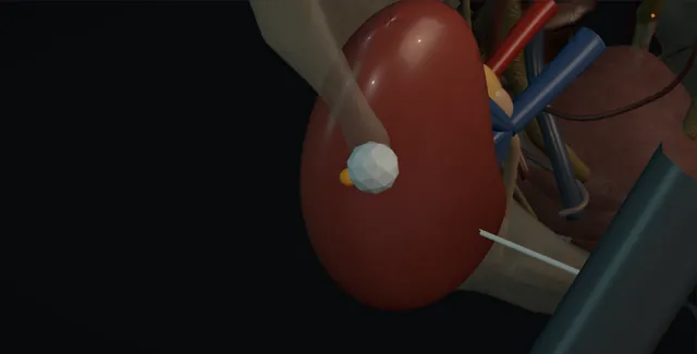
Allograft biopsy: how the structures relateUltrasound over the graft in the iliac fossa, needle from the lateral side. Cortex of the upper or lower pole: probe here; needle enters obliquely from lateral Lateral border under the abdominal wall. Cortex of the upper or lower pole: a deep or central throw tears them Hilum and segmental vessels in the sinus (danger). Cortex of the upper or lower pole: central; a core through a calyx bleeds and clots Collecting system (danger). Cortex of the upper or lower pole: a needle through the medial pole finds them External iliac artery and vein (danger). Cortex of the upper or lower pole: may lie over the graft; not seen, it is transfixed Bowel between skin and graft (danger). Cortex of the upper or lower pole: throw runs tangentially along the cortex Spring-loaded core needle.Cortex of the upper orlower poleLateral border underthe abdominal wall!Hilum and segmentalvessels in the sinus!Collecting system!External iliacartery and vein!Bowel between skinand graftSpring-loaded coreneedleprobe here; needleenters obliquelyfrom laterala deep or centralthrow tears themcentral; a corethrough a calyxbleeds and clotsa needle throughthe medial polefinds themmay lie over thegraft; not seen, itis transfixedthrow runstangentially alongthe cortexTargetDangerLandmarkContext
Ultrasound over the graft in the iliac fossa, needle from the lateral side. Schematic, not to scale; relationships only.
  • The graft sits in the right or left iliac fossa just under the abdominal wall, with its lateral convex border nearest the skin and its hilum turned medially toward the external iliac vessels.
  • The probe lies over the lateral border and the needle enters obliquely from the lateral side to run along the cortex of the upper or lower pole, its throw lying in the pale outer rim and away from the central sinus.
  • The bladder lies medial and below, the bowel medial above, and the iliac vessels behind the medial edge of the kidney.
  • ›The graft in the iliac fossa - extraperitoneal and superficial, its lateral surface a few centimetres under the skin lateral to the rectus.
  • ›The cortex of the poles - the target, taken tangentially so the throw of the needle stays in cortex; ten glomeruli and two arteries make the sample adequate.
  • ›The hilum and the segmental vessels - medial and deep; a needle that crosses the sinus tears an artery and makes a fistula or a pseudoaneurysm.
  • ›The collecting system - central; a needle through a calyx gives haematuria and a clot that can obstruct the transplant ureter.
  • ›The external iliac artery and vein - beneath and medial to the lower pole where the anastomoses lie; out of the path of a tangential polar biopsy.
  • ›Bowel - may lie between the skin and the graft, especially medially or in a thin patient; looked for on the ultrasound before the needle.
  • ›The needle - 16 gauge spring-loaded core, seen along its whole shaft in the plane of the probe, fired only with the throw mapped.
Click me next Section 3 of 8 Steps The operation in order, with the landmark that guards each move.

Operative steps

  1. Who: the creatinine that has risen A rise in creatinine of more than about twenty per cent from the patient's baseline, once the obvious causes have been excluded: dehydration, a calcineurin inhibitor level that is too high, a new drug, obstruction or a collection on ultrasound, infection in the urine, and a vascular problem on Doppler. New proteinuria, haematuria of graft origin, or delayed graft function that has not recovered within about a week of transplantation are the other indications. The differential the biopsy is answering is rejection, T-cell or antibody mediated, calcineurin inhibitor toxicity, BK virus nephropathy, recurrent or de novo glomerular disease, and acute tubular injury; none can be told apart reliably without tissue, and each is treated differently.
  2. Protocol and for-cause biopsies A for-cause biopsy is done for the dysfunction above and is done urgently, within a day or two, because rejection treated late costs nephrons. A protocol biopsy is done at fixed time points in a stable graft, in units that use them, typically at about three months and at a year, to find subclinical rejection or BK nephropathy in a kidney whose creatinine is normal; the case for them is strongest in the sensitised recipient and the marginal kidney. In delayed graft function the biopsy is repeated at intervals until the kidney works, because rejection hides behind acute tubular necrosis. The transplant team decides; the operator confirms the indication and that the result will change management before the needle goes in.
  3. Consent, clotting and the antiplatelets The consent names pain, bleeding into or around the kidney, haematuria, an arteriovenous fistula, the small chance of transfusion or embolisation, injury to bowel or bladder, an inadequate sample, and graft loss, which is rare but real. Blood pressure under about 160 over 90 on the day, platelets above about 100, an INR under about 1.5, and the drugs: aspirin is often continued in many units, clopidogrel stopped about seven days before, low molecular weight heparin omitted the evening before, warfarin bridged, direct oral anticoagulants held according to renal function. Uraemia flattens platelet function, so a patient with a high urea may be given desmopressin. A urine culture that is negative, and the patient is asked to empty the bladder.
    where it bites: a clopidogrel dose nobody asked about, or a blood pressure of 180 on the trolley, is the commonest avoidable cause of a major bleed; the operator checks the drugs and the pressure rather than assuming them.
  4. Ultrasound: the cortex and the vessels The patient lies supine, the graft is scanned with a curvilinear probe and the whole kidney mapped: its poles, its depth from the skin, the cortex thickness, the collecting system and whether it is dilated, any perinephric collection, and the hilum with its vessels on colour Doppler. Bowel loops between the skin and the kidney are looked for, and the iliac vessels beneath and medial to the graft are found. The target is the cortex of the upper or lower pole, whichever is more superficial and further from the hilum, with the needle path tangential to the surface so that it stays in cortex rather than heading for the medulla and the sinus. The entry point is marked and the depth measured.
    where it bites: a needle aimed at the centre of the kidney reaches the sinus, the segmental vessels and the pelvis; the target is the cortex of a pole, tangentially.
  5. The needle: the cortex, away from the hilum Skin cleaned and draped, local anaesthetic infiltrated down to the capsule under ultrasound, a small nick in the skin. A 16 gauge spring-loaded core needle, with or without a coaxial sheath, is advanced under continuous ultrasound guidance with the whole shaft visible in the plane of the probe, to sit on the capsule of the pole with the throw of the needle laid out to run through cortex only. The patient is asked to hold their breath if the graft moves with respiration, which it usually does not. The needle is fired, withdrawn, and the core inspected: cortex is pale and granular, medulla darker and striped, and a core of medulla means the trajectory was too deep.
    where it bites: the hilum and the iliac vessels: a needle that crosses the sinus tears a segmental artery, and a needle that passes through the lower medial pole can reach the external iliac vessels.
  6. Two cores and the pathologist Two cores are the standard; a third if the first two are short or if a transplant pathologist looking at them under the dissecting microscope asks for more. Adequacy for the Banff classification is ten or more glomeruli and at least two arteries, with seven glomeruli and one artery the minimum, and cortex, not medulla, is what counts. The cores are divided at the bedside according to the unit protocol: light microscopy in formalin, immunofluorescence in transport medium or fresh on saline for C4d and immunoglobulins, and a fragment in glutaraldehyde for electron microscopy where glomerular disease is suspected. The request form carries the clinical question, the drug levels, the donor-specific antibody status and the BK viraemia, because the pathologist reads the slides in that light.
  7. After: bed rest and the haematuria Pressure on the site for a few minutes, a dressing, and the patient lies flat for about four to six hours with observations by the early warning score, a check of the urine for blood, and a haemoglobin before discharge in a day-case unit. Microscopic haematuria is expected; frank haematuria in a few per cent settles with fluids in most. Pain that grows over the graft, a fall in blood pressure, a rising pulse or a drop in haemoglobin means bleeding into or around the kidney and is scanned at once. The patient goes home with the number to ring and the instruction to avoid heavy lifting for a week, and the antiplatelet or anticoagulant is restarted according to the plan written before the biopsy.
    where it bites: the bleed declares itself in the first hours; a patient sent home before the rest period is complete and the haemoglobin checked bleeds at home.
  8. The result and the treatment A preliminary read the same day or the next morning by a pathologist using the Banff classification: T-cell mediated rejection graded by tubulitis, interstitial inflammation and arteritis, antibody mediated rejection by microvascular inflammation, C4d staining and donor-specific antibody, borderline changes, interstitial fibrosis and tubular atrophy, calcineurin inhibitor toxicity, BK nephropathy on SV40 staining, and recurrent disease. Treatment follows the diagnosis: pulsed methylprednisolone for T-cell mediated rejection, anti-thymocyte globulin where it is steroid resistant or vascular, plasma exchange, immunoglobulin and rituximab for antibody mediated rejection by unit protocol, a reduction in immunosuppression for BK nephropathy, a dose change for toxicity. The result and the plan are discussed with the patient and recorded.
Click me next Section 4 of 8 Danger zones Where this operation bites, and why it bites there.
Critical relationship

Danger zones

The spatial relationships the animation is built to make automatic. Before you read each one, ask yourself: at this point in the operation, what is about to go wrong, and what is my first move? The save for each sits in the Complications tab.

  • RiskThe segmental vessels in the sinus - a deep or central trajectory tears them; the result is a haematoma today or an arteriovenous fistula in a month.
  • RiskThe hilum and the iliac vessels - medial and deep to the lower pole; a needle passed through the medial pole finds the anastomoses.
  • RiskThe collecting system - central; a core through a calyx bleeds into the urine and clots in the transplant ureter.
  • RiskBowel over the graft - between skin and kidney in the thin or previously operated patient; not seen, it is transfixed.
Practise it Take me into the scene Opens the animation at a danger moment and asks you to predict the mechanism and your first move before it reveals.
Click me next Section 5 of 8 Complications The save for each one: the first move, the escalation trigger, the op-note line.

Complications and immediate saves

Trigger each of these live in the Animation tab, then read the save: the first move, the escalation trigger, and the line that goes in the op note.

Perigraft haematoma and major haemorrhage

  • bleeding from the cortex or a torn segmental vessel into the kidney or around it: growing pain over the graft, a falling blood pressure and haemoglobin in the hours after the needle.
The save
  1. Observations by the early warning score for the whole rest period, pressure over the site, and a haemoglobin before discharge. first move
  2. Pain, tachycardia or a drop in haemoglobin: ultrasound with Doppler at once, and a CT if the picture is unclear.
  3. Correct the clotting and platelets, stop the antiplatelet or anticoagulant, cross-match, and transfuse as needed.
  4. Haemodynamic instability, a haematoma that grows on repeat imaging or a rising creatinine from compression: angiography and embolisation of the bleeding branch, or exploration if that fails or is unavailable. escalation trigger
  5. Procedure note: the needle, the number of passes, the trajectory, the clotting and drugs on the day, and the observation plan. document

Arteriovenous fistula and pseudoaneurysm

  • a needle through an artery and a vein together: a bruit and a colour Doppler mosaic found weeks later, with haematuria, hypertension or a creeping creatinine from steal.
The save
  1. Doppler at the first review after the biopsy if there was haematuria or pain, and at any later unexplained haematuria or rise in creatinine. first move
  2. A small fistula without symptoms is watched with serial Doppler; most close on their own within months.
  3. Control the blood pressure and stop anticoagulants where possible while it is watched.
  4. Persistent haematuria, a pseudoaneurysm, a fistula that enlarges or a graft losing function: selective embolisation by interventional radiology, sacrificing as little cortex as possible. escalation trigger
  5. Note: the Doppler findings, the size, the plan for surveillance and the trigger for embolisation. document

Haematuria and clot obstruction

  • a core through the collecting system: frank haematuria that settles in most, but a clot in the transplant ureter or the bladder that obstructs a single kidney and sends the creatinine up.
The save
  1. Fluids, and the urine watched until it clears; the patient is not sent home passing clots. first move
  2. A fall in urine output or pain over the graft: ultrasound for hydronephrosis of the graft and clot in the bladder.
  3. Clot retention: a large three-way catheter and irrigation, with cystoscopic evacuation if it will not clear.
  4. A dilated graft with a rising creatinine: nephrostomy of the transplant kidney or a retrograde stent, and an urgent Doppler for the source. escalation trigger
  5. Note: the haematuria, the catheter, the ultrasound findings and the creatinine trend. document

Bowel, bladder or iliac vessel injury

  • a loop of bowel between skin and graft, a full bladder medially or the iliac vessels behind the lower pole, transfixed by a needle that was not seen along its whole length.
The save
  1. Map the bowel, the bladder and the iliac vessels on the ultrasound before the needle, with the bladder emptied and the needle kept in the plane of the probe throughout. first move
  2. Bowel content on the needle or peritonism afterwards: antibiotics, nil by mouth, a CT and a surgical opinion.
  3. A bladder puncture is rarely consequential once emptied; a catheter for a day if there is haematuria from it.
  4. A pulsatile groin haematoma, a cold leg or a haemodynamic fall after a medial lower pole pass: an iliac vessel injury; pressure, a vascular surgeon and CT angiography now. escalation trigger
  5. Note: the structures seen on the pre-biopsy scan, the trajectory and any suspicion of transfixion. document

Inadequate or misallocated specimen

  • two cores of medulla, a crushed core, or the wrong fragment in the wrong pot: an invasive procedure with no diagnosis and a rejection treated blind or missed.
The save
  1. Look at each core as it comes out and take a third if it is short or dark; a pathologist or technician at the bedside where the unit has one. first move
  2. Handle cores with a needle tip, never forceps, and divide them for the three media by the written protocol before they dry.
  3. Label every pot at the bedside with a second person, with the clinical question and the drug levels on the form.
  4. A non-diagnostic result in a graft that is deteriorating: repeat the biopsy within days rather than treat empirically, unless the patient is too unstable to wait. escalation trigger
  5. Note: the number of cores, their length and appearance, and which medium each went into. document
Click me next Section 6 of 8 Troubleshooting When the case turns: the problem, then the next move.

Intra-operative troubleshooting

When the case turns: the problem, then the next move.

  • ActionThe graft is deep and the poles cannot be seen clearly - a lower frequency probe, more gel and gentle pressure; if the cortex cannot be seen along the needle path, the biopsy is not done blind and interventional radiology is asked.
  • ActionBowel lies over the lateral border - change the entry point, roll the patient to shift the loop, or scan again with the bladder empty; a needle never crosses bowel.
  • ActionThe core is dark and striped - medulla; the throw was too deep. Re-angle so the needle runs tangentially in the cortex and take another core.
  • ActionBlood pressure 175 over 100 on the trolley - not today, unless the indication is urgent and the team accepts it; treat the pressure and return.
  • ActionThe core is not adequate under the dissecting microscope - a third pass now, while the patient is still draped, rather than a second procedure next week.
  • ActionFrank haematuria as the patient stands to go home - the patient stays; fluids, urine watched, haemoglobin, and an ultrasound with Doppler if it does not clear.
Click me next Section 7 of 8 FRCS viva Say it out loud, then mark yourself against the model answer.

FRCS viva questions

The examiner stems this scene prepares you for. Try to answer out loud first, then reveal, and learn the discriminator, the detail that turns a pass into a strong pass.

QA transplant recipient's creatinine has risen from 120 to 160. What do you do before you biopsy?reveal answer
  • I would treat a rise of that size, more than twenty per cent from baseline, as graft dysfunction until proven otherwise, and I would exclude the causes that do not need tissue before I reached for the needle.
  • History and examination: fluid intake, diarrhoea or vomiting, new drugs including non-steroidals and anything that raises calcineurin inhibitor levels, pain or swelling over the graft, and adherence.
  • Bloods: the tacrolimus or ciclosporin level, electrolytes, a full blood count, CRP, and BK and cytomegalovirus viraemia.
  • Urine: dipstick, culture and a protein to creatinine ratio.
  • Ultrasound of the graft with Doppler: hydronephrosis, a perinephric collection or lymphocele compressing the kidney, and the resistive index and flow in the artery and vein.
  • Donor-specific antibodies would be sent, since they shape the reading of the biopsy.
  • If the level is high I would adjust the drug and repeat the creatinine; if the kidney is obstructed I would drain it; if there is a collection I would deal with it.
  • If none of those explains the rise, the differential is rejection, toxicity, BK nephropathy, recurrent disease or acute tubular injury, and only a biopsy separates them, so I would arrange it within a day or two.
Strong pass

Say all three

  • The calcineurin inhibitor level
  • Obstruction and a collection each excluded by name
  • Donor-specific antibodies sent before the biopsy so the pathologist reads the tissue with them
Examiner note
The calcineurin inhibitor level, obstruction and a collection each excluded by name, and donor-specific antibodies sent before the biopsy so the pathologist reads the tissue with them.
QDescribe how you would perform a percutaneous biopsy of a renal allograft.reveal answer
  • I would confirm the indication and consent, check the blood pressure was below about 160 over 90, platelets above 100 and INR below 1.5, and confirm the antiplatelet and anticoagulant plan had been followed.
  • With the patient supine and the bladder empty, I would scan the graft with a curvilinear probe to map its poles, depth, cortex, collecting system, hilum and vessels on Doppler, and to exclude bowel in the path and a perinephric collection.
  • I would choose the cortex of the more superficial pole, away from the hilum, with a tangential trajectory that keeps the throw of the needle in cortex.
  • After skin preparation and local anaesthetic down to the capsule, I would advance a 16 gauge spring-loaded needle under continuous ultrasound guidance with the whole shaft in the plane of the probe, rest it on the capsule, fire it, and inspect the core: pale granular cortex, not dark medulla.
  • I would take two cores, a third if needed for adequacy, divide them for light microscopy, immunofluorescence and electron microscopy according to protocol with a second person checking the labels, and send them with the clinical question and drug levels.
  • The patient would rest for four to six hours with observations and a urine check, and a haemoglobin before discharge.
Strong pass

Say both

  • The tangential polar trajectory with the reason for it
  • The core inspected for cortex before the second pass
Examiner note
The tangential polar trajectory with the reason for it, and the core inspected for cortex before the second pass.
QWhat are the complications of renal allograft biopsy and how are they managed?reveal answer
  • Bleeding is the main one.
  • Microscopic haematuria is almost universal and frank haematuria occurs in a few per cent, settling with fluids in most.
  • A perigraft or intrarenal haematoma presents with pain, a falling blood pressure and haemoglobin in the hours afterwards;
  • I would image with ultrasound and Doppler, correct clotting and platelets, transfuse if needed, and involve interventional radiology for embolisation if the patient is unstable or the haematoma grows or compresses the kidney.
  • Transfusion or intervention is needed in about one per cent, and graft loss is rare, well under one per cent, figures I would give as approximate.
  • An arteriovenous fistula or pseudoaneurysm follows a needle through an artery and vein together and is found on Doppler as a mosaic with a bruit;
  • most small ones close spontaneously, and those that persist with haematuria, hypertension or falling function are embolised selectively.
  • Clot in the collecting system can obstruct the transplant ureter, which is a single kidney, and needs a catheter, irrigation and sometimes a stent or nephrostomy.
  • Injury to bowel, the bladder or the iliac vessels follows a trajectory not seen along its whole length.
  • Infection is uncommon.
  • And an inadequate specimen, cortex under seven glomeruli, is a complication too, since it leaves the patient with the risk and without the answer.
Strong pass

Say both

  • The clot in a single-kidney ureter recognised as obstruction
  • The inadequate specimen counted as a complication with its Banff threshold
Examiner note
The clot in a single-kidney ureter recognised as obstruction, and the inadequate specimen counted as a complication with its Banff threshold.
QWhat will the pathologist tell you, and how does it change treatment?reveal answer
  • The pathologist reports against the Banff classification, which requires an adequate sample of ten or more glomeruli and at least two arteries.
  • T-cell mediated rejection is graded by the degree of tubulitis and interstitial inflammation, with intimal arteritis marking the vascular grades; borderline changes are inflammation short of the threshold.
  • Antibody mediated rejection needs the triad of microvascular inflammation in the glomeruli and peritubular capillaries, evidence of antibody interaction with the endothelium, classically C4d in the peritubular capillaries, and a circulating donor-specific antibody, which is why the serology is sent with the biopsy.
  • Chronic changes are scored as interstitial fibrosis and tubular atrophy, transplant glomerulopathy and arterial intimal fibrosis.
  • Calcineurin inhibitor toxicity shows isometric vacuolation and arteriolar hyalinosis;
  • BK nephropathy shows viral inclusions with SV40 staining;
  • recurrent or de novo glomerular disease is read with immunofluorescence and electron microscopy.
  • Treatment follows the category: pulsed methylprednisolone for T-cell mediated rejection with anti-thymocyte globulin for steroid-resistant or vascular grades;
  • plasma exchange, intravenous immunoglobulin and rituximab for antibody mediated rejection by unit protocol;
  • a reduction in immunosuppression for BK nephropathy;
  • a dose reduction or switch for toxicity;
  • and disease-specific treatment for recurrence.
  • The result is reviewed with the patient and the plan recorded.
Strong pass

Say both

  • The three-part definition of antibody mediated rejection with donor-specific antibody as the reason serology accompanies the tissue
  • The opposite direction of treatment for rejection and BK nephropathy
Examiner note
The three-part definition of antibody mediated rejection with donor-specific antibody as the reason serology accompanies the tissue, and the opposite direction of treatment for rejection and BK nephropathy.
Click me next Section 8 of 8 Evidence The guidance each of these answers is built on.

Evidence and guidance

Two minutes

Close the lap: recall it before you leave.

Say it aloud, then reveal. Reading is not remembering.

  1. 01 The relationship Say the critical spatial relationship out loud, then reveal. reveal
    Memory frame

    The graft sits in the right or left iliac fossa just under the abdominal wall, with its lateral convex border nearest the skin and its hilum turned medially toward the external iliac vessels.

    Why?
    The probe lies over the lateral border and the needle enters obliquely from the lateral side to run along the cortex of the upper or lower pole, its throw lying in the pale outer rim and away from the central sinus. The bladder lies medial and below, the bowel medial above, and the iliac vessels behind the medial edge of the kidney.
  2. 02 The first move Perigraft haematoma and major haemorrhage. Your first move? reveal
    First move

    Observations by the early warning score for the whole rest period, pressure over the site, and a haemoglobin before discharge.

  3. 03 The escalation trigger Perigraft haematoma and major haemorrhage. What makes you call for help? reveal
    Escalate when

    Haemodynamic instability, a haematoma that grows on repeat imaging or a rising creatinine from compression: angiography and embolisation of the bleeding branch, or exploration if that fails or is unavailable.

  4. 04 The discriminator What turns a pass into a strong pass? reveal
    Strong pass

    Say all three

    • The calcineurin inhibitor level
    • Obstruction and a collection each excluded by name
    • Donor-specific antibodies sent before the biopsy so the pathologist reads the tissue with them
    Examiner note
    The calcineurin inhibitor level, obstruction and a collection each excluded by name, and donor-specific antibodies sent before the biopsy so the pathologist reads the tissue with them.
  5. 05 The pitfall Where does this operation bite first? reveal
    Pitfall

    The segmental vessels in the sinus

    Why?
    a deep or central trajectory tears them; the result is a haematoma today or an arteriovenous fistula in a month.

Revision practice with self-marked answers, not an assessment of clinical competence. Kept in this browser only; it does not sync between devices.